Abstract Background Mobile upright PET devices have the potential to enable previously impossible neuroimaging studies. Currently available options are imagers with deep brain coverage that severely limit head/body movements or imagers with upright/motion enabling properties that are limited to only covering the brain surface. Methods In this study, we test the feasibility of an upright, motion-compatible brain imager, our Ambulatory Motion-enabling Positron Emission Tomography (AMPET) helmet prototype, for use as a neuroscience tool by replicating a variant of a published PET/fMRI study of the neurocorrelates of human walking. We validate our AMPET prototype by conducting a walking movement paradigm to determine motion tolerance and assess for appropriate task related activity in motor-related brain regions. Human participants (n = 11 patients) performed a walking-in-place task with simultaneous AMPET imaging, receiving a bolus delivery of F18-Fluorodeoxyglucose. Results Here we validate three pre-determined measure criteria, including brain alignment motion artifact of less than <2 mm and functional neuroimaging outcomes consistent with existing walking movement literature. Conclusions The study extends the potential and utility for use of mobile, upright, and motion-tolerant neuroimaging devices in real-world, ecologically-valid paradigms. Our approach accounts for the real-world logistics of an actual human participant study and can be used to inform experimental physicists, engineers and imaging instrumentation developers undertaking similar future studies. The technical advances described herein help set new priorities for facilitating future neuroimaging devices and research of the human brain in health and disease.
INTRODUCTION:Sentinel lymph node biopsy (SLNB) is a standard practice for staging cutaneous melanoma. High false-negative rates have an increased interest in adjunctive techniques for localizing SLNs. Mobile gamma cameras (MGCs) represent potential tools to enhance SLNB performance. METHODS:An institutional review board approval was obtained for this study (ClinicalTrials.gov ID NCT01531608). After obtaining informed consent, 20 eligible melanoma patients underwent 99mTc sulfur colloid injection and standard lymphoscintigraphy with a fixed gamma camera (FGC). A survey using a 20 cm square MGC, performed immediately preoperatively by the study surgeon, was used to establish an operative plan while blinded to the FGC results. Subsequently, SLNB was performed using a gamma probe and a novel 6 cm diameter handheld MGC. RESULTS:A total of 24 SLN basins were detected by FGC. Prior to unblinding, all 24 basins were identified with the preoperative MGC and the operative plan established by preoperative MGC imaging was confirmed accurate by review of the FGC images. All individual sentinel lymph nodes were identified during intraoperative MGC imaging, and in 5/24 (21%) cases, surgeon-reported additional clinically useful information was obtained from the MGC. CONCLUSIONS:Preoperative MGC images provide information consistent with FGC images for planning SLNB and in some cases provide additional information that aided in surgical decision-making.
Background Dedicated, breast-specific positron emission tomography (BPET)-cone-beam computed tomography (BPET/CT) systems have been developed to improve detection and diagnosis of cancer in women with indeterminate mammograms caused by radiodense breasts. The absorption of X-rays that often vexes mammography in this subset of women does not affect the detection of the high energy annihilation photons used in PET. PET imaging of the breast, however, is subject to limitations caused by their comparatively low spatial resolution (similar to 2 mm) and often moderate radiotracer uptake in lesions. Purpose The purpose of this investigation is to explore the PET-based lesion detection capabilities of a BPET/CT scanner developed by the Department of Radiology Instrumentation group at West Virginia University. Methods The PET component of the system consists of a rotating pair of 96 x 72 arrays of 2 x 2 x 15 mm(3) LYSO scintillator elements. The cone-beam-CT component utilized a pulsed X-ray source and flat panel detector operated in portrait orientation. The density maps created by the CT scanner were used to correct the BPET data for photon attenuation and Compton scattering. The nonuniform uptake of F-18-fluorodeoxyglucose (FDG) in normal breast tissue was emulated in a specially designed phantom consisting of an acrylic cylinder filled with a mixture of acrylic beads and liquid containing FDG. FDG-avid lesions were simulated with agar spheres (3, 4, 6, 8, and 10 mm diameters) containing vary amounts of FDG to produce target-to-background ratios (TBR) of 6:1, 8:1, and 10:1. The spheres also contained X-ray contrast agent to make even the smallest ones readily visible in CT images. Positions of all the lesions were identified in the CT images. These positions were used to extract signal present and signal absent sub-images from the PET images. The sub-images were then input to software that calculated areas-under-the-curve for two numerical model observers (Laguerre-Gauss channelized Hotelling observer and non-prewhitening matched filter). Results The results showed that the smallest detectable lesion with this system is no smaller than similar to 3 mm in diameter with a TBR of 6:1. Simulated lesions with diameters of 4 mm and greater were calculated to have good to excellent likelihood of detection for all TBRs tested. Conclusion The results from this investigation identified the detectability capabilities and limitations for a dedicated breast-PET/CT scanner. Its ability to detect relatively small simulated FDG-avid breast lesions for a range of TBRs indicates its potential for clinical application. Finally, the study used methodologies that could be applied to a detectability assessment of other PET/CT scanners.
We describe the design and performance of BRPET, a novel dedicated breast PET (dbPET) scanner designed to maximize visualization of posterior regions of the breast. BRPET uses prone imaging geometry and a 12-module detector ring built from pixelated LYSO crystals coupled to position sensitive photomultiplier tubes (PSPMTs). Optical coupling via slanted plastic fiber optic light guides permits partial insertion of the crystals into the exam table’s breast aperture. Image quality testing procedures were adapted from the NEMA NU4-2008 protocol. Two additional phantom tests quantified the posterior extent of the usable volume of view (VoV). BRPET axial, radial, and tangential FWHM spatial resolutions at the isocenter were 1.8, 1.7, and 1.9 mm, respectively. The peak absolute system sensitivity was 0.97% using an energy window of 460–562 keV. The peak noise equivalent counting rate was 5.33 kcps at 21.6 MBq. The scanner VoV extends to within ~6 mm of the plane defining the location of the chest wall. A pilot human study (n = 10) compared the diagnostic performance of FDG-BRPET to that of contrast enhanced MRI (CEMRI), with biopsy as ground truth. Averaged over three expert human observers, the sensitivity/specificity for BRPET was 0.93/1.0, compared to 1.0/0.25 for CEMRI.
PET scanners based on monolithic pieces of scintillator can potentially produce superior performance characteristics (high spatial resolution and detection sensitivity, for example) compared to conventional PET scanners. Consequently, we initiated development of a preclinical PET system based on a single 7.2 cm long annulus of LYSO, called AnnPET. While this system could facilitate creation of high-quality images, its unique geometry results in optics that can complicate estimation of event positioning in the detector. To address this challenge, we evaluated deep-residual convolutional neural networks (DR-CNN) to estimate the three-dimensional position of annihilation photon interactions. Monte Carlo simulations of the AnnPET scanner were used to replicate the physics, including optics, of the scanner. It was determined that a ten-layer-DR-CNN was most suited to application with AnnPET. The errors between known event positions, and those estimated by this network and those calculated with the commonly used center-of-mass algorithm (COM) were used to assess performance. The mean absolute errors (MAE) for the ten-layer-DR-CNN-based event positions were 0.54 mm, 0.42 mm and 0.45 mm along the x (axial)-, y (transaxial)- and z- (depth-of-interaction) axes, respectively. For COM estimates, the MAEs were 1.22 mm, 1.04 mm and 2.79 mm in the x-, y- and z-directions, respectively. Reconstruction of the network-estimated data with the 3D-FBP algorithm (5 mm source offset) yielded spatial resolutions (full-width-at-half-maximum (FWHM)) of 0.8 mm (radial), 0.7 mm (tangential) and 0.71 mm (axial). Reconstruction of the COM-derived data yielded spatial resolutions (FWHM) of 1.15 mm (radial), 0.96 mm (tangential) and 1.14 mm (axial). These findings demonstrated that use of a ten-layer-DR-CNN with a PET scanner based on a monolithic annulus of scintillator has the potential to produce excellent performance compared to standard analytical methods.
Time-of-flight (TOF) capability is becoming an important capability offered in both commercial and research PET scanners. Often commercial vendors and laboratory researchers develop and utilize proprietary electronics for their devices. Consequently, it is challenging for independent research groups to develop their own TOF-PET scanners. In this investigation, we tested a prototype scanner consisting of commercially available TOF-capable modules from Hamamatsu photonics that can be used as building blocks for PET scanners. The scanner consists of a ring of 16 modules, for a total diameter of 26.7 cm. Testing demonstrated that the scanner is capable of sustaining similar to 1 MHz single counting rate with a peak noise equivalent count rate of 117.5 kHz at 75.25 MBq measured with NEMA NU-4 "rat" phantom. Spatial resolution of 2.3-5 mm from the center of the scanner was measured. Energy resolution of 17.2% at 511 keV was measured. Peak sensitivity of 1.28% is reported. All the measurements were performed with energy cuts from 350 to 700 keV. Finally, scanner timing resolution was found to be 462 ps. Results from testing of a prototype scanner constructed using newly released TOF-capable detector modules produced by Hamamatsu demonstrated the promise for these devices to create high performance PET system with TOF capabilities.
PurposeApplication of advanced imaging techniques, such as PET and x ray CT, can potentially improve detection of breast cancer. Unfortunately, both modalities have challenges in the detection of some lesions. The combination of the two techniques, however, could potentially lead to an overall improvement in diagnostic breast imaging. The purpose of this investigation is to test the basic performance of a new dedicated breast-PET/CT. MethodsThe PET component consists of a rotating pair of detectors. Its performance was evaluated using the NEMA NU4-2008 protocols. The CT component utilizes a pulsed x ray source and flat panel detector mounted on the same gantry as the PET scanner. Its performance was assessed using specialized phantoms. The radiation dose to a breast during CT imaging was explored by the measurement of free-in-air kerma and air kerma measured at the center of a 16 cm-diameter PMMA cylinder. Finally, the combined capabilities of the system were demonstrated by imaging of a micro-hot-rod phantom. ResultsOverall, performance of the PET component is comparable to many pre-clinical and other dedicated breast-PET scanners. Its spatial resolution is 2.2 mm, 5 mm from the center of the scanner using images created with the single-sliced-filtered-backprojection algorithm. Peak NECR is 24.6 kcps; peak sensitivity is 1.36%; the scatter fraction is 27%. Spatial resolution of the CT scanner is 1.1 lp/mm at 10% MTF. The free-in-air kerma is 2.33 mGy, while the PMMA-air kerma is 1.24 mGy. Finally, combined imaging of a micro-hot-rod phantom illustrated the potential utility of the dual-modality images produced by the system. ConclusionThe basic performance characteristics of a new dedicated breast-PET/CT scanner are good, demonstrating that its performance is similar to current dedicated PET and CT scanners. The potential value of this system is the capability to produce combined duality-modality images that could improve detection of breast disease. The next stage in development of this system is testing with more advanced phantoms and human subjects.
Abstract. Development of advanced preclinical imaging techniques has had an important impact on the field of biomedical research, with positron emission tomography (PET) imaging the most mature of these efforts. Developers of preclinical PET scanners have joined the recent multimodality imaging trend by combining PET imaging with other modalities, such as magnetic resonance imaging (MRI). Our group has developed a combined PET-MRI insert for the imaging of animals up to the size of rats in a clinical 3T MRI scanner. The system utilizes a sequential scanner configuration instead of the more common coplanar geometry. The PET component of the system consists of a ring of 12 liquid-cooled, SiPM-based detector modules (diameter = 15.2 cm). System performance was evaluated with the NEMA NU 4-2008 protocol. Spatial resolution is ∼1.71 mm 5 cm from the center of the field-of-view measured from single-slice rebinned filtered backprojection-reconstructed images. Peak noise equivalent count rate is 17.7 kcps at 8.5 MBq; peak sensitivity is 2.9%. The MRI component of the system is composed of a 12-cm-diameter birdcage transmit/receive coil with a dual-preamplifier interface possessing very low noise preamplifiers. System performance was evaluated using American College of Radiology-based methods. Image homogeneity is 99%; the ghosting ratio is 0.0054. The signal-to-noise ratio is 95 and spatial resolution is ∼0.25 mm. There was no discernable cross-modality interference.
The advent of hybrid scanners, combining complementary modalities, has revolutionized the application of advanced imaging technology to clinical practice and biomedical research. In this project, we investigated the melding of two complementary, functional imaging methods: positron emission tomography (PET) and electron paramagnetic resonance imaging (EPRI). PET radiotracers can provide important information about cellular parameters, such as glucose metabolism. While EPR probes can provide assessment of tissue microenvironment, measuring oxygenation and pH, for example. Therefore, a combined PET/EPRI scanner promises to provide new insights not attainable with current imagers by simultaneous acquisition of multiple components of tissue microenvironments. To explore the simultaneous acquisition of PET and EPR images, a prototype system was created by combining two existing scanners. Specifically, a silicon photomultiplier (SiPM)-based PET scanner ring designed as a portable scanner was combined with an EPRI scanner designed for the imaging of small animals. The ability of the system to obtain simultaneous images was assessed with a small phantom consisting of four cylinders containing both a PET tracer and EPR spin probe. The resulting images demonstrated the ability to obtain contemporaneous PET and EPR images without cross-modality interference. Given the promising results from this initial investigation, the next step in this project is the construction of the next generation pre-clinical PET/EPRI scanner for multi-parametric assessment of physiologically-important parameters of tissue microenvironments.
Several applications exist for a whole brain positron-emission tomography (PET) brain imager designed as a portable unit that can be worn on a patient's head. Enabled by improvements in detector technology, a lightweight, high performance device would allow PET brain imaging in different environments and during behavioral tasks. Such a wearable system that allows the subjects to move their heads and walk-the Ambulatory Microdose PET (AM-PET)-is currently under development. This imager will be helpful for testing subjects performing selected activities such as gestures, virtual reality activities and walking. The need for this type of lightweight mobile device has led to the construction of a proof of concept portable head-worn unit that uses twelve silicon photomultiplier (SiPM) PET module sensors built into a small ring which fits around the head. This paper is focused on the engineering design of mechanical support aspects of the AM-PET project, both of the current device as well as of the coming next-generation devices. The goal of this work is to optimize design of the scanner and its mechanics to improve comfort for the subject by reducing the effect of weight, and to enable diversification of its applications amongst different research activities.
Positron emission tomography (PET) scanners designed for imaging of small animals have transformed translational research by reducing the necessity to invasively monitor physiology and disease progression. Virtually all of these scanners are based on the use of pixelated detector modules arranged in rings. This design, while generally successful, has some limitations. Specifically, use of discrete detector modules to construct PET scanners reduces detection sensitivity and can introduce artifacts in reconstructed images, requiring the use of correction methods. To address these challenges, and facilitate measurement of photon depth-of-interaction in the detector, we investigated a small animal PET scanner (called AnnPET) based on a monolithic annulus of scintillator. The scanner was created by placing 12 flat facets around the outer surface of the scintillator to accommodate placement of silicon photomultiplier arrays. Its performance characteristics were explored using Monte Carlo simulations and sections of the NEMA NU4-2008 protocol. Results from this study revealed that AnnPET's reconstructed spatial resolution is predicted to be [Formula: see text] full width at half maximum in the radial, tangential, and axial directions. Peak detection sensitivity is predicted to be 10.1%. Images of simulated phantoms (mini-hot rod and mouse whole body) yielded promising results, indicating the potential of this system for enhancing PET imaging of small animals.
Mice are the perhaps the most common species of rodents used in biomedical research, but many of the current generation of small animal PET scanners are non-optimal for imaging these small rodents due to their relatively low resolution. Consequently, a number of researchers have investigated the development of high-resolution scanners to address this need. In this investigation, the design of a novel, high-resolution system based on the dual-detector, virtual-pinhole PET concept was explored via Monte Carlo simulations. Specifically, this system, called TandemPET, consists of a 5 cm × 5 cm high-resolution detector made-up of a 90 × 90 array of 0.5 mm × 0.5 × 10 mm (pitch = 0.55 mm) LYSO detector elements in coincidence with a lower resolution detector consisting of a 68 × 68 array of 1.5 mm × 1.5 mm × 10 mm LYSO detector elements (total size = 10.5 cm × 10.5 cm). Analyses indicated that TandemPET's optimal geometry is to position the high-resolution detector 3 cm from the center-of-rotation, with the lower resolution detector positioned 9 cm from center. Measurements using modified NEMA NU4-2008-based protocols revealed that the spatial resolution of the system is ~0.5 mm FWHM, after correction of positron range effects. Peak sensitivity is 2.1%, which is comparable to current small animal PET scanners. Images from a digital mouse brain phantom demonstrated the potential of the system for identifying important neurological structures.
A new low profile gamma camera is being developed for use in a dual modality (x-ray transmission and gamma-ray emission) tomosynthesis system. Compared to the system’s current gamma camera, the new camera has a larger field of view (~20x25 cm) to better match the system’s x-ray detector (~23x29 cm), and is thinner (7.3 cm instead of 10.3 cm) permitting easier camera positioning near the top surface of the breast. It contains a pixelated NaI(Tl) array with a crystal pitch of 2.2 mm, which is optically coupled to a 4x5 array of Hamamatsu H8500C position sensitive photomultiplier tubes (PSPMTs). The manufacturer-provided connector board of each PSPMT was replaced with a custom designed board that a) reduces the 64 channel readout of the 8x8 electrode anode of the H8500C to 16 channels (8X and 8Y), b) performs gain non-uniformity correction, and c) reduces the height of the PSPMT-base assembly, 37.7 mm to 27.87 mm. The X and Y outputs of each module are connected in a lattice framework, and at two edges of this lattice, the X and Y outputs (32Y by 40X) are coupled to an amplifier/output board whose signals are fed via shielded ribbon cables to external ADCs. The camera uses parallel hole collimation. We describe the measured camera imaging performance, including intrinsic and extrinsic spatial resolution, detection sensitivity, uniformity of response, energy resolution for 140 keV gamma rays, and geometric linearity.
1952 Objectives One challenge of pre-clinical PET imaging is the amount of scan time required to produce high quality, high-resolution images. This issue is important because many pre-clinical studies require the rapid scanning of numerous animals. To create a very high sensitivity and high resolution pre-clinical PET scanner, one based on a single scintillator crystal formed into an annulus is proposed. The objective of this study was to evaluate, via simulation, the potential performance of this system. Methods The simulated scanner consisted of a 72.7mm long cylindrical piece of LYSO crystal with a diameter of 75 mm. The cylinder was formed into an annulus by machining a hole (diameter of 50mm) in the center of the scintillator. Twelve flat surfaces were formed on the outer surface of the annulus to accommodate light detection devices. Specifically, an array of 5 by 17 discrete SiPMs with a size of 3mmx3mm (pitch of 4.1mm, similar to the SensL C-series SiPMs) was attached to each facet. Evaluation of scanner performance was carried out in three steps. First, GATE simulation software was used to obtain a list mode distribution of detected 511keV photons. Next, a trial GATE data set was used in conjunction with the DETECT2000 optical simulation software to explore light transport in the detector. From these results, the three-dimensional positions of the simulated positron annihilations in the scintillator were calculated from the detected optical photons distribution detected at the surfaces of the SiPMs (depth of the events were estimated by the shape of the light distribution). Calculated positions were compared to original emission points to determine spatial accuracy of the event reconstruction. Finally, the data were histogrammed into 100 radial and 180 angular bins and reconstructed using the SSRB-FBP algorithm. Images from a point source and the brain section of the MOBY phantom were created. Data from the point source were used to measure spatial resolution and detection sensitivity. Results Event positioning was accurate to within 1.9mm in the tangential and radial directions. Interaction depth resolution was ~4.4mm. Detection sensitivity at the center of the scanner was 10.3% which is more than twice of any pre-clinical scanner of comparable axial size and diameter. Spatial resolution at the center of the scanner was 1.04mm FWHM. Finally, images of the MOBY mouse brain phantom resulted in good results for FBP reconstruction. Conclusions The results from this initial study demonstrated the promise of a solid, annular pre-clinical PET scanner.
The desire to understand normal and disordered human brain of upright, moving persons in natural environments motivates the development of an ambulatory micro-dose brain PET imager (AMPET) [1]. An ideal system would be light weight and have high sensitivity and spatial resolution. These requirements are often in conflict with each other. Therefore, we performed simulation studies to search for the optimal system configuration and to evaluate the improvement in performance over existing scanners. An intuitive design to achieve high sensitivity is to use a tight geometry that covers the brain. However, a tight geometry also increases parallax error in peripheral lines of response, which may increase the variance in ROI quantification.
Background: Positron Emission Tomography (PET) is traditionally used to image patients in restrictive positions, with few devices allowing for upright, brain-dedicated imaging. Our team has explored the concept of wearable PET imagers which could provide functional brain imaging of freely moving subjects. To test feasibility and determine future considerations for development, we built a rudimentary proof-of-concept prototype (Helmet_PET) and conducted tests in phantoms and four human volunteers.Methods: Twelve Silicon Photomultiplier-based detectors were assembled in a ring with exterior weight support and an interior mechanism that could be adjustably fitted to the head. We conducted brain phantom tests as well as scanned four patients scheduled for diagnostic F(18-)FDG PET/CT imaging. For human subjects the imager was angled such that field of view included basal ganglia and visual cortex to test for typical resting-state pattern. Imaging in two subjects was performed similar to 4hr after PET/CT imaging to simulate lower injected F(18-)FDG dose by taking advantage of the natural radioactive decay of the tracer (F-18 half-life of 110min), with an estimated imaging dosage of 25% of the standard.Results: We found that imaging with a simple lightweight ring of detectors was feasible using a fraction of the standard radioligand dose. Activity levels in the human participants were quantitatively similar to standard PET in a set of anatomical ROIs. Typical resting-state brain pattern activation was demonstrated even in a 1min scan of active head rotation.Conclusion: To our knowledge, this is the first demonstration of imaging a human subject with a novel wearable PET imager that moves with robust head movements. We discuss potential research and clinical applications that will drive the design of a fully functional device. Designs will need to consider trade-offs between a low weight device with high mobility and a heavier device with greater sensitivity and larger field of view.
1540 Objectives As part of the BRAIN initiative, we are designing a mobile molecular brain imager that can be used on healthy subjects to study the functioning of the human brain during motion. The Ambulatory Micro-Dose, Wearable PET Brain Imager (AMPET) must be as light as possible while still providing adequate sensitativity and resolution. We report here on initial simulations studies to quantify the AMPET design trade-offs. Methods We used the PET simulation system SimSET to model effects of AMPET design. Although SimSET provides fast and accurate simulations, it is limited to cylindrical volumes and surfaces for PET scanners. We added a new processing layer wherein an outer 9virtual target9 cylinder is used to capture all potential annihiliation photon detection events. These events are then projected back to the AMPET detector surface, which is modelled as a truncated hemisphere. After verification we evaluated the relative coicidence efficiency of three designs for AMPET (radius 12 cm, axial extent of truncated hemisphere of 4 6 and 8 cm, no confounding effects, 100,000 decays). For input we used the 3D Hoffman brain digital reference object (DRO). The DRO mimics 18F-FDG uptake in normal brain, but can be modified to represent uptake of other neuro radiotracers. Results The new approach of simulation of non-cylindrical scanners was verified. Using this approach the base efficiency of the AMPET designs were estimated. Doubling the axial extent of truncated hemisphere from 4 to 8 cm would more than triple the coincidence effeciency (3.5% to 11.4%), but would also double the weight, unless detector thickness is reduced. Conclusions Using simulations of non-cylindrical scanners using the 3D Hoffman brain digital reference object (DRO), we are able to evaluate the performance and design trade-offs for AMPET to optimze the trade-offs between mobility, sensitivity, and resolution. Research Support Suporrted by grant R24 MH106057
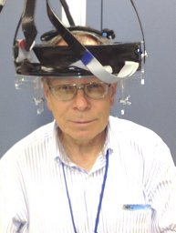 Stan Majewski合作论文数Department of Physics, University of Florida37
 Raymond Raylman合作论文数26
 James Proffitt合作论文数18Andrew G. Weisenberger合作论文数thomas jefferson national accelerator facility17Peter F Martone合作论文数13McKisson, J.E.合作论文数12
 Mark B Williams合作论文数12Jaliparthi, G.合作论文数10
 Julie A. Brefczynski-Lewis合作论文数10S. Majewski合作论文数9John Mckisson合作论文数8
 Christopher Edward Bauer合作论文数Department of Neuroscience, Lexington, KY, USA.7m f smith合作论文数6Brian Kross合作论文数6
 Calvin R. Howell合作论文数Trinity College of Arts & Sciences, Duke University5
 Wenze Xi合作论文数5Mark F. Smith合作论文数5James S. Goddard合作论文数5Benjamin L. Welch合作论文数5
 J.s. Baba合作论文数4
 Pablo Conde合作论文数4Lewis James W合作论文数4Lee Seungjoon合作论文数4
 José María Benlloch Baviera合作论文数Instituto De Instrumentación Para Imagen Molecular4Alexander S. Crowell合作论文数4
 Antonio Javier Gonzalez Martinez合作论文数4C. Zorn合作论文数4
 Jinyi Qi (祁锦毅)合作论文数Department of Biomedical Engineering, College of Engineering, University of California, Davis;Lawrence Berkeley National Laboratory4c d reid合作论文数3s j lee合作论文数3
 V. Popov合作论文数University of Iowa3
 Liczandro Hernandez合作论文数3Judy, P.G.合作论文数3Filomeno Sánchez合作论文数Nuclear Medical Physics Group, IFIC Institute3
 Vishwesh Patil合作论文数3Neal H. Clinthorne合作论文数3
 Ban-An Khaw合作论文数Center for Drug Targeting and Analysis, Bouvé College of Pharmacy and Health Sciences3Mitali J. More合作论文数Department of Radiology, University of Virginia3R. Pani合作论文数2B. K. Kundu合作论文数2
 Andrew Polemi合作论文数2Mb Williams合作论文数2Mary Beth Mandich合作论文数2keith vaigneur合作论文数2John W. Murphy合作论文数2
 Samantha Melroy合作论文数2
 Kuang Gong合作论文数Univ Calif Davis, Dept Biomed Engn, Davis, CA 95616 USA2
 Marco Bettiol合作论文数Dept Med Phys, IRCCS Ist Tumori Giovanni Paolo II2




