The introduction of silicon photomultipliers (SiPM) has facilitated construction of compact, efficient and magnetic field-hardened positron emission tomography (PET) scanners. To take full advantage of these devices, methods for using them to produce large field-of-view PET scanners are needed. In this investigation, we explored techniques to combine two SiPM arrays to form the building block for a small animal PET scanner. The module consists of a 26 × 58 array of 1.5 × 1.5mm2 LYSO elements (spanning 41 × 91mm2) coupled to two SensL SiPM arrays. The SiPMs were read out with new multiplexing electronics developed for this project. To facilitate calculation of event position with multiple SiPM arrays it was necessary to spread scintillation light amongst a number of elements with a small light guide. This method was successful in permitting identification of all detector elements, even at the seam between two SiPM arrays. Since the performance of SiPMs is enhanced by cooling, the detector module was fitted with a cooling jacket, which allowed the temperature of the device and electronics to be controlled. Testing demonstrated that the peak-to-valley contrast ratio of the light detected from the scintillation array was increased by ∼45% when the temperature was reduced from 28 °C to 16 °C. Energy resolution for 511 keV photons improved slightly from 18.8% at 28 °C to 17.8% at 16 °C. Finally, the coincidence timing resolution of the module was found to be insufficient for time-of-flight applications (∼2100 ps at 14 °C). The first use of these new modules will be in the construction of a small animal PET scanner to be integrated with a 3T clinical magnetic resonance imaging scanner.
This paper describes the development of a hand-held gamma camera for intraoperative surgical guidance that is based on silicon photomultiplier (SiPM) technology. The camera incorporates a cerium doped lanthanum bromide ( LaBr3:Ce) plate scintillator, an array of 80 SiPM photodetectors and a two-layer parallel-hole collimator. The field of view is circular with a 60 mm diameter. The disk-shaped camera housing is 75 mm in diameter, approximately 40.5 mm thick and has a mass of only 1.4 kg, permitting either hand-held or arm-mounted use. All camera components are integrated on a mobile cart that allows easy transport. The camera was developed for use in surgical procedures, including determination of the location and extent of primary carcinomas, detection of secondary lesions, and sentinel lymph node biopsy (SLNB). Here, we describe the camera design and its principal operating characteristics, including spatial resolution, energy resolution, sensitivity uniformity, and geometric linearity. The gamma camera has an intrinsic spatial resolution of 4.2 mm FWHM, an energy resolution of 21.1% FWHM at 140 keV, and a sensitivity of 481 and 73 cps/MBq when using the single- and double-layer collimators, respectively.
Recent advances in the development of silicon photomultipliers (SiPM) offer new opportunities for medical imaging applications. Specifically, novel imaging devices for positron emission (PET) and single photoemission computed tomography (SPECT) are becoming feasible. In this investigation, we tested a monolithic array of new generation SiPMs, an SB-8 array from SensL. 8×8 array of 6-mm square SiPMs was studied with two different multiplexing readouts, 4- and 16-channels. SB-8 detector was coupled to various scintillation arrays and resulting prototypes were evaluated. Testing demonstrated that the new device is capable of resolving 1 mm LYSO and 1.5 mm NaI crystals with 16-channel readout and 1.57 mm LYSO and 3 mm NaI crystals with 4-channel readout. Energy resolution of approximately 15 % at 511 keV and 19 % at 122 keV were obtained with LYSO and NaI crystals respectively. Timing resolution of 1.52 ns was measured with 1.57 mm LYSO array and the 16-channel readout. It is concluded that new B-series SiPMs from SensL are suitable for use in high performance nuclear medicine particle detectors.
Gamma ray detectors merging continuous and pixelated crystal arrays are described. We show a pilot study about the detector performance of an innovative scintillation design that simultaneously uses both a continuous and a crystal array. This novel idea is the base of a high-resolution and high-sensitivity gamma ray detector. Alternatively monolithic or crystal arrays can be mounted on the front or in between this and the photosensor to improve spatial or photon depth of interaction resolution, without sensitivity detriment. We have successfully tested a total scintillation thickness of ~20 mm. A new photosensor and readout electronics were developed for this purpose. We built a large array of 12X12 SiPMs (SensL) and also 16X16 MPPCs (Hamamatsu). Two readout electronics have been used, one based on 3 scalable ASICs for the array of 144 SiPMs and one based on a diode circuit that provides signals for each row and column (16+16). A center of gravity algorithm is used in this pilot experiment. We have already tested several configurations (1.5X1.5 mm 2 and 2X2 mm 2 ) with crystal arrays in the front, followed by a monolithic painted black scintillator of 12 mm thickness (50X50 mm 2 base). An energy resolution as good as 13% was found for single pixels.
Our research group constructed a 12-module PET detection ring composed of Hamamatsu multi-pixel photon counter (MMPC) silicon photomultiplier (SiPM) detectors placed in a ring that is fully MRI compatible. This brain imager can be placed around the head of a patient (clinical setting) or subject (research setting) and allow for comfortable upright imaging. However, an alternative way to use this device, as enabled by the technology, is to indeed scan individuals in the supine position in conjuncture with current MRI systems. This PET prototype is able to image simultaneously as the MRI scan is occurring, thus maximizing co-registration and the accuracy of the assignment of metabolically active voxels to their anatomically correct counterparts as identified by the MR image. In this study, we first conducted some basic instrumentation tests to ensure the device was functioning properly outside of the MRI, and additionally scan some phantoms (Derenzo; Hoffman Brain) to assess the quality in which our brain imager is able to produce adequate images. After these initial studies, we conducted multiple different experiments inside a 3 Tesla MRI in order to see how the magnetic field would influence the operation of the PET imager and vice versa. Through simultaneous PET/MRI scanning of a Hoffman brain phantom filled with F-18 radioactivity and water, it was shown that the quality of the MR image was largely unaffected by the PET imager. Furthermore, although the quality of PET imaging was affected by the RF pulsing, an acceptable PET image was nevertheless produced. As we discuss following the results, the success of this study shows that our brain imager is indeed MR compatible, and that next generation devices based on its concepts will continue to improve combined PET/MRI functionality. As two of the major advantages of this imager are the potential for low-dose scanning and its adaptability to any MRI scanner, this study suggests that PET/MRI brain imaging with low dose is in principle possible using an insert which could be adapted to any MRI scanner, using standard RF coils for that scanner.
We are developing the charge division (“resistive”) readout for several arrangements of Silicon Photomultiplier (SiPM) arrays, based on devices from Hamamatsu and SensL. The challenge with the SiPM arrays, as opposed to position sensitive photomultipliers (PSPMTs), is that the noise level is known to be high, and signal to noise ratio (S/N) is lower than in PMTs. In addition, the S/N decreases quickly with the increasing size of the module and with increasing temperatures. Key parameters to optimize are: size and coverage of the SiPM arrays, operational temperature (potential necessity of introducing system cooling), and bias voltage. All these parameters have impact on the S/N, and in consequence on the spatial resolution and the energy resolution of the detector modules. Our somewhat arbitrary but practical goal is to achieve operation similar to the one offered by H8500/H9500 flat panel PSPMTs when using LYSO scintillation arrays in applications to small PET imaging modules. Ultimately we would like to use the reduced channel number readout in the depth-of interaction (DOI) modules. Our first application is to construct ~5cm×5cm compact PET modules for the HelmetPET brain imager prototype under construction at WVU.
We are developing a dedicated high resolution (sub-mm), high efficiency, and very fast (with live reconstruction) prostate PET imager composed of an endorectal PET probe and two PET panel modules placed close to the patient on the opposite side of the prostate, and operating in coincidence with the probe. The immediate live image feedback will be primarily useful in biopsy guidance. PET images will be co-registered with the images from the Transrectal Ultrasound (TRUS) probe that will provide the usual structural 2D or 3D information, while the PET imager will provide the metabolic information related to the biological state of the prostate. We are reporting on preliminary data acquired with the prototype imager. The major highlight is that we are achieving ~1mm FWHM DOI resolution with the PET probe, using new monolithic MPPC arrays from Hamamatsu. But even with a non-DOI probe, we obtained good performance in this limited angle tomography problem.
We have tested a concept of a mini gamma camera based on monolithic arrays of MPPCs from Hamamatsu. CsI(Tl), and Cs(Na) arrays and a thin scintillation GSO plate were tested with 122 keV gammas from 57Co sources. The planned application requires placement of this mini-camera in an endorectal probe and thus needs to be very compact and possess high spatial resolution. The high sensitivity and high granularity collimator and gamma shield made out composite material (tungsten powder with epoxy) completes the detector package. We are developing the dual modality (hybrid) imaging prostate probe combining in one compact device a high resolution and high efficiency single gamma imager with an Ultrasound (US) sensor. The US component will typically provide not only the usual structural 3D information, as the standard TransRectal Ultrasound (TRUS) probe, but also the tissue differentiating information through proper US signal analysis, such as elastography. The mini gamma probe will provide the direct metabolic information related to the biological state of the prostate and specifically about the presence of any cancerous structures exhibiting increased metabolic activity, when used with the single gamma labeled dedicated imaging agents for prostate cancer. In addition to cancer diagnosis, the dual-modality Gamma/US prostate probe can be used in biopsy and in surgical guidance.
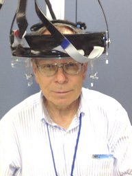
We are developing the HelmetPET, a wearable human PET brain imager which has the potential application of evaluating brain function utilizing PET based radiopharmaceuticals in standing, balancing or moving patients. The HelmetPET is composed of two rings of radiation detectors together providing a cylindrical reconstructed volume with an axial length of 5 cm. Each ring is composed of twenty 2.5 cm2 silicon photomultiplier (SiPM) based detector modules. Each detector module is composed of a 5×5 array of twenty-five Hamamatsu S10362-33-050P Multi Pixel Photon Counters (MPPCs). The 3 mm2 MPPCs are arranged on a 5mm step. Coupled to each of the MPPC modules is a LYSO scintillator crystal array coupled to the MPPC array using to two different LYSO pixel arrays: 1.0×1.0×10 mm3 and 1.5×1.5×10 mm3. The current phase of the project is to equip the forty 2.5 cm2 detector modules with resistive readout and assemble them in a helmet type head support and suspend from a flexible mechanical mount.
Introduction: We describe a compact, portable dual-gamma camera system (named "MONICA" for MObile Nuclear Imaging CAmeras) for visualizing and analyzing the whole-body biodistribution of putative diagnostic and therapeutic single photon emitting radiotracers in animals the size of mice.Methods: Two identical, miniature pixelated Na(TI) gamma cameras were fabricated and installed "looking up" through the tabletop of a compact portable cart. Mice are placed directly on the tabletop for imaging. Camera imaging performance was evaluated with phantoms and field performance was evaluated in a weeklong In-111 imaging study performed in a mouse tumor xenograft model.Results: Tc-99m performance measurements, using a photopeak energy window of 140 keV +/- 10%, yielded the following results: spatial resolution (FWHM at 1 cm), 2.2 mm; sensitivity, 149 cps (counts per seconds)/MBq (5.5 cps/mu Ci); energy resolution (FWHM, full width at half maximum). 10.8%; count rate linearity (count rate vs. activity), r(2)=0.99 for 0-185 MBq (0-5 mCi) in the field of view (FOV); spatial uniformity, <3% count rate variation across the FOV. Tumor and whole-body distributions of the In-111 agent were well visualized in all animals in 5-min images acquired throughout the 168-h study period.Conclusion: Performance measurements indicate that MONICA is well suited to whole-body single photon mouse imaging. The field study suggests that inter-device communications and user-oriented interfaces included in the MONICA design facilitate use of the system in practice. We believe that MONICA may be particularly useful early in the (cancer) drug development cycle where basic whole-body biodistribution data can direct future development of the agent under study and where logistical factors, e.g., limited imaging space, portability and, potentially, cost are important. (C) 2010 Elsevier Inc. All rights reserved.
 Stan Majewski合作论文数Department of Physics, University of Florida27
 Andrew G. Weisenberger合作论文数thomas jefferson national accelerator facility19Alexander V. Stolin合作论文数18
 Raymond Raylman合作论文数12Vladimir Popov合作论文数8McKisson, J.E.合作论文数8John Mckisson合作论文数7Hammond William T合作论文数6
 Steven R. Majewski合作论文数Department of Astronomy, University of Virginia5
 V. Popov合作论文数University of Iowa4S. Majewski合作论文数4Brian Kross合作论文数4
 Mark B Williams合作论文数4Peter F Martone合作论文数3jonathan sutton合作论文数3
 Wenze Xi合作论文数3Judy, P.G.合作论文数3Mark F. Smith合作论文数3Benjamin L. Welch合作论文数3
 Julie A. Brefczynski-Lewis合作论文数3Mitali J. More合作论文数Department of Radiology, University of Virginia3Amir Yazdi合作论文数2m f smith合作论文数2Lewis James W合作论文数2Jianguo Qian合作论文数2
 Michael Green合作论文数2R. Wojcik合作论文数2
 Margaret Saha合作论文数Department of Biology, School of Arts and Sciences, College of William and Mary2Jurgen Seidel合作论文数2Robert E. Welsh合作论文数2Kyle M Champley合作论文数2
 John W Kakareka合作论文数2Steven W. Falen合作论文数2
 Peter L. Choyke合作论文数National Cancer Institute, National Institutes of Health2
 D. Kieper合作论文数Thomas Jefferson National Accelerator Facility2
 Paul E. Kinahan合作论文数Department of Bioengineering, College of Engineering and UW Medicine, University of Washington;UW Medical Center, University of Washington2Eric L. Bradley合作论文数2
 J.s. Baba合作论文数1R Hoefer合作论文数1
 Srikanth Ramasubramanian合作论文数1keith vaigneur合作论文数1s j lee合作论文数1st saviour合作论文数1Roman Janer合作论文数1
 Christopher Edward Bauer合作论文数Department of Neuroscience, Lexington, KY, USA.1
 Liczandro Hernandez合作论文数1
 Pablo Conde合作论文数1
 Albert Aguilar合作论文数1




