Aims: Bone mineral density (BMD) is the diagnostic standard for osteoporosis but reflects only part of bone mass change. Testing bone yield strength is a better measure of biomechanical competence, yet requires destructive testing. We developed two clinically feasible approaches for assessing yield strength to improve osteoporosis identification: an intraoperative model based on surgical mechanical data, and a preoperative model using demographic and noninvasive measures. Methods: Axial force and torque during reaming/tapping were integrated to compute process energy in synthetic bone analogues (SBA). This computation was calibrated with finite element analysis (FEA) by modelling the same processes and aligning calculated and simulated energies. A relationship between destruction energy and yield strength was then derived in SBA. The protocol was applied to ex vivo human cancellous bone, where the computed energies were converted to predicted yield strength via the SBA mapping, and calibrated against paired uniaxial compression. Intraoperative mechanical data were then collected across multiple centres to estimate vertebral yield strength and to train a preoperative model from demographic and noninvasive variables. Results: Data were acquired for six SBA densities. Computed energies correlated with finite element energies (R² = 0.95), and the SBA energy to yield strength relationship showed excellent fit (R² = 0.99). In ex vivo human cancellous bone (n = 14), predicted yield strengths were corrected against uniaxial compression with high agreement (R² = 0.96) and validated in an independent sample (n = 6). Intraoperative data from 200 procedures supported a high-performing preoperative model (R² = 0.99). SHapley Additive exPlanations (SHAP) indicated positive contributions from T-score and Hounsfield units (HU) and negative contributions from BMI, female sex, and age. Conclusion: This study proposes two clinically applicable models for bone yield strength prediction, circumventing destructive testing. The preoperative model enables risk stratification, while the intraoperative model provides accurate vertebral assessment. Cite this article: Bone Joint Res 2026;15(5):437–450.
Background Lumbar interbody fusion at L4-5 is commonly performed for degenerative lumbar disorders. Patients with Roussouly type II sagittal alignment may have a relatively flat lumbar profile and altered lower-lumbar load transfer, which may influence postoperative adjacent-segment mechanics and implant loading. Most finite element studies of lumbar fusion have focused on the lumbar spine alone, whereas the pelvis and hip joints are rarely included as part of the load-transfer pathway. Methods A three-dimensional finite element model of the lumbar spine, pelvis, and bilateral proximal femora was developed from computed tomography data obtained from a healthy adult male volunteer with Roussouly type II alignment. PLIF, TLIF, and LLIF constructs at L4-5 were simulated with bilateral pedicle screw fixation. Under flexion, extension, left and right lateral bending, and left and right axial rotation, von Mises stress distributions were evaluated in the adjacent L3-4 intervertebral disc, the L5 superior endplate, the pedicle screw-rod system, and acetabular cartilage. Results All three constructs provided immediate stabilization at the operated segment. Peak stress in the L3-4 nucleus pulposus showed only modest inter-construct differences, whereas stress distribution differed under lateral bending and axial rotation. TLIF produced the highest stress concentration in the posterior fixation system, particularly during lateral bending and axial rotation. Endplate stress was more concentrated in the posterior constructs, while LLIF showed a broader and more homogeneous stress distribution across the L5 superior endplate. Acetabular cartilage stress showed construct-dependent distribution patterns, suggesting altered lumbopelvic load transfer after L4-5 fusion. Conclusions In a Roussouly type II finite element model, PLIF, TLIF, and LLIF achieved comparable immediate stabilization at L4-5 but differed in load-sharing behavior. LLIF reduced mechanical demand on posterior instrumentation and produced more homogeneous endplate loading, while TLIF showed greater posterior implant stress concentration. These findings should be interpreted as comparative biomechanical evidence rather than direct proof of clinical superiority.
Maintaining the balance and safety of the exoskeleton human-robot coupling system is a prerequisite for realizing the rehabilitation training function. Therefore, research on the balance of lower limb exoskeleton robots has attracted much attention. When the exoskeleton human-robot coupling system reaches the critical state of falling, there is an issue with inaccurate detection in the extrapolated center of mass (XCoM) balance index. This paper firstly establishes an inverted pendulum model after the system is disturbed, fully considering that the external environment may exert a disturbance force on the system at any time, and proposes an improved extrapolated center of mass(PXCoM) balance index based on XCoM. Secondly, we aim to the problem of balance recovery in the critical state of the human-exoskeleton system falling. Using an ankle joint under-actuated exoskeleton robot as a research object, we study the balance recovery method of the exoskeleton based on active stepping strategies under large disturbance states. Finally, a lower limb exoskeleton robot experimental platform was built, on which the balance sensing method and balance recovery mechanism studied in this article were transplanted, and its feasibility was verified. The experiment results show that the PXCoM balance evaluation index proposed in this paper has a better perceptual effect than the XCoM balance evaluation index in the critical state of the human-exoskeleton system falling. The proposed gait recovery strategy can effectively restore the balance of the human-exoskeleton system under significant disturbances, thereby ensuring safety under disturbances while wearing the exoskeleton.
Scoliosis is a 3-D deformity of the spine that severely affects the healthy growth of adolescents. This disease leads to impaired balance, severely affecting the quality of life. Although X-ray examinations can only assess the Cobb angle or other radiologic parameters and cannot perform integrated dynamic and static evaluations, they remain the most commonly used method in clinical practice. Force platforms can capture force signals, calculate spatial, mechanical, and gait cycle parameters and assess the changes in both dynamic and static balance in scoliosis patients. In this study, we collected force platform data from patients with scoliosis and healthy participants under four dynamic and static postures. We extracted spatial, mechanical, and gait features, applied a random forest algorithm to determine the feature importance and classification changes. Even in mild scoliosis (a Cobb angle range of 10 degrees-25 degrees), balance ability in all postures is altered. In static postures, balance declined in scoliosis patients is primarily concentrated on mechanical parameters, whereas in dynamic postures, the changes are more evident in spatial and gait parameters. This decline in balance ability (such as work, stance-to-swing ratio, center of plantar pressure trajectory length, etc.) is correlated with the Cobb angle measured on full-spine X-rays in the standing position ( R-2=0.5063 -0.6989). The clinical application of plantar force examination can provide valuable information for the combined dynamic and static evaluation of balance in scoliosis.
The clinical efficacy of 3D ultrasound for spinal curvature assessment was evaluated in adolescent idiopathic scoliosis (AIS) patients through comparative analysis with conventional radiography. A prospective cohort of 230 preoperative and 22 postoperative AIS patients was evaluated over a 24-month period. Spinal curvature assessments were performed using a standardized 3D ultrasound system (SCN801), with reliability quantified via intraclass correlation coefficients (ICC) and validity assessed through Spearman correlation analysis. Ultrasound measurements demonstrated good reliability (ICC > 0.70), particularly for transverse process (TP)-based angle measurements, which showed superior consistency (ICC > 0.80). Strong correlations were observed between radiographic Cobb angles and TP-derived ultrasound curve angles (UCA) in mild-to-moderate curves (R2 > 0.70), though correlations diminished in severe deformities (R2 > 0.6). Mean scanning time measured 37.3 ± 6.8 s, with diagnostic-quality images obtained in 81 % of initial acquisitions. Multivariate regression analysis identified thoracic kyphosis magnitude (B = 3.334, p = 0.012) and vertebral rotation severity (B = 1.862-2.209, p < 0.05) as significant predictors of measurement variability in thoracic and (thoraco) lumbar curves. The findings demonstrate that TP-based 3D ultrasound enables accurate radiation-free spinal assessments in mild-to-moderate AIS, with measurement accuracy significantly influenced by vertebral rotation and thoracic kyphosis severity. Despite reduced performance in complex deformities, the method demonstrates operational efficiency, diagnostic precision, and an enhanced safety profile, supporting its clinical utility as a screening alternative to conventional radiography.
Bone tissue engineering materials are crucial for bone repair, but existing repair materials still face many challenges, including poor biocompatibility and bioactivity, slow self-repair processes, limited adaptability, inability to promote angiogenesis and so on. To address these issues, the development of third-generation bone repair materials, which are being designed to stimulate specific cellular responses at the molecular level, such as borate and borosilicate bioactive glasses (BBGs/BSBGs) that activate cells and genes, offers new potential for promoting bone tissue self-renewing. Their unique characteristic lies in a flow of life-giving energy, releasing beneficial ions such as boron, calcium and silicon to stimulate cell proliferation and differentiation, accelerating the regeneration of bones. Through this dynamic repair mechanism, these VitaFlux glasses operate like a "living system" within the body, not only speeding up the healing of damaged tissues but also interacting seamlessly with surrounding tissues during the repair process. In this review, we provide a comprehensive analysis of the current understanding of the osteogenesis mechanisms of BBGs/BSBGs, emphasizing their interactions with cells, including ion release and exchange, protein adsorption, and cell adhesion. We also examine key osteogenic signaling pathways related to the alkaline and ionic microenvironments of BBGs/BSBGs, such as the cell cycle, Wnt, MAPK, and BMP signaling pathways, along with macrophage polarization and angiogenesis. Additionally, strategies and future prospects for advancing BBGs/BSBGs research are discussed. Special attention is given to the NaBC1 and GPCR-mediated signaling pathways, which require further investigation.
Fracture is a common type of traumas and alternative therapies to boost bone fracture healing is necessary. The aim of this study is to elucidate the role of sulfasalazine in bone fracture healing by using MC3T3-E1 cells in vitro and murine femoral fracture model in vivo. Western blotting, flow cytometry, RNA sequencing, Calcein AM/PI staining, Alizarin-Red-S staining, ALP activity assay, transmission electron microscope, histological staining, immunohistochemistry, immunofluorescence and Surface plasmon resonance analysis were performed in this study. Sulfasalazine failed to elicit ferroptosis in osteoblasts within acceptable dose manner while promoted osteogenic differentiation. Furthermore, sulfasalazine was identified to inhibit inflammation by declination of inflammatory biomarkers. Besides, TNFα was verified as a potential downstream target for sulfasalazine and the adverse effect of TNFα on osteogenic differentiation could be largely salvaged by sulfasalazine due to direct binding between these two molecules. RNA-seq further implied decreased transcription of genes related to NF-κB pathway. Murine study showed sulfasalazine promotes fracture healing as evidenced by increased bone remodeling both histologically and radiologically. Overall, sulfasalazine accelerates osteogenic differentiation and promotes bone healing by direct binding to and thus inhibiting TNFα, which subsequently suppresses NF-κB signaling. Therefore, sulfasalazine shows a promising outcome for the treatment of bone fracture.
Interbody fusion is a primary surgical procedure for conditions associated with intervertebral disc degeneration and nucleus pulposus herniation. However, osteointegration at the bone-implant interface is often hindered by the limited vascularization within the intervertebral space and the low biological activity of traditional cages. To address this limitation, we developed a composite cage system (Alg-Zn/Lap/Ta) by incorporating laponite (Lap) and alginate (Alg-Zn) with a tantalum (Ta) cage. The results of our experiments demonstrate that the Alg-Zn/Lap/Ta cage exhibits suitable physicochemical properties and excellent biocompatibility. In vitro, it effectively promoted the osteogenic differentiation of rabbit bone marrow mesenchymal stem cells (BMSCs) without showing significant toxic effects on their adhesion and morphology. After implantation into the lumbar intervertebral space of rabbits, the composite cage significantly promoted new bone formation and achieved superior osteointegration at the interface. Therefore, this composite cage system, possessing excellent biocompatibility and a high capacity for osteointegration, represents a promising strategy for the treatment of spinal degenerative diseases.
BACKGROUND:The accurate diagnosis of fresh vertebral fractures (VFs) was critical to optimizing treatment outcomes. Existing studies, however, demonstrated insufficient accuracy, sensitivity, and specificity in detecting fresh fractures using magnetic resonance imaging (MRI), and fall short in localizing the fracture sites. METHODS:This prospective study comprised 716 patients with fresh VFs. We obtained 849 Short TI Inversion Recovery (STIR) image slices for training and validation of the AI model. The AI models employed were yolov7 and resnet50, to detect fresh VFs. RESULTS:The AI model demonstrated a diagnostic accuracy of 97.6% for fresh VFs, with a sensitivity of 98% and a specificity of 97%. The performance of the model displayed a high degree of consistency when compared to the evaluations by spine surgeons. In the external testing dataset, the model exhibited a classification accuracy of 92.4%, a sensitivity of 93%, and a specificity of 92%. CONCLUSIONS:Our findings highlighted the potential of AI in diagnosing fresh VFs, offering an accurate and efficient way to aid physicians with diagnosis and treatment decisions.
Periarticular fracture of the shoulder is a common type of fractures in the elderly. Postoperative adverse events such as internal fixation failure, humeral head ischemic necrosis and upper limb dysfunction occur frequently, which seriously endangers the exercise and health of the elderly. Compared with the fracture with normal bone mass, the osteoporotic periarticular fracture of the shoulder is complicated with slow healing and poor rehabilitation, so the clinical management becomes more difficult. At present, there is no targeted guideline or consensus for this type of fracture in China. In such context, experts from Youth Osteoporosis Group of Chinese Orthopedic Association, Orthopedic Expert Committee of Geriatrics Branch of Chinese Association of Gerontology and Geriatrics, Osteoporosis Group of Youth Committee of Chinese Association of Orthopedic Surgeons and Osteoporosis Committee of Shanghai Association of Chinese Integrative Medicine developed the Chinese expert consensus on the diagnosis and treatment of osteoporotic periarticular fracture of the shoulder in the elderly ( version 2023). Nine recommendations were put forward from the aspects of diagnosis, treatment strategies and rehabilitation of osteoporotic periarticular fracture of the shoulder, hoping to promote the standardized, systematic and personalized diagnosis and treatment concept and improve functional outcomes and quality of life in elderly patients with osteoporotic periarticular fracture of the shoulder.
Osteoporotic fractures are the most severe complications of osteoporosis, characterized by poor bone quality, difficult realignment and fixation, slow fracture healing, and a high risk of recurrence. Clinically managing these fractures is relatively challenging, and in the context of rapid aging, they pose significant social hazards. The rapid advancement of disciplines such as biophysics and biochemistry brings new opportunities for future medical diagnosis and treatment. However, there has been limited attention to precision diagnosis and treatment strategies for osteoporotic fractures both domestically and internationally. In response to this, the Chinese Medical Association Orthopaedic Branch Youth Osteoporosis Group, Chinese Geriatrics Society Geriatric Orthopaedics Committee, Chinese Medical Doctor Association Orthopaedic Physicians Branch Youth Committee Osteoporosis Group, and Shanghai Association of Integrated Traditional Chinese and Western Medicine Osteoporosis Professional Committee have collaborated to develop this consensus. It aims to elucidate emerging technologies that may play a pivotal role in both diagnosis and treatment, advocating for clinicians to embrace interdisciplinary approaches and incorporate these new technologies into their practice. Ultimately, the goal is to improve the prognosis and quality of life for elderly patients with osteoporotic fractures.
Lower extremity deep vein thrombosis (DVT) is one of the main complications in patients with traumatic fractures, and for severe patients, the DVT can even affect arterial blood supply, resulting in insufficient limb blood supply. If the thrombus breaks off, pulmonary embolism may occur, with a high mortality. The treatment and rehabilitation strategies of thrombosis in patients with lower extremity fractures have its particularity. DVT in traumatic fractures patients has attracted extensive attention and been largely studied, and the measures for prevention and treatment of DVT are constantly developing. In recent years, a series of thrombosis prevention and treatment guidelines have been updated at home and abroad, but there are still many doubts about the prevention and treatment of DVT in patients with different traumatic fractures. Accordingly, on the basis of summarizing the latest evidence-based medical evidence at home and abroad and the clinical experience of the majority of experts, the authors summarize the clinical treatment and prevention protocols for DVT in patients with traumatic fractures, and make this consensus on the examination and assessment, treatment, prevention and preventive measures for DVT in patients with different fractures so as to provide a practicable approach suitable for China ′s national conditions and improve the prognosis and the life quality of patients.
TSCI have dyskinesia and sensory disturbance that can cause various life-threaten complications. The patients with traumatic spinal cord injury (TSCI), seriously affecting the quality of life of patients. Based on the epidemiology of TSCI and domestic and foreign literatures as well as expert investigations, this expert consensus reviews the definition, injury classification, rehabilitation assessment, rehabilitation strategies and rehabilitation measures of TSCI so as to provide early standardized rehabilitation treatment methods for TSCI.
 Yan Hu (胡衍)合作论文数5Feng Niu (牛丰)合作论文数5LieHu Cao (曹烈虎)合作论文数5
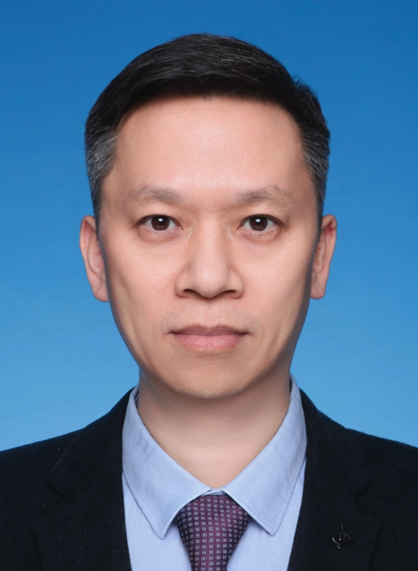 Jiacan Su (苏佳灿)合作论文数Changhai Hospital;Institute of Translational Medicine, Shanghai University5
 Yunfei Zhang (张云飞)合作论文数第四军医大学口腔医学院5
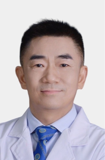 Guohui Liu (刘国辉)合作论文数Union Hospital, Tongji Medical College, Huazhong University of Science and Technology5
 Zhongmin Shi (施忠民)合作论文数Shanghai Sixth People's Hospital Affiliated to Shanghai Jiao Tong University5
 Peijian Tong (童培建)合作论文数Zhejiang Provincial Hospital of Chinese Medicine5weiguo yang合作论文数4Lei Yang合作论文数4Yong Zhang (张勇)合作论文数4Xiao-tao CHEN (陈晓涛)合作论文数4DongLiang Wang (王栋梁)合作论文数4Tao Shen (沈涛)合作论文数4Yong Wang (王勇)合作论文数4Yanxi Chen (陈雁西)合作论文数上海东方医院4
 Baoqing Yu (禹宝庆)合作论文数Department of Orthopaedics,Changhai Hospital,Second Military Medical University,Shanghai ,China4
 Dianying Zhang (张殿英)合作论文数Peking University People's Hospital4
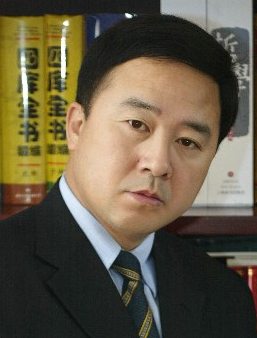 Yingze Zhang (张英泽)合作论文数Hebei Medical University4
 Liming Xiong (熊蠡茗)合作论文数Union Hospital, Tongji Medical College, Huazhong University of Science and Technology4Wencai Zhang合作论文数3Kuo Sun (孙廓)合作论文数3XiMing Liu (刘曦明)合作论文数3SiCheng Wang (王思成)合作论文数3
 Hongjian Liu (刘宏建)合作论文数Henan Provincial People's Hospital3
 Lei Zhang (张磊)合作论文数Shanghai Innovation Center of TCM Health Service, Shanghai University of Traditional Chinese Medicine3
 Jianzheng Zhang (张建政)合作论文数解放军第七医学中心创伤科3
 Zhiyong Hou (侯志勇)合作论文数The Third Hospital of Hebei Medical University3
 Bin Yu (余斌)合作论文数Nanfang Hospital, Southern Medical University3Qiang Zhou合作论文数3Zhengrong Gu (顾峥嵘)合作论文数2Jian-fei WANG (王剑飞)合作论文数2Qinglin HAN (韩庆林)合作论文数2Guo-dong Liu合作论文数2BiaoTong Huang (黄标通)合作论文数2ChengDong Hu (胡成栋)合作论文数2HaoDong Lin (林浩东)合作论文数2
 Wenming Chen (陈文明)合作论文数Academy for Engineering and Technology, Fudan University2
 Shijie Chen (陈世杰)合作论文数The Third Xiangya Hospital of Central South University2
 Zhiwei Wang (王志伟)合作论文数中国人民解放军第二军医大学2
 Shiwu Dong (董世武)合作论文数中国人民解放军第三军医大学2
 Zhimin Ying (应志民)合作论文数2Bobin Mi (米博斌)合作论文数2
 Yong Feng (冯勇)合作论文数Union Hospital, Tongji Medical College, Huazhong University of Science and Technology2
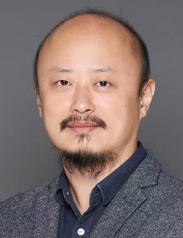 Qining Wang (王启宁)合作论文数School of Advanced Manufacturing and Robotics, Peking University;Department of Industrial Engineering and Management, College of Engineering, Peking University2Jun Miao合作论文数2
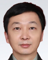 Jinpeng Jia (贾金鹏)合作论文数Chinese PLA General Hospital2




