Myelin protein zero (MPZ) is a member of the immunoglobulin gene superfamily with single extracellular, transmembrane and cytoplasmic domains. Homotypic interactions between extracellular domains of MPZ adhere adjacent myelin wraps to each other. MPZ is also necessary for myelin compaction since mice which lack MPZ develop severe dysmyelinating neuropathies in which compaction is dramatically disrupted. MPZ mutations in humans cause the inherited demyelinating neuropathy CMT1B. Some mutations cause the severe neuropathies of infancy designated as Dejerine-Sottas disease, while others cause a 'classical' Charcot-Marie-Tooth (CMT) disease Type 1B (CMT1B) phenotype with normal early milestones but development of disability during the first two decades of life. Still other mutations cause a neuropathy that presents in adults, with normal nerve conduction velocities, designated as a 'CMT2' form of CMT1B. To correlate the phenotype of patients with MPZ mutations with their genotype, we identified and evaluated 13 patients from 12 different families with eight different MPZ mutations. In addition, we re-analysed the clinical data from 64 cases of CMT1B from the literature. Contrary to our expectations, we found that most patients presented with either an early onset neuropathy with signs and symptoms prior to the onset of walking or a late onset neuropathy with signs and symptoms at around age 40 years. Only occasional patients presented with a 'classical' CMT phenotype. Correlation of specific MPZ mutations with their phenotypes demonstrated that addition of either a charged amino acid or altering a cysteine residue in the extracellular domain caused a severe early onset neuropathy. Severe neuropathy was also caused by truncation of the cytoplasmic domain or alteration of an evolutionarily conserved amino acid. Taken together, these data suggest that early onset neuropathy is caused by MPZ mutations that significantly disrupt the tertiary structure of MPZ and thus interfere with MPZ-mediated adhesion and myelin compaction. In contrast, late onset neuropathy is caused by mutations that more subtly alter myelin structure and which probably disrupt Schwann cell-axonal interactions.
Recombinant adenovirus (AVR) promises to be an efficient vector in gene therapy for neuromuscular diseases, but in preclinical experiments the expression of therapeutic genes is shorter lived in immunocompetent animals than in immunocompromised hosts. Interferons (IFN), which are known to have a role both in early antiviral activity and in late cytotoxic immunoreaction against the virus or transduced cells, may influence the efficiency of gene transfer. In this study we investigated the role of IFNs in determining the efficiency of gene transfer by AVR. AVRs expressing β-galactosidase (β-gal) from either a cytomegalovirus (CMV) or a troponin-I promoter were used. Muscle cells were infected by AVR after exposure to various IFNs. The αIFN treatment significantly reduced (up to fivefold) the CMV promoter-driven gene expression in muscle cells in vitro and in immature muscles in vivo, while the least effective inhibitor was βIFN. The decrease in gene expression by IFNs was more pronounced with the CMV-driven transgene than troponin-I promoter-driven one and was due to a decrease in transcript level. Intrinsic IFNs that are triggered by AVR administration can decrease the efficiency of gene transfer in muscle cells. Therefore the use of muscle specific promoters in AVR and/or IFN inhibitory agents will likely improve the prospects of effective gene therapy by AVR.
We demonstrated different transduction efficiency in several major organs of the immature (newborn) versus mature (adult) mice using adenoviral recombinants containing expression cassettes for either firefly luciferase or bacterial β-galactosidase reporter genes. The studied tissues included skeletal muscle, heart, brain, lung, kidney, and liver. The transduction efficiency in all tissues, especially skeletal muscle, was significantly less in adults than in newborns, with two exceptions. In the heart, transduction efficiency was the same in newborns and adults, while in brain, it was greater in the adult than in the newborn. The cited differences in transduction efficiencies between newborn and adult tissues applied approximately equally to both reporter genes. The αv integrin level showed the same trend as the transduction efficiency in all tissues, except the heart. Polymerase chain reaction showed a specific adenoviral product in proportion to the reporter gene expression in muscle, heart, and brain. The results of this study should be considered in designing gene therapy strategies in genetic diseases.
Plasmid pRSVL persisted and expressed luciferase for at least 19 months in mouse skeletal muscle after intramuscular injection. Other injected plasmids also stably expressed long-term suggesting that any plasmid DNA could stably persist and express in muscle. Plasmid DNA was demonstrated by quantitative PCR in some of the muscle DNA samples for at least 19 months after injection. The methylation pattern of the plasmid DNA remained in its bacterial form indicating that the foreign DNA did not replicate in the muscle cells. The electroporation of total cellular DNA from injected muscles into bacteria indicated that the plasmid DNA was extrachromosomal. Chromosomal integration of plasmid DNA was searched for by electroporating the injected muscle DNA into bacteria after restriction enzyme digestion and ligation. No plasmids containing plasmid/chromosome junctions were observed in over 1800 colonies examined. Lack of integration increases the theoretical safety of this gene transfer technique. Long-term stability of plasmid DNA in muscle indicates that muscle is an attractive target tissue for the introduction of extrachromosomal plasmid or viral DNA for the purpose of gene therapy.
Plasmid DNA constructs containing either a human full length dystrophin cDNA (G. Dickson, London, UK) or a deleted, Becker type, dystrophin cDNA (K.E.Davies, DR. Love, Oxford, UK.) were injected into hearts and quadriceps of dystrophin deficient MDX mice. Seven days later, analysis of immunohistochemical staining for dystrophin showed expression of dystrophin proteins in ∼1 % of quadriceps muscle cells localized mostly in the sarcolemma. 10-15 dystrophin positive cardiocyte were also seen in sections of injected hearts. Western-blot of quadriceps muscles has also identified the dystrophin expressions. Peripherally localized nuclei of muscle cells were shown in about 50 percent of dystrophin positive myofibers compared with 20 percent of dystrophin negativ myofibers suggesting the the functional effect of expressed human dystrophin protein. This direct gene transfer method has an alternative application for genetherapy of inherited myopathies but the efficiency must be increased.
The purpose of this study was to determine whether plasmid DNA is able to persist in nondividing or slowly dividing brain cells in vivo. A new cationic lipid formulation which contains 70 mol% of DOTMA {N[1-(2,3-dioleyloxy)propyl]-N, N, N-trimethylammonium} and 30 mol% of cholesterol was used to transfect reporter genes into fetal brain cells in culture that were then transplanted into adult host brains. Gene expression was localized both to glial and neuronal cells after transfection of fetal brain cells with pRSVLac-Z, the gene coding for Escherichia coli β-galactosidase protein. After the transfection of pRSVL plasmid which contains the firefly luciferase gene into fetal brain cells that were transplanted, substantial amounts of luciferase and pRSVL DNA were present in the host brains for 1 to 2 months. These results have implications for intracerebral viral infections and gene therapy of brain disorders.
This report extends our previous findings that mouse muscle cells in situ can take up naked DNA injected intramuscularly in vivo. Various conditions such as needle type, speed of injection, volume of injection fluid, tonicity of injection fluid, type of solute, type of muscle, physiologic condition of the muscle and age of the animals were appraised for their effect on the levels of luciferase activity expressed from the pRSVL plasmid. Specific conditions such as the use of normal saline as an injection fluid increased the efficiency of expression. The implantation of DNA pellets was an effective way to deliver DNA to muscle, especially for smaller muscle groups. Also, newborn and adult rat muscles expressed plasmid DNA delivered intramuscularly.
Duchenne's muscular dystrophy (DMD), which affects one in 3,500 males, causes progressive myopathy of skeletal and cardiac muscles and premature death. One approach to treatment would be to introduce the normal dystrophin gene into diseased muscle cells. When pure plasmid DNA is injected into rodent skeletal or cardiac muscle, the cells express reporter genes. We now show that a 12-kilobase full-length human dystrophin complementary DNA gene and a 6.3-kilobase Becker-like gene can be expressed in cultured cells and in vivo. When the human dystrophin expression plasmids are injected intramuscularly into dystrophin-deficient mdx mice, the human dystrophin proteins are present in the cytoplasm and sarcolemma of approximately 1% of the myofibres. Myofibres expressing human dystrophin contain an increased proportion of peripheral nuclei. The results indicate that transfer of the dystrophin gene into the myofibres of DMD patients could be beneficial, but a larger number of genetically modified myofibres will be necessary for clinical efficacy.
Skeletal and cardiac muscles of rodents are able to take up and express directly injected plasmids containing reporter genes such as luciferase (pRSVL), E. coli B-galactosidase (pRSVLac-z). After injection of pRSVLac-z DNA into heart or skeletal muscle muscle, gene expression was localized histochemically to cardiocytes or myocytes, respectively. Seven days after injection of pRSVL DNA, luciferase activity (mean±SE×103 light units per ug DNA injected) was 1325±287 in normal mouse skeletal muscle, 1543±407 in mdx skeletal muscle, 3015±1295 in rat cardiac muscle. After injection of pRSVL, gene expression was present in cardiac and mdx skeletal muscle for only 1 month while it persisted in normal skeletal muscle for one year. Gene expression was stable in cardiac muscle of athymic and ciclosporin treated rats for at least 2 months that suggest the role of immune response in heart. Rapid turnover of myofibers in mdx muscle can explain the instability of gene expression. Direct transfer of genes into muscle has applications for gene therapy.(*Pres.inst.:Dept. of Ped. Univ. of Pecs, Hungary)
We found previously that genes injected into skeletal muscle can be taken up by myofibers and expressed. In the present study we found that myocardial cells can also express a variety of reporter genes injected into myocardium as efficiently as skeletal myofibers, while the cells of several other tissues cannot. The inability of tissues other than striated muscle to express injected DNA is not due to technical difficulties of injection because injected DNA was detected in these other tissues by PCR analysis. These results suggest that skeletal and cardiac muscle cells have unique features such as T tubules that may play a critical role in DNA uptake. Expression in cardiac muscle was stable for only two weeks, possibly because of an immune response against the transfected cells. The ability to directly transfer genes into myocardial cells raises the possibility of gene therapy for both acquired and genetic heart diseases.
 Gyula Acsadi合作论文数13
 Jon A. Wolff合作论文数Department of Pediatrics, School of Medicine and Public Health, University of Wisconsin7
 P Williams合作论文数5George Karpati合作论文数Neuromuscular Research Group, Montreal Neurological Institute, McGill University5
 Hanns Lochmüller合作论文数CHEO Research Institute;Faculty of Medicine, University of Ottawa;Department of Medicine, Division of Neurology, The Ottawa Hospital4S Jiao合作论文数3W Chong合作论文数3
 Bernard Massie合作论文数3
 Josephine Nalbantoglu合作论文数Department of Neurology & Neurosurgery, Faculty of Medicine and Health Sciences, McGill University;Montreal Neurological Institute3Ja Wolff合作论文数2
 Eric Shoubridge合作论文数The Neuro Montreal Neurological Institute-Hospital;McGill University2
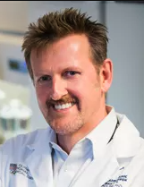 Johnny Huard合作论文数Center for Regenerative Sports Medicine, Steadman Philippon Research Institute;Department of Clinical and Biomedical Sciences, College of Veterinary Medicine, Colorado State University2Seiichi Tsujino合作论文数2Janne Balsamo合作论文数1Karen Krajewski合作论文数1
 Jim Ludtke合作论文数1Ba Kakulas合作论文数1
 Sue Fletcher合作论文数1
 Robert Malone合作论文数1KL Blaschuk合作论文数1
 Salvatore Dimauro合作论文数Vagelos College of Physicians & Surgeons, Columbia University Irving Medical Center;H. Houston Merritt Clinical Research Center, Columbia University1Stephen Prescott合作论文数1P HOLLAND合作论文数1JM Howell合作论文数1C GUERIN合作论文数1Paul Holland合作论文数1
 Heather D Durham合作论文数McGill University1
 Philip L. Felgner合作论文数Protein Microarray Laboratory, School of Medicine, University of California, Irvine;Dept. of Epidemiology, School of Medicine, University of California, Irvine1
 Kay Davies合作论文数Department of Physiology, Anatomy and Genetics, University of Oxford1
 Teresa Collins合作论文数1Dickson George合作论文数1John Kamholz合作论文数1
 Michael Shy合作论文数Department of Neurology, Roy J. and Lucille A. Carver College of Medicine, University of Iowa;Division of Neuromuscular Medicine, Neurology, Roy J. and Lucille A. Carver College of Medicine, University of Iowa1
 Jun Li合作论文数Stanley H. Appel Department of Neurology, Houston Methodist1
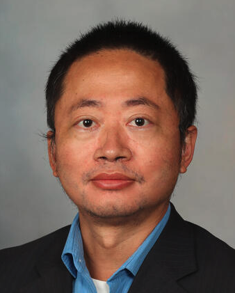 Chen Wang合作论文数Division of Computational Biology, Department of Quantitative Health Sciences, Mayo Clinic;Department of Health Sciences Research, Mayo Clinic College of Medicine1
 Richard A. Lewis合作论文数Cedars-Sinai Medical Center1Lee A. Davies合作论文数1
 Marina Grandis合作论文数Department of Neuroscience, Ophthalmology and Genetics, University of Genova1Rosemary Shy合作论文数1Sara Shanske合作论文数1Shoushu Jiao合作论文数1
 Frank S Walsh合作论文数Ossianix1
 James Garbern合作论文数School of Medicine, Wayne State University1J.Mcc. Howell合作论文数1
 Donald R. Love合作论文数LabPlus, Auckland City Hospital1Nancy Larochelle合作论文数1Jack Lilien合作论文数1Cyula Acsadi合作论文数1
 Steve D. Wilton合作论文数QEII Medical Centre, Australian Neuromuscular Research Institute1




