Brain cancer therapeutics, particularly for glioblastoma, face significant challenges due to the restrictive nature of the blood-brain barrier, tumor heterogeneity, and resistance to conventional treatments. Recent advancements in nanotechnology have introduced graphene oxide (GO) as a promising candidate for enhancing treatment efficacy and improving patient outcomes. This review discusses the anticancer properties of GO including applications in targeted drug delivery, increasing the effectiveness of photothermal and photodynamic therapies, and highlighting its ability to induce cell death in glioblastoma cells. The experimental findings from in vitro and in vivo studies on the anticancer activity of GO and the potential use of this material as a nanocarrier for drug delivery in glioblastoma therapy are presented. Possible challenges related to its safety and the synthesis and functionalization of GO nanoparticles are discussed. The findings of the reviewed studies highlight the need for future research on combination therapies with GO and other treatments, including chemotherapy, radiation therapy, and immunotherapy.
In Parkinson's disease (PD), the degeneration of dopaminergic neurons markedly reduces both dopamine release and its downstream signaling. The present study aims to design a new electrochemical sensor for detecting dopamine release in vitro. First, a screen-printed carbon electrode (SPCE) was coated with a layer of carboxylated MWCNTs using a chronoamperometric deposition process. Subsequently, an Au-dendrimer solution was electrodeposited on the MWCNT/SPCE via cyclic voltammetry (CV), yielding the Au/Den/MWCNT/SPCE electrode. The enhancement in redox peak currents demonstrated that the Au/Den/MWCNTs significantly improved conductivity and expanded the active surface area of the modified electrode compared with both the bare SPCE and the MWCNT/SPCE. The nanosensor exhibited a detection limit of 5.8 nM and a linear response range from 10 nM to 6 μM. The high selectivity of the sensor was verified by examining its response to ascorbic acid (AA) and uric acid (UA). The effect of 3-acetyl-11-keto-β-boswellic acid (AKBA) on dopamine release was evaluated by fabricated nanosensor in the SH-SY5Y neuroblastoma cells following 6-hydroxydopamine (6-OHDA) insult. The findings indicated that treatment with AKBA at concentrations of 2.5 and 5 μM significantly enhanced dopamine secretion by SH-SY5Y cells after exposure to 6-OHDA, with the difference reaching high statistical significance (p < 0.001). Therefore, the developed nanosensor demonstrated high efficiency and accuracy for dopamine detection, highlighting its potential applications in medical diagnostics, neuroscience research, and drug screening.
Background: In recent decades, numerous herbal products have been shown to have antihyperglycemic and beta cell-regenerative effects in animal studies. However, there is no clinical evidence that those products completely cure patients with type-1 diabetes (T1D). Therefore, it seems that most of the phytochemicals do not have a significant impact on human beta cells, and the results of experimental studies conducted on them may not be generalizable to the clinic. Purpose: The present work aims to review extensively the methods and results of preclinical studies on phytotherapy of T1D published in the last 10 years. Methods: This paper critically analyzes the designs of studies, treatment protocols, methods of diabetes induction, characteristics of the studied animals, clinical relevance, reproducibility of research, and other aspects related to conducting preclinical studies on T1D. We discussed limitations that make many of the results of these studies not generalizable to the clinic. Finally, some recommendations were given to improve studies on the phytotherapy of T1D to avoid misleading interpretations about the antidiabetic effect of herbal compounds. Conclusion: This paper can be considered a practical guide for researchers interested in the field of phytotherapy of T1D to increase the reliability, reproducibility, and validity of their preclinical studies.
Background:: Consumption of a high-fat diet (HFD) is one of the main causes of nonalcoholic fatty liver disease (NAFLD), which is increasing due to lifestyle changes and is still an important global health issue. Despite the efforts, there is still no common treatment for this disease. Studies have shown that the root of Rheum turkestanicum Janisch has a hypolipidemic effect and a significant antioxidant effect on liver tissue in diabetic rats. However, no experimental study has been performed on the hepatoprotective effects of this herb on HFD-induced NAFLD have been proven. background: Consumption of a high-fat diet (HFD) is one of the main causes of non-alcoholic fatty liver disease (NAFLD), which is increasing due to lifestyle changes and is still an important global health issue. Despite the efforts, there is still no common treatment for this disease. Studies have shown that the root of Rheum turkestanicum Janisch has a hypolipidemic effect and a significant antioxidant effect on liver tissue in diabetic rats. However, no experimental study has been performed on which the hepatoprotective effects of this herb on HFD-induced NAFLD have been proven. Objective:: This study aimed to evaluate the effect of Rheum turkestanicum Janisch extract (RTE) on HFD-induced NAFLD in BALB/c mice. Materials and Methods:: The study was performed with two models of prevention and therapeutic effect of RTE. Serum biochemical markers, histopathology, oxidative stress indicators, and qRTPCR were measured to evaluate the effects of RTE on lipid metabolism disorders in mice feeding with HFD. Results:: In the prevention model, compared to the HFD group, RTE treatment decreased the levels of glucose, triglyceride, and cholesterol and improved liver profile markers, oxidative stress, and expression of genes involved in lipid metabolism. Conclusion:: The results of this study suggest that RTE has hepatoprotective effects against HFDinduced liver damage by reducing oxidative stress, lipogenesis, and increasing beta-oxidation of free fatty acids.
PURPOSE:Ferula gummosa Boiss. is a well-known and valuable medicinal plant in Iran. Research has shown that this plant has several pharmacological properties, including anti-bacterial, anti-cancer and etc. In the present study, we investigated the cytotoxic properties of F. gummosa Boiss. extract in MCF-7 breast adenocarcinoma cells.METHODS:The cytotoxicity and pro-apoptotic properties of the extract were assessed by 3-(4,5-dimethylthiazol-2-yl)-2,5-diphenyl tetrazolium bromide (MTT) test and propidium iodide (PI) stained cells, respectively. Apoptosis and necrosis were evaluated by annexin V-PI staining. The levels of reactive oxygen species (ROS),malondialdehyde (MDA), glutathione (GSH), and superoxide dismutase (SOD) was determined to evaluate oxidative stress. The cell migration and the gene expression were assessed by scratch assay and quantitative real-time polymerase chain reaction (q-RT-PCR), respectively.RESULTS:The extract of F. gummosa decreased the viability and cell cycle progression of MCF-7 cells by inducing apoptosis and necrosis, increasing ROS and MDA levels, and decreasing GSH levels and SOD activity. It also lowered the cells' migration capability by enhancing p53 mRNA levels and reducing MMP-9 mRNA expression.CONCLUSION:F. gummosa exhibited pro-apoptotic, anti-proliferative, and anti-metastatic effects on MCF-7 cells. It is therefore recommended that detailed future research be done on different parts of the plant or its secondary metabolites to find anti-cancer lead compounds.
Temozolomide (TMZ) is the first-line chemotherapy for glioblastoma, a deadly brain cancer. Doxorubicin (DOX) is another cancer chemotherapeutic drug, but low blood–brain barrier permeability limits its use for glioblastoma. This limitation may be overcome using nanocarriers as new delivery strategies. This study aimed to load DOX and TMZ on cyclodextrin-graphene oxide nanocarrier and evaluate their combined cytotoxic effect against glioblastoma. The α-, β-, and γ-cyclodextrins (CDs) were separately conjugated to graphene oxide (GO) sheets to synthesize nanocarriers for TMZ and DOX. The entrapment and drug loading efficiency were determined by UV–visible spectrophotometry analysis at 480 nm for TMZ and 330 nm for DOX. The MTT assay and annexin V/PI staining were used to assess cytotoxicity of TMZ, DOX, and their nanocarriers on viability and apoptosis of U87 cells. Drug-content evaluation showed that the loading efficiency of α-, β-, and γ-CD-GO-TMZ was 29
Objectives:Experimental studies reported that some plants in the genus of Moraea (Iridaceae family) show anticancer potential. This study aimed to evaluate the effects of Moraea sisyrinchium on U87 glioblastoma multiforme and HepG2 liver cancer cells.Materials and Methods:The cells were incubated for 24 hr with hydroalcoholic extract of the stem, flower, and bulb of M. sisyrinchium. Then, the cell proliferation (MTT) assay, cell cycle analysis (propidium iodide staining), cell migration test (scratch), Western blotting (Bax and Bcl-2 expression), and gelatin zymography (for matrix metalloproteinases, MMPs) were performed. Oxidative stress was evaluated by determining the levels of reactive oxygen species and lipid peroxidation. Angiogenesis was evaluated on chick embryo chorioallantoic membrane.Results:The extracts of the flower, stem, and bulb significantly decreased the proliferation of HepG2 and U87 cells. This effect was more for U87 than HepG2 and for the bulb and stem than the flower. In U87 cells, the bulb extract increased oxidative stress, cell cycle arrest, and the Bax/Bcl-2 ratio. Also, this extract suppressed the migration ability of HepG2 and U87 cells, which was associated with the inhibition of MMP2 activity. In addition, it significantly reduced the number and diameter of vessels in the chorioallantoic membrane. Liquid chromatography-mass spectrometry revealed the presence of xanthones (bellidifolin and mangiferin), flavonoids (quercetin and luteolin), isoflavones (iridin and tectorigenin), and phytosterols (e.g., stigmasterol) in the bulb.Conclusion:M. sisyrinchium bulb decreased the proliferation and survival of cancer cells by inducing oxidative stress. It also reduced the migration ability of the cells and inhibited angiogenesis.
In the past few years, there has been notable advancement in nanotechnology, leading to the development of new materials with potential uses in the medical field, especially in cancer diagnosis, imaging, and therapy. Black phosphorus quantum dots (BPQDs) are one of the emerging nanomaterials that have generated interest due to their unique properties and potential in biomedical applications. This review aims to give a detailed overview of how BPQDs are synthesized, characterized, and utilized. The synthesis methods of BPQDs are discussed, with a focus on obtaining size-controlled and high-quality BPQDs. Two main approaches, top-down exfoliation and bottom-up techniques, are described. Despite advancements in synthesis, there are challenges hindering the practical application of BPQDs, such as poor dispersion and short durability. To address these issues, techniques to enhance biocompatibility and reduce potential toxicity, such as surface modifications, are discussed. BPQDs have potential in bioimaging as they offer higher resolution and sensitivity compared with traditional imaging agents. Their small size and expansive surface area make them suitable for drug delivery systems, enabling the effective incorporation of therapeutic substances. By functionalizing BPQDs with targeting ligands, they can selectively bind to cancer cells or tissue, making them ideal for targeted therapies. Moreover, BPQDs can serve as biosensors to detect biomarkers in bodily fluids, further expanding their biomedical applications. However, before they can be successfully translated into clinical settings, further research is needed to optimize the synthesis methods of BPQDs and evaluate their long-term safety profiles. Nonetheless, with ongoing research and development, the medical uses of BPQDs are expected to expand.
Introduction: Glioblastoma is a common malignant brain tumor, with limited therapeutic options. In our previous study, the Moraea sisyrinchium plant showed cytotoxicity against glioblastoma and hepatocellular carcinoma cells. Among different parts of this plant (flower, stem, and bulb), the bulb showed better anticancer potential. The present work aimed to test the anticancer activity of different fractions of the bulb extract, to determine its phytochemicals, and to study its mechanism action on glioblastoma. Methods: The bulb extract was partitioned into different fractions using immiscible solvents. The U87 glioblastoma cells were incubated with the obtained fractions. Then, the cell proliferation assay (MTT), cell migration test (scratch), cell cycle analysis (propidium iodide staining), apoptosis/necrosis assay (annexin V/propidium iodide staining), and real-time PCR (PTEN, Akt, mTOR, BAX and BCL-2 genes) were performed. Phytochemicals were determined using liquid chromatography-mass spectroscopy. Results: The chloroform fraction showed more antiproliferative effect than n-hexane, ethyl acetate, and n-butanol fractions. Also, chloroform fraction induced cell cycle arrest, increased apoptosis, and inhibited cell migration ability (P < 0.05). The expression of PTEN, mTOR, and BAX genes was significantly up-regulated, while the expression of Akt and Bcl-2 showed down-regulation. The phytochemicals identified in the chloroform fraction were mainly xanthones, phytosterols, and isoflavones. Conclusion: The chloroform fraction of Moraea sisyrinchium bulb inhibits the proliferation and migration of glioblastoma cells by inducing cell cycle arrest and apoptosis by upregulation of the PTEN gene and Bax/Bcl-2 ratio. The identified compounds in the chloroform fraction are potential candidates for further investigation as anticancer agents against glioblastoma.
Mephedrone, a synthetic derivative of cathinone, is a commonly used psychoactive substance. Our previous study showed that exposure to mephedrone during pegnancy induced antiproliferative and pro-apoptotic effects in hippocampus of mice delivered pups. However, its effects on neural stem/progenitor cells (NS/PC) remain unexplored. The aim of this study is to investigate the effects of mephedrone exposure on the proliferation, differentiation, and apoptosis of rat embryonic NS/PC. NS/PC were isolated from rat fetal ganglionic eminence region at embryonic day 14.5. The effects of mephedrone on cell proliferation, neurosphere formation (colonies of NS/PC), neuronal differentiation, and apoptosis of NS/PC were assessed using MTT, immunocytochemistry, and flow cytometry. Mephedrone at concentrations of 20-640 mu M significantly decreased the proliferation of NS/PC, induced cell cycle arrest, and enhanced the percent of apoptotic and necrotic cells. Neurosphere assays revealed a significant reduction in the number and diameter of neurosphere-forming cells. In addition, mephedrone significantly decreased the expressions of DCX and NeuN neuronal markers. Taken together, our results suggeste that exposure to mephedrone decreases the viability and neuronal differentiation of embryonic NS/PC. This study showed that mephedrone exposure during fetal or neonatal life may impair neurogenesis and subsequent brain development.
Patients with diabetes are at risk for liver disorders including glycogen hepatopathy, non-alcoholic fatty liver disease, cirrhosis, and hepatic fibrosis. The pathophysiological mechanisms behind diabetic hepatopathy are complex, some of them include fatty acid accumulation, increased reactive oxygen species, increased advanced glycation end-products, hyperactivity of polyol pathways, increased apoptosis and necrosis, and promotion of fibrosis. A growing number of studies have shown that herbal extracts and their active phytochemicals have antihyperglycemic properties and beneficial effects on diabetic complications. The current review, for the first time, focused on herbal agents that showed beneficial effects on diabetic hepatopathy. For example, animal studies have shown that Moringa oleifera and Morus alba improve liver function in both type-1 and type-2 diabetes. Also, evidence from clinical trials suggests that Boswellia serrata, Juglans regia, Melissa officinalis, Portulaca oleracea, Silybum marianum, Talapotaka Churna, and Urtica dioica reduce serum liver enzymes in diabetic patients. The main active ingredient of these plants to protect the liver seems to be phenolic compounds such as niazirin, chlorogenic acid, resveratrol, etc. Mechanisms responsible for the hepatoprotective activity of herbal agents include improving glucose metabolism, restoring adipokines levels, antioxidant defense, and anti-inflammatory activity. Several signaling pathways are involved in hepatoprotective effects of herbal agents in diabetes, such as phosphoinositide 3-kinase, adenosine monophosphate-activated protein kinase, mitogen-activated protein kinase, and c-Jun NH2-terminal kinase.
Objective: Hepatocellular carcinoma is one of the leading causes of cancer-related death worldwide. Experimental studies reported that some plants in the genus of Psoralea (Fabaceae family) show anticancer potential. The present study aimed to evaluate the effects of Psoralea drupacea extract (PDE) on HepG2 liver cancer cells. Methods: The proliferation, cell cycle, and migration of HepG2 cells were determined by thiazolyl blue tetrazolium bromide test, propidium iodide staining, and scratch assay, respectively. The effects of PDE on the activity of matrix metalloproteinases (MMPs) and angiogenesis were evaluated by the gelatin zymography method and chicken chorioallantoic membrane model, respectively. Results: The culture of HepG2 cells in the presence of PDE (24 hr and 48 hr) significantly reduced their viability (at a concentration of ≥ 50 µg/mL) and increased the percentage of cells in the sub-G1 stage. PDE also increased the antiproliferative and proapoptotic activities of doxorubicin (3 and 6 µg/mL). The extract significantly decreased the generation of reactive oxygen species and lipid peroxidation in the cells. Moreover, PDE (25 and 50 µg/mL) significantly suppressed the migration ability of HepG2 cells, which was associated with inhibition in the activity of MMP2 and MMP9 (50 µg/mL). Furthermore, treatment with PDE significantly reduced the number and diameter of vessels in the chick chorioallantoic membrane. Conclusion: PDE decreased the survival and cell cycle progression of liver cancer cells through a mechanism other than oxidative stress. This extract also showed an anti-angiogenesis effect and diminished the migration ability of HepG2 cells by inhibiting MMP activity
Background:The possible toxicity of natural products must be tested before being used in the market. The present work aimed to evaluate acute, subacute, and subchronic toxicity of an herbal formulation containing Anethum graveolens, Cynara scolymus, Citrus aurantium, Portulaca oleracea, and Silybum marianum.Material and methods:Acute toxicity (2000 mg/kg, single dose) and sub-acute toxicity (600 and 1200 mg/kg/day, 4 weeks) tests were performed on female and male rats according to OECD 423 and OECD 407 guidelines, respectively. In the subchronic study (12 weeks), the animals were divided into three groups (6 females and 6 males per group): control, low-dose group (food supplemented with 300 mg/kg of the herbal product), and high-dose group (600 mg/kg).Results:The herbal product at a single dose of 2000 mg/kg did not induce mortality for 14 days. In the sub-acute study, administration of the product for 28 days at 1200 mg/kg/day had no effect on survival, appetite (water and food consumption), body weight, serum biochemical parameters (BUN, creatinine, AST, ALT, ALP, bilirubin, albumin), histology of vital organs (liver, kidney, heart, brain), and hematological markers related to erythrocyte, platelet, and leukocyte. Similarly, in the subchronic study, the product did not induce mortality, change in histology of the vital organs, or alteration in hematological or biochemical parameters (except for an increase in ALP in female rats received 600 mg/kg).Conclusion:The formulated product shows no signs of toxicity in rats up to 2000 mg/kg, 1200 mg/kg, and 600 mg/kg in acute, subacute, and subchronic phases, respectively. It is suggested to monitor ALP levels in females in case of long-term use of the product.
Abstract Objective Experimental studies reported that some plants in the genus of Moraea (Iridaceae family) show anticancer multiform and HepG2 liver cancer cells. Methods The cells were incubated potential. This study aimed to evaluate the effects of Moraea sisyrinchium on U87 Glioblastoma for 24 with hydro alcoholic extract of the stem, flower, and bulb of Moraea sisyrinchium. The cell proliferation, cell cycle, and migration were determined by MTT assay, propidium iodide staining, and scratch assay, respectively. Oxidative stress was evaluated by determining the levels of reactive oxygen species and lipid peroxidation. The activity of matrix metalloproteinases (MMPs), the expression of Bax and Bcl-2 proteins, and angiogenesis were evaluated by the gelatin zymography, Western blotting, and model of chick embryo chorioallantoic membrane, respectively. Results The extracts of the flower, stem, and bulb significantly decreased the proliferation of HepG2 and U87 cells. This effect was more for U87 than for HepG2 and for bulb and stem than for flower. In U87 cells, the bulb extract increased oxidative stress, cell cycle arrest, and the Bax/Bcl-2 ratio. Also, this extract suppressed the migration ability of HepG2 and U87 cells, which was associated with the inhibition of MMP2 activity. In addition, it significantly reduced the number and diameter of vessels in the chorioallantoic membrane. Liquid chromatography-mass spectrometry revealed the presence of xanthones (bellidifolin, mangiferin), flavonoids (quercetin, luteolin), isoflavones (iridin,tectorigenin),and phytosterols (e.g., stigmasterol) in the bulb extract. Conclusion The extract of Moraea sisyrinchium bulb decreased the proliferation and survival of cancer cells by inducing oxidative stress. The extract also reduced the migration ability of the cells and inhibited angiogenesis.
Objective: The present randomized clinical trial assessed the antihyperglycemic and hypolipidemic effects of hydro-ethanolic extract of Ribes khorassanicum. Materials and Methods: Eighty type 2 diabetic patients were randomly allocated to placebo or intervention groups and respectively received placebo or extract capsules (700 mg, bid) beside their conventional medication for 3 months. Patients' blood pressure and blood levels of fasting blood glucose (FBS), glycosylated hemoglobin (HbA1c), 2 hr postprandial glucose (2hPPG), triglyceride, total cholesterol, low-density lipoprotein cholesterol (LDL-C), and high-density lipoprotein cholesterol (HDL-C) were measured at the beginning of the study and after 3 months of treatment. For determination of plant safety, liver enzymes (SGOT and SGPT) and kidney function (in terms of urea, creatinine, and microalbumin levels) were assessed and patients were asked to report adverse effects. Results: The R. khorasanicum hydro-ethanolic extract supplementation significantly decreased the levels of FBS, total cholesterol, triglyceride, and LDL-C in the extract group compared to the placebo group (p<0.05-p<0.01). However, 2hPPG, HbA1c, HDL-C, SGOT, SGPT, urea, creatinine, and urine microalbumin values were not significantly different between the placebo and the extract groups. No adverse effects were reported by the patients. Conclusion: Co-supplementation of diabetic patients with R. khorasanicum extract ameliorated hyperglycemia and hyperlipidemia without causing any adverse effects; therefore, the plant extract may be recommended as a complementary therapy to improve diabetes-induced metabolic disturbances.
Hepatoprotective effects of many herbal agents have been reported in animal studies and clinical trials. In this study, five hepatoprotective plants with potent antioxidant, anti-inflammatory, and hypolipidemic effects were chosen to prepare a polyherbal compound for managing NAFLD. Sixty patients with NAFLD were randomly divided into treatment and control groups (2:1 ratio). Both group were advised to take healthy diet and exercise. The treatment group also received herbal capsules containing 400 mg of the mixture of Anethum graveolens, Citrus aurantium, Cynara scolymus, Portulaca oleracea, and Silybum marianum (2 capsules, thrice daily, for two months). The liver ultrasound and biochemical markers including the serum lipids, liver enzymes, and glucose were evaluated before starting the study and at the end of the treatment. Thirty patients in the treatment group and sixteen patients in the control group completed the study. The herbal compound significantly decreased the serum level of alanine transaminase (ALT), aspartate transaminase (AST), and total cholesterol. Treatment with the herbal compound significantly improved the grade of the fatty liver, but no significant change was found in the control group. In conclusion, the formulated herbal compound appeared to be effective in biochemical improvement and decreasing the grade of the fatty liver in the patients with NAFLD.
Colorectal cancer (CRC) is the second cause of cancer-associated death globally. Recently, herbal medicinal products and, in particular, zerumbone have been widely studied and used for cancer treatment as they induce significant anti-cancer effects. However, there is limited information about the anti-cancer effects of zerumbone in CRC. Therefore, we aimed to investigate the in vitro anti-cancer effects of the zerumbone in CRC, focusing on cell apoptosis and migration. Anti-proliferative and anti-migratory effects of zerumbone on HT-29 cells were evaluated using MTT and scratch wound healing assay, respectively. Quantitative real-time PCR (qRT-PCR) was performed to determine the mRNA expression levels of migration and apoptosis-related genes. Apoptosis and cell cycle distribution were evaluated by flow cytometry. The intracellular level of reactive oxygen species (ROS) was measured using a ROS assay kit. Additionally, matrix metalloproteinase-2/-9 (MMP-2/-9) activity was determined using gelatin zymography. Zerumbone suppressed the viability of the HT-29 cells dose-dependently while having less cytotoxicity on normal NIH/3T3 cells. Zerumbone induced apoptosis in HT-29 cells and arrested the cell cycle in the G2/M phase. These effects were associated with alteration in the expression of apoptosis-related genes (up-regulation of Bax and down-regulation of Bcl-2 genes). Zerumbone also enhanced the generation of ROS in HT-29 cells. Furthermore, zerumbone significantly inhibited the migration of HT-29 cells and decreased MMP-2/-9 mRNA expression and activity. Our findings provide a potential use for zerumbone to induce apoptosis and suppress metastasis in HT-29 cells; thus, it could be developed as a promising natural agent for future CRC therapy.
AIMS:Levetiracetam (LEV) is a broad-spectrum antiepileptic drug with neuroprotective properties and novel mechanisms of action. Some evidence suggests that LEV may impact adult neurogenesis, but the results are controversial. The present study was aimed to evaluate the effects of LEV on the proliferation and differentiation of rat embryonic neural stem cells (NSCs) and to explore the role of GABAB or NMDA receptors.MAIN METHODS:NSCs were isolated from rat fetal ganglionic eminence at embryonic day 14.5. The effects of LEV on viability, proliferation, neurosphere formation, and neuronal or astroglial differentiation of NSCs were assessed using resazurin, BrdU incorporation, immunocytochemistry, quantitative real-time PCR, and western blotting. Additionally, we addressed the relationship between treatment with NMDA and GABAB receptor antagonists (MK801 and saclofen, respectively) in combination with LEV on these parameters.KEY FINDINGS:The data showed that LEV (50 μM) significantly increased the number (p < 0.01) and diameter of neurospheres (p < 0.05), enhanced proliferation (p < 0.01), and promoted neuronal differentiation, as revealed by significantly increased expressions of DCX and NeuN. The expressions of astroglial markers, GFAP and Olig2, were markedly reduced. The addition of MK801 (10 μM) significantly diminished neurospheres growth (p < 0.001), decreased the number of proliferating cells (p < 0.01), and reduced the number of new neurons (p < 0.001) but increased the astroglial cells (p < 0.001) induced by LEV. Co-treatment with saclofen (25 μM) did not significantly affect LEV-induced NSCs proliferation and differentiation.SIGNIFICANCE:Our findings suggest that LEV may enhance rat embryonic neurogenesis mainly through an NMDA receptor-mediated mechanism.
Objective: Previous clinical trials have suggested that herbal medicines can improve the quality of life (QOL) and survival of cancer patients. This study was aimed to evaluate the effects of a polyherbal compound (PHC, formulated as syrup) consisting of Allium sativum, Curcuma longa, Panax ginseng, and Camellia sinensis on the quality of life (QOL) and survival in patients with upper gastrointestinal cancers. Materials and Methods: A randomized placebo-controlled trial was carried out on patients with esophageal or gastric cancer who had finished their oncological treatments. The patients were randomly assigned to PHC (n=20) or placebo (n=20) group. The PHC group was treated with the PHC for 12 weeks, while the placebo group received 70% sucrose syrup. The QOL was assessed at baseline and after 12 weeks. The patients were followed for up to 24 months to determine overall survival. Results: PHC significantly improved cancer-related symptoms, physical performance, and psychological and social functions of the patients (p<0.05 for all cases). Death occurred in 33 and 22% of cases in the placebo and PHC group, respectively. The mean survival time was 16.8 months (95% CI: 12.8-20.9) in the placebo group and 21.4 months (95% CI: 19.1-23.6) in the PHC group but the difference was not statistically significant. Conclusion: The PHC improved cancer-related symptoms, physical performance, and psychological and social functions in patients with gastrointestinal cancers. It seems that this herbal compound has the potential to be used as a supplement in the management of cancer.
 Sadeghnia Hamid Reza合作论文数Dept Pharmacol, Mashhad Univ Med Sci47
 Soukhtanloo Mohammad合作论文数23
 Hassan Rakhshandeh合作论文数Department of Pharmacology and Pharmacological Research Center of Medicinal Plants, Mashhad University of Medical Sciences23
 Azar Hosseini合作论文数School of Medicine, Mashhad University of Medical Sciences16
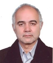 Shafiee-Nick Reza合作论文数School of Medicine, Mashhad University of Medical Sciences;School of Medicine, Mashhad University of Medical Sciences;Mashhad University of Medical Sciences, Mashhad University of Medical Sciences15
 Seyed Hadi Mousavi合作论文数11
 M.M. Hosseini合作论文数10
 Amiri Mohammad Sadegh合作论文数Department of Biology, Payame Noor University10
 Elham Asadpour合作论文数MUMS),, Mashhad University of Medical Science10
 Mohammad Jalili-Nik合作论文数Student Research Committee, Mashhad University of Medical Sciences9
 Roghayeh Rashidi合作论文数Pharmacological Research Center of Medicinal Plants, Mashhad University of Medical Sciences8Forouzanfar Fatemeh合作论文数Faculty of Medicine, Mashhad University of Medical Sciences8
 Hooshmand Sara合作论文数Department of Chemistry, Payame Noor University8
 Elham Einafshar合作论文数School of Chemistry, Damghan University;;School of Chemistry, Damghan University7Hadjzadeh Mousa-Al-Reza合作论文数7
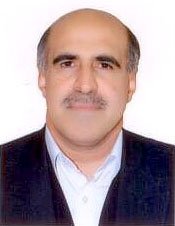 Mohammad Taher Boroushaki合作论文数Department of Pharmacology;Ghaem Medical Centre;University of Medical;Ghaem Medical Centre, University of Medical7
 Mohaddeseh Sadat Alavi合作论文数Faculty of Medicine, Mashhad University of Medical Sciences6
 Maede Hasanpour合作论文数Biotechnol Res Ctr, Mashhad Univ Med Sci6
 Baratali Mashkani合作论文数School of Medicine, Mashhad University of Medical Sciences;School of Biomedical Sciences and Pharmacy, University of Newcastle;Mashhad University of Medical Sciences, University of Newcastle6
 Faezeh Ghasemi合作论文数Faculty of Medicine, Mashhad University of Medical Sciences;Faculty of Medicine, Mashhad University of Medical Sciences6
 Hamed Ghazavi合作论文数School of Medicine, Mashhad University of Medical Sciences6Masoumeh Varedi合作论文数Department of Physiology .6Mortazavian Seyed Mohsen合作论文数6Taraz Jamshidi Shirin合作论文数6Majid Ghayour-Mobarhan合作论文数5
 Leila Mobasheri合作论文数5
 Reza Mohebbati合作论文数Applied Biomedical Research Center, Mashhad University of Medical Sciences5
 Seyed javad Hoseini合作论文数Dept Med Technol, Mashhad Univ Med Sci5Ziba Rajaei合作论文数School of Medicine, Mashhad University of Medical Sciences5
 Jafarian Amir Hossein合作论文数Department of Pathology, Ghaem Hospital5
 Maliheh Moradzadeh合作论文数Faculty of Medicine, Mashhad University of Medical Science5
 Mahmood Abedinzade合作论文数5
 Hamid Sadeghian合作论文数Faculty of Sciences, Ferdowsi University of Mashhad5
 seyyed abbas Zojaji合作论文数4
 Gholamreza Naseri合作论文数4
 Amir R. Afshari合作论文数School of Medicine, North Khorasan University of Medical Sciences4
 Farshad Mirzavi合作论文数School of Medicine, Gonabad University of Medical Sciences4
 Mohammad Vahedi合作论文数Dept Pharmacol, Zahedan Univ Med Sci4
 Tabarraei Alijan合作论文数Golestan University of Medical Sciences4
 Hamid Mollazadeh合作论文数Faculty of Medicine, Mashhad University of Medical Sciences4Alinejad Bagher合作论文数4
 Esmaeilizadeh Mahdi合作论文数Faculty of Medicine, Ahvaz Jundishapur University of Medical Sciences4
 Mojarrad Majid合作论文数4Mehrdad Iranshahi合作论文数4Shirin Ghasemian合作论文数3Solmaz Pourgonabadi合作论文数3
 Amirali Jahani Yazdi合作论文数Dept Lab Sci, Mashhad Univ Med Sci3
 Sajad Javanshir合作论文数Student Res Comm, Mashhad Univ Med Sci3Mohammad Reza Mahdinezhad合作论文数Faculty of Medicine, Mashhad University of Medical Sciences3




