Introduction In addition to spatially organized excitatory forebrain inputs along its mediolateral, dorsoventral, and anteroposterior axes, the dorsal striatum (caudoputamen) relies on cellular diversity to subserve its myriad processing functions. Distinct GABAergic interneuron subtypes, including somatostatin (SST), parvalbumin (PV), and tyrosine hydroxylase (TH) interneurons likely subserve complementary computational roles. However, a detailed understanding of how these microcircuit components are distributed across the caudoputamen remains lacking.Methods To address this gap, we generated a comprehensive three-dimensional atlas of genetically-defined SST-, PV-, and TH- labeled interneuron populations across the mouse caudoputamen using genetic labeling, caudoputamen-wide imaging, and voxel-wise quantification.Results We found that genetically-defined SST and TH interneurons were relatively enriched in the ventral caudoputamen, whereas PV interneurons were enriched dorsally. In addition, PV and TH interneurons exhibited opposing anteroposterior distribution patterns, with PV interneurons enriched posteriorly and TH interneurons showing a marked decline in density toward the tail of the caudoputamen. Consequently, while the three interneuron subtypes displayed comparable densities in the functionally defined lateral caudoputamen and anterior ventromedial caudoputamen, PV interneurons predominated in the dorsomedial caudoputamen and tail of the caudoputamen. While some statistically significant sex differences were detected, the overall spatial distribution patterns of interneurons were similar across sexes.Discussion Together, these findings reinforce the view that the caudoputamen is not a monolithic structure: in addition to excitatory and neuromodulatory inputs, inhibitory microcircuits themselves are differentially distributed across the caudoputamen, providing region-specific constraints on circuit computation. By integrating interneuron organization into existing anatomical frameworks, this atlas provides a foundation for linking dorsal striatal anatomy to function across behavioral domains.
The cortical mechanisms that actively suppress consciousness remain poorly understood. Here, we identify a specific subset of prefrontal cortical (PFC) neurons preferentially active under anesthesia while most neurons are suppressed. Chemogenetic activation of these excitatory neurons enhances anesthetic potency and deepens NREM sleep, whereas their inhibition blunts anesthetic effects. We identify these NREM and Anesthesia Promoting (NAP) neurons as PFC Layer 5 extratelencephalic (L5 ET) neurons. Remarkably, NAPs have sparse cortical projections and predominately communicate with subcortical nuclei including anterior and reticular thalamic nuclei, hypothalamus, and claustrum. We identify a transgenic mouse line that labels L5 ET neurons and verify that PFC L5 ET neurons are uniquely activated under anesthesia. Furthermore, we show that activation of PFC L5 ET neurons promotes deep NREM sleep. These findings identify a unique excitatory PFC circuit that promotes both naturally-occurring and drug-induced unconsciousness, with implications for both sleep regulation and anesthetic action.
Ketamine is a unique anesthetic agent that induces dissociative anesthesia, characterized by perceptual detachment, analgesia, and altered states of consciousness. Beyond its widespread use in anesthesia, subhypnotic ketamine dosing has emerged as a rapid-acting antidepressant and a valuable model for probing the neural mechanisms underlying consciousness and neuropsychiatric disorders. At the core of its effects are actions on cortical circuits, primarily through NMDA receptor and HCN1 channel antagonism, disinhibition of pyramidal neurons, and altered thalamocortical connectivity. This review brings together emerging findings from ketamine pharmacology, cell type-resolved and region-specific in vivo imaging, and systems neuroscience to define how ketamine alters cortical circuit dynamics to drive dissociation. We further explore the intriguing possibility that ketamine freely diffuses into and concentrates within intracellular compartments and, in doing so, modulates neuronal excitability, intracellular signaling, and an epigenetic state, even following a single dose. A deeper mechanistic understanding of these cortical and cellular processes will not only advance our knowledge of ketamine's complex pharmacology but may also inform new therapeutic strategies for treatment-resistant depression and facilitate the study of diverse states of consciousness.
Understanding the neurophysiological changes underlying conscious-unconscious transitions is a key goal in neuroscience. Using magnetic resonance neuroimaging, we investigate the network connectivity and neurovascular changes occurring as the human brain transitions from wakefulness to dexmedetomidine-induced hypnosis, and recovery. Hypnosis led to widespread decreases in functional connectivity strength and increased structure-function coupling, indicating functional patterns more constrained by the underlying anatomical connectivity. As individuals began to regain consciousness, both connectivity markers returned towards awake levels, with particularly prominent coupling changes across the cerebellum. Neurovascular dynamics were disrupted during hypnosis as well: cerebral blood flow decreased globally-most notably in the brainstem, thalamus, and cerebellum-and continued decreasing even as recovery commenced, except within the cerebellum. Notably, regions with higher functional connectivity strength during wakefulness exhibited greater blood flow reductions during hypnosis. Hypnosis also heightened the amplitude of low-frequency fluctuations in the hemodynamic signal, especially in visual and somatomotor regions. Critically, individuals who regained consciousness faster displayed higher baseline levels of both neurovascular, but not connectivity, markers. Together, these results reveal that the induction of, and emergence from, dexmedetomidine-induced unconsciousness involve widespread, coordinated changes in brain connectivity and neurovascular function; across our findings, we also highlight the recurrent role of cerebellum in conscious-unconscious transitions.
During wakefulness, external stimuli elicit conscious experiences. In contrast, dreams and drug-induced dissociated states are characterized by vivid internally generated conscious experiences and reduced ability to perceive external stimuli. Understanding the physiological distinctions between normal wakefulness and dissociated states may therefore disambiguate signatures of responsiveness to external stimuli from those that underlie conscious experience. The hypothesis that conscious experiences are associated with brain criticality has received considerable theoretical and experimental support. Consistent with this hypothesis, statistical signatures of criticality are similar in normal wakefulness and dissociative states but are abolished in dreamless sleep and under anesthesia. Thus, while statistical measures of criticality are associated with the ability to have conscious experience, they do not readily distinguish between perception of the external world from internally generated percepts. Here, we investigate distinct, dynamical, signatures of criticality during escalating ketamine doses in high-density EEG in human male volunteers. We show that during normal wakefulness, EEG is found at a critical point between damped and exploding oscillations. With increasing doses of ketamine, as dissociative symptoms intensify, activity is progressively stabilized-most prominently at higher frequencies. We also show that stabilization is a more reliable marker of the effects of ketamine than conventional measures such as power spectra. These findings suggest that stabilization of cortical dynamics correlates with decreased ability to respond to and perceive external stimuli rather than the ability to have conscious experiences per se. Altogether, these results suggest that combining statistical and dynamical criticality measures may distinguish wakefulness, dissociation, and unconsciousness.
BACKGROUND:Time to emergence from general anaesthesia is highly variable between individuals. This variability has been attributed to individual differences in anaesthetic sensitivity. However, this hypothesis has not been verified experimentally. We explicitly test this hypothesis by quantifying emergence from anaesthesia repeatedly in the same individuals over time. METHODS:Genetically identical adult (12-24 weeks old) male (n=40) and female (n=20) C57BL/6J mice were exposed to 2 h of isoflurane (0.90 vol%) on 10 separate occasions. Time to emergence was measured using the return of the righting reflex. Predictions of the standard effect-site pharmacokinetic-pharmacodynamic (PK-PD) model and neuronal dynamics model of stochastic fluctuations between the awake and anaesthetised states were fit to observed emergence times. Repeated steady-state assessments of the righting reflex obtained during the last 2 h of a 4-h exposure to 0.3, 0.4, 0.6, or 0.7 vol% isoflurane (n=20 per concentration) were used to determine individual probabilities of losing the righting reflex, which was defined as an individual's anaesthetic sensitivity. RESULTS:Emergence times varied by at least two orders of magnitude after identical anaesthetic exposure. We did not find consistent inter-individual differences in emergence times. Instead, we found that variability in emergence times across trials in each individual was as large as that between two different individuals. Emergence times were not correlated across time. Consistent with previous work, we identified large individual differences in anaesthetic sensitivity which persisted on a time scale of at least 1 week. A standard PK-PD model failed to reproduce inter-trial variability. In contrast, the neuronal dynamics model reproduced both population- and individual-level variability in emergence times. CONCLUSIONS:Stochastic state switching contributes to inherent variability in emergence from general anaesthesia. Delayed emergence occurred in a small proportion of anaesthetic exposures in a genetically homogeneous population. The neuronal dynamics model predicts that anaesthetic emergence times will be probabilistically long, which might explain delayed emergence observed in clinical settings.
Elucidating the mechanisms of the central nervous system necessitates the ability to precisely visualize, record, and manipulate specific neuronal populations within the living brain. Here, we report a fast and efficient technique for delivering adeno-associated virus (AAV) in neonatal pups for the robust co-expression of reporters and sensors in cortical neuronal subpopulations under different promoters and transgenic mouse lines. This approach has lower training requirements than adult AAV injections, and it enables a 40- to 50-fold increase in injection throughput per animal with high reproducibility. Pup-injected adult mice show reduced signals of neuroinflammation compared to adult-injected mice. Importantly, adult pup-injected mice have stable and high expression of multiple AAVs suitable for structure-function two-photon imaging in local cortical circuits of awake, behaving adult animals. This approach offers a fast, efficient, and robust method of introducing multiple AAVs for investigating neuronal structure and function in defined cortical neuronal populations in the living brain.
The relationship between sensory stimuli and perceptions is brain-state dependent: in wakefulness stimuli evoke perceptions; under anesthesia perceptions are abolished; during dreaming and in dissociated states, percepts are internally generated. Here, we exploit this state dependence to identify brain activity associated with internally generated or stimulus-evoked perception. In awake mice, visual stimuli phase reset spontaneous cortical waves to elicit 3-6 Hz feedback traveling waves. These stimulus-evoked waves traverse the cortex and entrain visual and parietal neurons. Under anesthesia and during ketamine-induced dissociation, visual stimuli do not disrupt spontaneous waves. Uniquely in the dissociated state, spontaneous waves traverse the cortex caudally and entrain visual and parietal neurons, akin to stimulus-evoked waves in wakefulness. Thus, coordinated neuronal assemblies orchestrated by traveling cortical waves emerge in states in which perception can manifest. The awake state is privileged in that this coordination is elicited by specifically by external visual stimuli.
General anesthesia-a pharmacologically induced reversible state of unconsciousness-enables millions of life-saving procedures. Anesthetics induce unconsciousness in part by impinging upon sexually dimorphic and hormonally sensitive hypothalamic circuits regulating sleep and wakefulness. Thus, we hypothesized that anesthetic sensitivity should be sex-dependent and modulated by sex hormones. Using distinct behavioral measures, we show that at identical brain anesthetic concentrations, female mice are more resistant to volatile anesthetics than males. Anesthetic sensitivity is bidirectionally modulated by testosterone. Castration increases anesthetic resistance. Conversely, testosterone administration acutely increases anesthetic sensitivity. Conversion of testosterone to estradiol by aromatase is partially responsible for this effect. In contrast, oophorectomy has no effect. To identify the neuronal circuits underlying sex differences, we performed whole brain c-Fos activity mapping under anesthesia in male and female mice. Consistent with a key role of the hypothalamus, we found fewer active neurons in the ventral hypothalamic sleep-promoting regions in females than in males. In humans, we demonstrate that females regain consciousness and recover cognition faster than males after identical anesthetic exposures. Remarkably, while behavioral and neurocognitive measures in mice and humans point to increased anesthetic resistance in females, cortical activity fails to show sex differences under anesthesia in either species. Cumulatively, we demonstrate that sex differences in anesthetic sensitivity are evolutionarily conserved and not reflected in conventional electroencephalographic-based measures of anesthetic depth. This covert resistance to anesthesia may explain the higher incidence of unintended awareness under general anesthesia in females.
DNA transposable elements and transposase-derived genes are present in most living organisms, including vertebrates, but their function is largely unknown. PiggyBac Transposable Element Derived 5 (PGBD5) is the oldest evolutionarily conserved DNA transposase-derived gene with retained nuclease activity in vertebrates. Vertebrate brain development is known to be associated with prominent neuronal cell death and DNA breaks, but their causes and functions are not well understood. Here, we show that PGBD5 contributes to normal brain development in mice and humans, where its deficiency causes disorder of intellectual disability, movement and seizures. In mice, Pgbd5 is required for the developmental induction of post-mitotic DNA breaks and recurrent somatic genome rearrangements in neurons. Together, these studies nominate PGBD5 as the long-hypothesized neuronal DNA nuclease required for brain function in mammals.
Most cognitive functions require the brain to maintain immediately preceding stimuli in working memory. Here, using a human working memory task with multiple delays, we test the hypothesis that working memories are stored in a discrete set of stable neuronal activity configurations called attractors. We show that while discrete attractor dynamics can approximate working memory on a single time scale, they fail to generalize across multiple timescales. This failure occurs because at longer delay intervals the responses contain more information about the stimuli than can be stored in a discrete attractor model. We present a modeling approach that combines discrete attractor dynamics with activity-dependent plasticity. This model successfully generalizes across all timescales and correctly predicts intertrial interactions. Thus, our findings suggest that discrete attractor dynamics are insufficient to model working memory and that activity-dependent plasticity improves durability of information storage in attractor systems.
Background: Connected consciousness, assessed by response to command, occurs in at least 5% of general anaesthetic procedures and perhaps more often in young people. Our primary objective was to establish the incidence of connected consciousness after tracheal intubation in young people aged 18-40 yr. The secondary objectives were to assess the nature of these responses, identify relevant risk factors, and determine their relationship to postoperative outcomes.Methods: This was an international, multicentre prospective cohort study using the isolated forearm technique to assess connected consciousness shortly after tracheal intubation.Results: Of 344 enrolled subjects, 338 completed the study (mean age, 30 [standard deviation, 6.3] yr; 232 [69%] female). Responses after intubation occurred in 37/338 subjects (11%). Females (13%, 31/232) responded more often than males (6%, 6/106). In logistic regression, the risk of responsiveness was increased with female sex (odds ratio [ORadjusted]=2.7; 95% confidence interval [CI], 1.1-7.6; P=0.022) and was decreased with continuous anaesthesia before laryngoscopy (ORadjusted=0.43; 95% CI, 0.20-0.96; P=0.041). Responses were more likely to occur after a command to respond (and not to nonsense, 13 subjects) than after a nonsense statement (and not to command, four subjects, P=0.049).Conclusions: Connected consciousness occured after intubation in 11% of young adults, with females at increased risk. Continuous exposure to anaesthesia between induction of anaesthesia and tracheal intubation should be considered to reduce the incidence of connected consciousness. Further research is required to understand sex-related differences in the risk of connected consciousness.
The relationship between neuronal activity and computations embodied by it remains an open question. We develop a novel methodology that condenses observed neuronal activity into a quantitatively accurate, simple, and interpretable model and validate it on diverse systems and scales from single neurons in C. elegans to fMRI in humans. The model treats neuronal activity as collections of interlocking 1-dimensional trajectories. Despite their simplicity, these models accurately predict future neuronal activity and future decisions made by human participants. Moreover, the structure formed by interconnected trajectories-a scaffold-is closely related to the computational strategy of the system. We use these scaffolds to compare the computational strategy of primates and artificial systems trained on the same task to identify specific conditions under which the artificial agent learns the same strategy as the primate. The computational strategy extracted using our methodology predicts specific errors on novel stimuli. These results show that our methodology is a powerful tool for studying the relationship between computation and neuronal activity across diverse systems.
Sensory processing is distributed among many brain regions that interact via feedforward and feedback signaling. Neuronal oscillations have been shown to mediate intercortical feedforward and feedback interactions. Yet, the macroscopic structure of the multitude of such oscillations remains unclear. Here, we show that simple visual stimuli reliably evoke two traveling waves with spatial wavelengths that cover much of the cerebral hemisphere in awake mice. 30-50 Hz feedforward waves arise in primary visual cortex (V1) and propagate rostrally, while 3-6 Hz feedback waves originate in the association cortex and flow caudally. The phase of the feedback wave modulates the amplitude of the feedforward wave and synchronizes firing between V1 and parietal cortex. Altogether, these results provide direct experimental evidence that visual evoked traveling waves percolate through the cerebral cortex and coordinate neuronal activity across broadly distributed networks mediating visual processing.
The brain can become transiently disconnected from the environment while maintaining vivid, internally generated experiences. This so-called 'dissociated state' can occur in pathological conditions and under the influence of psychedelics or the anesthetic ketamine (KET). The cellular and circuit mechanisms producing the dissociative state remain poorly understood. We show in mice that KET causes spontaneously active neurons to become suppressed while previously silent neurons become spontaneously activated. This switch occurs in all cortical layers and different cortical regions, is induced by both systemic and cortical application of KET and is mediated by suppression of parvalbumin and somatostatin interneuron activity and inhibition of NMDA receptors and HCN channels. Combined, our results reveal two largely non-overlapping cortical neuronal populations-one engaged in wakefulness, the other contributing to the KET-induced brain state-and may lay the foundation for understanding how the brain might become disconnected from the surrounding environment while maintaining internal subjective experiences.
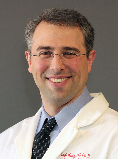 Max B. Kelz合作论文数Department of Anesthesiology and Critical Care, Perelman School of Medicine, University of Pennsylvania35
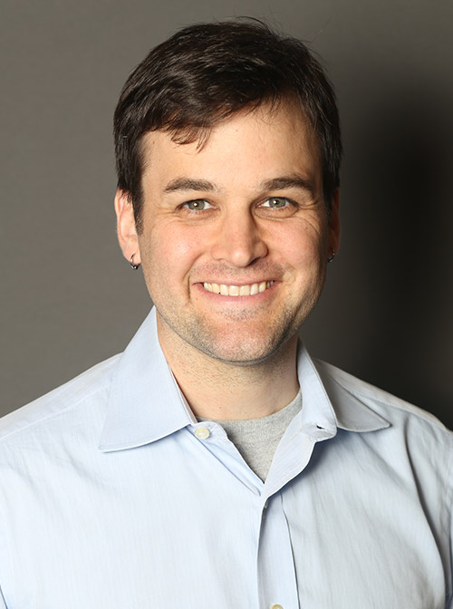 Andrew R. McKinstry-Wu合作论文数Department of Anesthesiology and Critical Care, Perelman School of Medicine, University of Pennsylvania14
 Andrzej Z Wasilczuk合作论文数Dept Anesthesiol & Crit Care, Univ Penn13Klaudiusz R. Weiss合作论文数Department of Physiology and Biophysics, Mount Sinai School of Medicine12
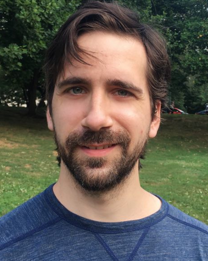 Connor Brennan合作论文数Ego9Adeeti Aggarwal合作论文数Saratoga High School9Shortal Brenna合作论文数8
 Diego Contreras合作论文数Department of Neuroscience, Perelman School of Medicine, University of Pennsylvania7
 Jian Jing (景键)合作论文数School of Life Sciences, Nanjing University7Vladimir Brezina合作论文数7
 Hugh C. Hemmings合作论文数Department of Anesthesiology, Weill Cornell Medical College;Weill Medical College, Cornell University4
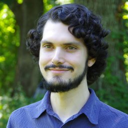 Ethan Blackwood合作论文数University of Minnesota4
 Leandro M Alonso合作论文数Department of Physics, UBA4
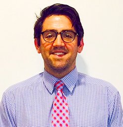 Cichon Joseph合作论文数4
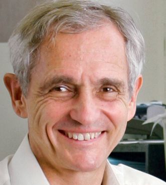 Donald W. Pfaff合作论文数Laboratory of Neurobiology and Behavior, The Rockefeller University4
 Guillermo Cecchi合作论文数IBM Research;PREDiCTOR Center4Yuriy Zhurov合作论文数4
 Michael Avidan合作论文数Division of Education, Department of Anesthesiology, School of Medicine, Washington University in St. Louis;Institute of Clinical and Translational Sciences, School of Medicine, Washington University in St. Louis;Roy and Diana Vagelos Division of Biology & Biomedical Sciences, School of Medicine, Washington University in St. Louis4
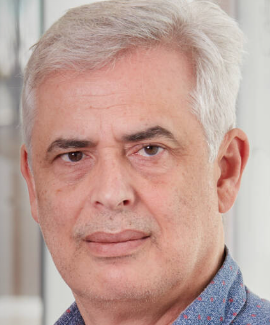 Marcelo O. Magnasco合作论文数Laboratory of Integrative Neuroscience, Rockefeller University4Jennifer Luo合作论文数3Benjamin A Harrison合作论文数3
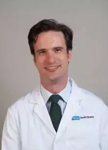 Andrew E. Hudson合作论文数University of California, Los Angeles Health3
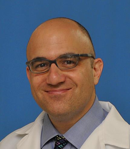 George A. Mashour合作论文数Michigan Neuroscience Institute, University of Michigan3
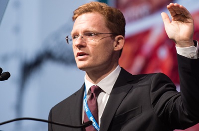 Timothy H. Lucas合作论文数Translational Neuromodulation Lab in the Perelman School of Medicine at the University of Pennsylvania3
 Kane O Pryor合作论文数Weill Medical College, Cornell University3
 Toru Yanagawa合作论文数Institute of Clinical Medicine, University of Tsukuba3
 Guillermo Solovey合作论文数3
 Andrew Richardson合作论文数School of Informatics, Computing, and Cyber Systems, Northern Arizona University3
 Elizabeth C Cropper合作论文数Puerto Rico Medical Science Campus;Fishberg Research Center for Neurobiology, Mount Sinai School of Medicine;Fishberg Research Center for Neurobiology, Mount Sinai School of Medicine3Adarli Romero合作论文数Institute of Neurobiology, University of Puerto Rico3Charles W. Carspecken合作论文数Department of Pediatrics, University of Colorado Health Sciences Center3Paula Kwasniewska合作论文数2Cole Rinehart合作论文数2Eric Hudgins合作论文数2Cameron Brandon合作论文数2Nataliya Kozlova合作论文数2Colin G Evans合作论文数2Helen Chung合作论文数2Bo Ku合作论文数2Connor Brennen合作论文数2Andrezj Wasilczuk合作论文数2
 L Brian Hickman合作论文数David Geffen School of Medicine, UCLA2Sarah L Reitz合作论文数2Vivek Buch合作论文数Dept Neurosurg, Stanford Univ2
 Diany Paola Calderon合作论文数Department of Anesthesiology, Weill Cornell Medicine, Cornell University2
 Joel Stein合作论文数Department of Rehabilitation Medicine, Weill Cornell Medicine, Cornell University2
 Aeyal Raz合作论文数Anesthesia Department, Rambam Medical Center;Department of Anesthesiology and Intensive Care, Rappaport School of Medicine, Technion2Rosen S C合作论文数2
 Ramayya Ashwin G合作论文数Department of Psychology, University of Pennsylvania2




