The thalamus comprises multiple nuclei that support higher-order cognitive functions. However, its internal architecture remains difficult to delineate using conventional T1- or T2-weighted MRI because of limited tissue contrast. Diffusion-weighted MRI provides richer microstructural detail, yet accurate segmentation is still challenged by low anisotropy and tissue heterogeneity. To address these challenges, we present a modified spectral clustering framework for thalamic segmentation. Our approach jointly leverages voxel-wise information and fiber orientation distribution (FOD) features derived from multi-shell multi-tissue constrained spherical deconvolution. When evaluated using spatial probabilistic maps that capture across-subject spatial variability in labels, k-means and spectral clustering exhibit broadly similar group-level variability patterns. However, the spectral clustering framework accommodates smaller thalamic subdivisions, including the lateral and medial geniculate nuclei (LGN and MGN), which required exclusion from the k-means configuration for stable parcellation. Under these conditions, spectral clustering achieved Dice scores of 0.73 for the mediodorsal-parafascicular (MD-Pf) complex and 0.51 for the ventral posterolateral (VPL) nucleus and produce a cluster corresponding to LGN. Furthermore, by combining structural and diffusion information, our approach enabled subdivision of the pulvinar into four distinct regions. These result position our modified spectral clustering as a robust and anatomically informed tool for thalamic clustering and pulvinar sub-segmentation.
Leveraging diffusion tractography, connectivity-based parcellation (CBP) is one of the oldest methods for thalamic nuclei segmentation. The goal of this work was to reassess CBP using higher spatial resolution diffusion MRI data and reconstruction algorithms, and to compare it with recent state-of-the-art methods for thalamic nuclei segmentation. Furthermore, these methods were systematically evaluated against three histological atlases and one functional MRI-based atlas to examine their relative anatomical similarities and differences. High resolution diffusion and T1-weighted MRI data from 67 healthy individuals in the Human Connectome Project Young Adult database were analyzed. CBP was performed using probabilistic tractography with cortical targets derived from combining labels of the Human Connectome Project Multi-Modal Parcellation 1.0 atlas into 8, 11, and 23 regions. Results were compared against three recent methods: orientation distribution function clustering (ODF), track density imaging (TDI), and structural MRI-based segmentation. Group level analyses were conducted in the Montreal Neurological Institute space, and Dice overlap coefficients were calculated using four atlases (three histological, one functional). CBP results using newer data and methods were still remarkably similar to the original CBP parcellation results. Across atlases, a consistent hierarchy was observed: HIPS-THOMAS performed best, followed by TDI, ODF, and CBP (Kendall's W = 1.00, p = 0.007). Histological atlases showed strong mutual agreement (Pearson r = 0.71-0.85), whereas the Zhang atlas demonstrated lower concordance (Pearson r = 0.51-0.63). Despite methodological advances, CBP remains constrained in its ability to delineate thalamic nuclei with histological accuracy. By contrast, structural and diffusion microstructural approaches provided better nuclear localization. These findings highlight the need for hybrid workflows that integrate structural and diffusion-based information to enable more reliable thalamic segmentation for neuroscience research.
AIMS:Purpose: Dictionary matching is a standard tool in quantitative MRI (qMRI), but typically lacks uncertainty quantification (UQ). This is critical when advanced reconstructions (e.g., compressed sensing, deep learning) introduce complex-valued, spatially varying, and temporally correlated noise that violates standard assumptions of independent and identically distributed (iid) noise. METHODS:Two voxel-wise uncertainty methods: a frequentist Likelihood Ratio Test (LRT) and a Bayesian marginal posterior approach, are introduced. Noise is modeled as spatially varying and temporally correlated using the covariance estimated from background regions. Methods were validated via simulations and phantom experiments using radial turbo spin-echo ( T 2 mapping) and radial Look-Locker ( T 1 mapping). In vivo experiments characterized uncertainty under varying acceleration factors. RESULTS:Simulations confirmed both methods achieve nominal coverage rates (e.g., 95% intervals containing the true value 95% of the time) where standard iid assumptions fail. Phantom results showed excellent agreement with gold-standard spin-echo references. In vivo experiments highlighted that higher acceleration factors widen uncertainty intervals for T 1 and T 2 . The LRT method proved more computationally efficient than the Bayesian approach while providing comparable interval estimates. CONCLUSION:A robust framework is presented for UQ in dictionary-matched qMRI. By modeling the non-iid noise inherent in modern reconstructions, these methods provide statistically interpretable UQ that assesses the reliability of parameter maps in clinical settings.
This work describes and evaluates an efficient radial gradient- and spin-echo (RADGRASE) pulse sequence and reconstruction algorithm for concurrent measurement of proton-density weighted fat fraction (FF) and water component T2 (T2w) within breast tissues. The ability to estimate T2w in breast tissues, where fat can be highly abundant, is demonstrated using oil/gel phantoms across a wide range of FF values (0.1-0.7). Successful T2w mapping of breast tissues is also demonstrated in vivo by comparison with fat suppressed T2 values. The sensitivity of RADGRASE to detect changes in the breast was assessed by tracking T2w in 3 healthy volunteers through their menstrual cycle, demonstrating T2w values in the late luteal phase to be 18-29 ms higher than in the follicular phase. The technique is also applied to a cohort of 68 patients taking tamoxifen for breast cancer risk reduction, where significant positive correlation between the FF parameter Frac50 and T2w (p = 0.035) was observed in premenopausal subjects (n = 20). Our findings demonstrate the ability and efficacy of RADGRASE for simultaneously mapping FF and T2w within breast tissues, and the potential utility of the technique in studying breast tissue changes in clinical applications.
T2 mapping allows for the classification of focal liver lesions, differentiating malignancies from the most common benign liver lesions, hemangiomas, and bile duct hamartomas (BDH). Partial volume (PV) due to the presence of liver and lesion within the same voxel confounds the classification of small lesions. Our objective is to develop a robust two-component T2 estimation technique (SEPG2-SP) to enable accurate T2 estimation in the presence of PV. T2 estimation accuracy was evaluated using computer simulations, physical phantom data, and in vivo in 27 subjects with focal liver lesions (16 males, 62.4 ± 14.3 years old; 11 females, 66.8 ± 5.8 years old) imaged at 1.5 T with a radial turbo spin-echo (RADTSE) technique. The SEPG2-SP model was compared to a single-component model, which does not account for PV. The area under the receiver operator characteristic curve (AUROC) was used to analyze lesion classification. Phantom data showed that the SEPG2-SP model had a T2 estimation error of 2–9
Accurate thalamic nuclei segmentation is critical for neuroscience research and clinical interventions such as deep brain stimulation and magnetic resonance guided focused ultrasound. Connectivity based parcellation has been widely used for two decades, yet its anatomical validity remains uncertain compared with newer imaging approaches. Methods:We analyzed high resolution diffusion magnetic resonance imaging (MRI) and T1 weighted data from 67 healthy young adults in the Human Connectome Project. Connectivity based parcellation was performed using probabilistic tractography with cortical targets derived from the HCP MMP1 atlas, generating 8, 11, and 23 region parcellations. Results were compared against three state of the art methods: orientation distribution function (ODF) clustering, track density imaging (TDI), and the structural MRI based segmentation. Group level analyses were conducted in Montreal Neurological Institute and Hospital (MNI) space, and Dice overlap coefficients were calculated against the histology based Morel atlas. Results:Connectivity based parcellation demonstrated limited anatomical precision, with increasing cortical target counts introducing greater variability and noise without improving nuclear boundary definition. ODF clustering and TDI recovered subdivisions consistent with cytoarchitectonic patterns, particularly in the pulvinar and mediodorsal nuclei. Structural MRI based segmentation achieved the highest overall Dice coefficients, closely approximating Morel defined boundaries, while Connectivity based parcellation consistently underperformed across nuclei. Conclusion:Despite methodological advances, Connectivity based parcellation remains constrained in its ability to delineate thalamic nuclei with histological accuracy. By contrast, structural and diffusion microstructural (ODF, TDI) approaches provide superior nuclear localization. These findings highlight the need for hybrid workflows that integrate structural and diffusion based information to enable more reliable thalamic segmentation for research and clinical targeting applications.
Objective.Deep neural networks have been shown to be very effective at artifact reduction tasks such as magnetic resonance imaging (MRI) reconstruction from undersampled k-space data. In recent years, attention-based vision transformer models have been shown to outperform purely convolutional models at a wide variety of tasks, including MRI reconstruction. Our objective is to investigate the use of different transformer architectures for multi-channel cascaded MRI reconstruction.Approach.In this work, we explore the effective use of cascades of small transformers in multi-channel undersampled MRI reconstruction. We introduce overlapped attention and compare it to hybrid attention in shifted-window (Swin) transformers. We also investigate the impact of the number of Swin transformer layers in each architecture. The proposed methods are compared to state-of-the-art MRI reconstruction methods for undersampled reconstruction on standard 3T and low-field (0.3T) T1-weighted MRI images at multiple acceleration rates.Main results.The models with overlapped attention achieve significantly higher or equivalent quantitative test metrics compared to state-of-the-art convolutional approaches. They also show more consistent reconstruction performance across different acceleration rates compared to their hybrid attention counterparts. We have also shown that transformer architectures with fewer layers can be as effective as those with more layers when used in cascaded MRI reconstruction problems.Significance.The feasibility and effectiveness of cascades of small transformers with overlapped attention for MRI reconstruction is demonstrated without incorporating pre-training of the transformer on ImageNet or other large-scale datasets.
Diffusion tensor imaging (DTI) is essential for assessing brain microstructure but requires long acquisition times, limiting clinical use. Recent deep learning (DL) approaches, such as SuperDTI or deepDTI, improve DTI metrics but demand large, high-quality datasets for training. We propose a self-supervised deep learning with fine-tuning (SSDLFT) framework to reduce training data requirements. SSDLFT involves self-supervised pretraining, which denoises data without clean labels, followed by fine-tuning with limited high-quality data. Experiments using Human Connectome Project data show that SSDLFT outperforms traditional methods and other DL approaches in qualitative and quantitative assessments of DWI reconstructions and tensor metrics. SSDLFT’s ability to maintain high performance with fewer training subjects and DWIs presents a significant advancement, enhancing DTI’s practical applications in clinical and research settings.
PURPOSE:To accelerate respiratory triggered free-breathing T2 mapping of the abdomen while maintaining high-quality anatomical images, accurate T2 maps, and fast reconstruction times. METHODS:We developed a flexible deep learning framework that can be trained in a fully supervised manner to improve T2-weighted images or in a self-supervised manner to reconstruct T2 maps. RESULTS:For retrospectively undersampled data, anatomical images and T2 maps reconstructed by the proposed deep learning method demonstrated reduced voxel-wise error compared to existing traditional and compressed sensing techniques. Reconstruction times were approximately 1 s per slice, significantly faster than existing compressed sensing techniques. Prospectively undersampled data were also acquired to assess the model. CONCLUSION:The proposed deep-learning framework reconstructed high-quality anatomical images and accurate T2 maps from datasets undersampled to only 160 total radial views (5 views per echo time), enabling full liver coverage in under three minutes on average with per-slice reconstruction times of approximately one second.
Purpose The purpose of this study was to develop DiffDL, a generative diffusion probabilistic model designed to produce high-quality diffusion tensor imaging (DTI) and diffusion kurtosis imaging (DKI) metrics from a reduced set of diffusion-weighted images (DWIs). This model addresses the challenge of prolonged data acquisition times in diffusion MRI while preserving metric accuracy. Methods DiffDL was trained using data from the Human Connectome Project, including 300 training/validation subjects and 50 testing subjects. High-quality DTI and DKI metrics were generated using many DWIs and combined with subsets of DWIs to form training pairs. A UNet architecture was used for denoising, trained over 500 epochs with a linear noise schedule. Performance was evaluated against conventional DTI/DKI modeling and a reference UNet model using normalized mean absolute error (NMAE), peak signal-to-noise ratio (PSNR), and Pearson correlation coefficient (PCC). Results DiffDL showed significant improvements in the quality and accuracy of fractional anisotropy (FA) and mean diffusivity (MD) maps compared to conventional methods and the baseline UNet model. For DKI metrics, DiffDL outperformed conventional DKI modeling and the UNet model across various acceleration scenarios. Quantitative analysis demonstrated superior NMAE, PSNR, and PCC values for DiffDL, capturing the full dynamic range of DTI and DKI metrics. The generative nature of DiffDL allowed for multiple predictions, enabling uncertainty quantification and enhancing performance. Conclusion The DiffDL framework demonstrated the potential to significantly reduce data acquisition times in diffusion MRI while maintaining high metric quality. Future research should focus on optimizing computational demands and validating the model with clinical cohorts and standard MRI scanners.
BACKGROUND:Contrastive learning, a successful form of representational learning, has shown promising results in pretraining deep learning (DL) models for downstream tasks. When working with limited annotation data, as in medical image segmentation tasks, learning domain-specific local representations can further improve the performance of DL models.PURPOSE:In this work, we extend the contrastive learning framework to utilize domain-specific contrast information from unlabeled Magnetic Resonance (MR) images to improve the performance of downstream MR image segmentation tasks in the presence of limited labeled data.METHODS:The contrast in MR images is controlled by underlying tissue properties (e.g., T1 or T2) and image acquisition parameters. We hypothesize that learning to discriminate local representations based on underlying tissue properties should improve subsequent segmentation tasks on MR images. We propose a novel constrained contrastive learning (CCL) strategy that uses tissue-specific information via a constraint map to define positive and negative local neighborhoods for contrastive learning, embedding this information in the representational space during pretraining. For a given MR contrast image, the proposed strategy uses local signal characteristics (constraint map) across a set of related multi-contrast MR images as a surrogate for underlying tissue information. We demonstrate the utility of the approach for downstream: (1) multi-organ segmentation tasks in T2-weighted images where a DL model learns T2 information with constraint maps from a set of 2D multi-echo T2-weighted images (n = 101) and (2) tumor segmentation tasks in multi-parametric images from the public brain tumor segmentation (BraTS) (n = 80) dataset where DL models learn T1 and T2 information from multi-parametric BraTS images. Performance is evaluated on downstream multi-label segmentation tasks with limited data in (1) T2-weighted images of the abdomen from an in-house Radial-T2 (Train/Test = 30/20), (2) public Cartesian-T2 (Train/Test = 6/12) dataset, and (3) multi-parametric MR images from the public brain tumor segmentation dataset (BraTS) (Train/Test = 40/50). The performance of the proposed CCL strategy is compared to state-of-the-art self-supervised contrastive learning techniques. In each task, a model is also trained using all available labeled data for supervised baseline performance.RESULTS:The proposed CCL strategy consistently yielded improved Dice scores, Precision, and Recall metrics, and reduced HD95 values across all segmentation tasks. We also observed performance comparable to the baseline with reduced annotation effort. The t-SNE visualization of features for T2-weighted images demonstrates its ability to embed T2 information in the representational space. On the BraTS dataset, we also observed that using an appropriate multi-contrast space to learn T1+T2, T1, or T2 information during pretraining further improved the performance of tumor segmentation tasks.CONCLUSIONS:Learning to embed tissue-specific information that controls MR image contrast with the proposed constrained contrastive learning improved the performance of DL models on subsequent segmentation tasks compared to conventional self-supervised contrastive learning techniques. The use of such domain-specific local representations could help understand, improve performance, and mitigate the scarcity of labeled data in MR image segmentation tasks.
Efficient abdominal coverage with T1-mapping methods currently available in the clinic is limited by the breath hold period (BHP) and the time needed for T1 recovery. This work develops a T1-mapping framework for efficient abdominal coverage based on rapid T1 recovery curve (T1RC) sampling, slice-selective inversion, optimized slice interleaving, and a convolutional neural network (CNN)-based T1 estimation. The effect of reducing the T1RC sampling was evaluated by comparing T1 estimates from T1RC ranging from 0.63 to 2.0 s with reference T1 values obtained from T1RC = 2.5-5 s. Slice interleaving methodologies were evaluated by comparing the T1 variation in abdominal organs across slices. The repeatability of the proposed framework was demonstrated by performing acquisition on test subjects across imaging sessions. Analysis of in vivo data based on retrospectively shortening the T1RC showed that with the CNN framework, a T1RC = 0.84 s yielded T1 estimates without significant changes in mean T1 (p > 0.05) or significant increase in T1 variability (p > 0.48) compared to the reference. Prospectively acquired data using T1RC = 0.84 s, an optimized slice interleaving scheme, and the CNN framework enabled 21 slices in a 20 s BHP. Analyses across abdominal organs produced T1 values within 2% of the reference. Repeatability experiments yielded Pearson's correlation, repeatability coefficient, and coefficient of variation of 0.99, 2.5%, and 0.12%, respectively. The proposed T1 mapping framework provides full abdominal coverage within a single BHP.
Radial trajectories are appealing for efficient parameter mapping due to their robustness to undersampling compared to Cartesian trajectories. However, streaks due to scanner imperfections can significantly affect image quality and accuracy of parameter maps. Previously, we developed a streak reduction technique named CACTUS. CACTUS was demonstrated in cross-sectional (axial) abdominal parameter imaging. The other two imaging planes (coronal and sagittal) in radial MRI are more vulnerable to these anomaly streaks. In this work, we demonstrate the benefits of CACTUS in all three imaging orientations and investigate the effect of undersampling on the presence of streaks and associated parameter maps.
Radial imaging is appealing for quantitative parameter mapping, due to their inherently robustness to undersampling compared to Cartesian trajectories and thus, its ability to yield parameter maps with high spatial resolution from highly undersampled data. Two-dimensional radial imaging typically acquires equiangular spaced lines, resulting in a circular field-of-view (FOV). Circular FOVs are not ideal for imaging applications with an anisotropic region-of-support (e.g., spine, leg). Larson et al. proposed an algorithm for designing fully sampled radial trajectories matching the prescribed anisotropic FOV. In this work we investigate the benefits of anisotropic FOV in radial MRI in the context of parameter mapping.
Obtaining manual annotations for large datasets for supervised training of deep learning (DL) models is challenging. The availability of large unlabeled datasets compared to labeled ones motivate the use of self-supervised pretraining to initialize DL models for subsequent segmentation tasks. In this work, we consider two pre-training approaches for driving a DL model to learn different representations using: a) regression loss that exploits spatial dependencies within an image and b) contrastive loss that exploits semantic similarity between pairs of images. The effect of pretraining techniques is evaluated in two downstream segmentation applications using Magnetic Resonance (MR) images: a) liver segmentation in abdominal T2-weighted MR images and b) prostate segmentation in T2-weighted MR images of the prostate. We observed that DL models pretrained using self-supervision can be finetuned for comparable performance with fewer labeled datasets. Additionally, we also observed that initializing the DL model using contrastive loss based pretraining performed better than the regression loss.
Background T2 mapping is of great interest in abdominal imaging but current methods are limited by low resolution, slice coverage, motion sensitivity, or lengthy acquisitions. Purpose Develop a radial turbo spin‐echo technique with refocusing variable flip angles (RADTSE‐VFA) for high spatiotemporal T2 mapping and efficient slice coverage within a breath‐hold and compare to the constant flip angle counterpart (RADTSE‐CFA). Study Type Prospective technical efficacy. Subjects Testing performed on agarose phantoms and 12 patients. Focal liver lesion classification tested on malignant ( N = 24) and benign ( N = 11) lesions. Field Strength/Sequence 1.5 T/RADTSE‐VFA, RADTSE‐CFA. Assessment A constrained objective function was used to optimize the refocusing flip angles. Phantom and/or in vivo data were used to assess relative contrast, T2 estimation, specific absorption rate (SAR), and focal liver lesion classification. Statistical Tests t ‐Tests or Mann–Whitney Rank Sum tests were used. Results Phantom data did not show significant differences in mean relative contrast ( P = 0.10) and T2 accuracy ( P = 0.99) between RADTSE‐VFA and RADTSE‐CFA. Adding noise caused T2 overestimation predominantly for RADTSE‐CFA and low T2 values. In vivo results did not show significant differences in mean spleen‐to‐liver ( P = 0.62) and kidney‐to‐liver ( P = 0.49) relative contrast between RADTSE‐VFA and RADTSE‐CFA. Mean T2 values were not significantly different between the two techniques for spleen (T2 VFA = 109.2 ± 12.3 msec; T2 CFA = 110.7 ± 11.1 msec; P = 0.78) and kidney‐medulla (T2 VFA = 113.0 ± 8.7 msec; T2 CFA = 114.0 ± 8.6 msec; P = 0.79). Liver T2 was significantly higher for RADTSE‐CFA (T2 VFA = 52.6 ± 6.6 msec; T2 CFA = 60.4 ± 8.0 msec) consistent with T2 overestimation in the phantom study. Focal liver lesion classification had comparable T2 distributions for RADTSE‐VFA and RADTSE‐CFA for malignancies ( P = 1.0) and benign lesions ( P = 0.39). RADTSE‐VFA had significantly lower SAR than RADTSE‐CFA increasing slice coverage by 1.5. Data Conclusion RADTSE‐VFA provided noise‐robust T2 estimation compared to the constant flip angle counterpart while generating T2‐weighted images with comparable contrast. The VFA scheme minimized SAR improving slice efficiency for breath‐hold imaging. Level of Evidence 2 Technical Efficacy Stage 1
In recent years, significant research has been performed on developing powerful and efficient Convolutional Neural Network (CNN) architectures. To utilize these architectures in pixel-level regression tasks such as tomographic image reconstruction, a feature extraction encoder is often combined with a symmetrical decoder to generate an encoder-decoder structure, such as a U-Net. However, a more powerful decoder focusing on high-frequency features can provide higher pixel-level accuracy. In this work, we investigate the use of asymmetrical encoder-decoder architectures in medical image reconstruction tasks. The state-of-the-art EfficientNet architecture utilizes depthwise convolutions and channel attention within inverted residual bottleneck blocks to generate highly compressed features while maintaining a significant FLOPS efficiency advantage compared to regular convolutional encoders. We develop an asymmetric encoder-decoder architecture, which uses the EfficientNet as an encoder. The proposed decoder architecture combines the multi-resolution features generated by the EfficientNet encoder using an incremental feature expansion strategy, which leads to better preservation of the structural details in reconstructed images. We have tested our asymmetrical encoder-decoder approach on undersampled MRI reconstruction tasks using the Calgary Campinas multi-channel brain MR dataset. Results demonstrate that the proposed asymmetric approach vastly outperforms a symmetric Efficient U-Net, achieving a 3dB improvement in PSNR. SSIM was also improved, and the asymmetric network was found to recover small structural details more effectively. Furthermore, the proposed asymmetric Efficient U-Net provides a four-fold reduction in inference time when compared to the conventional U-Net architecture.
Purpose In radial abdominal imaging, it has been commonly observed that signal from the arms cause streaks due to system imperfections. We previously introduced a streak removal technique (B‐STAR), which is inherently spatially variant and limited to work in image space. In this work, we propose a spatially invariant streak cancellation technique (CACTUS), which can be applied in either image space or k‐space and is compatible with iterative reconstructions. Theory and Methods Streak sources are typically spatially localized and can be represented using a low‐dimensional subspace. CACTUS identifies the streak subspace by leveraging the spatial redundancy of receiver coils and projects the data onto the streak null space to eliminate the streaks. When applied in k‐space, CACTUS can be combined with iterative reconstructions. CACTUS was tested in phantoms and in vivo abdominal imaging using a radial turbo spin‐echo pulse sequence. Results In phantoms, CACTUS improved T2 estimation in comparison to previous de‐streaking methods. In vivo experiments showed that CACTUS reduced streaks and yielded T2 estimation, in regions affected by streaks, closer to a streak‐free reference. Evaluation using a clinical abdominal dataset ( n = 20) showed that CACTUS is comparable to B‐STAR and yields significantly better signal preservation and streak cancellation than coil removal and suppression methods. Conclusion CACTUS provides superior signal preservation and streak reduction performance compared to coil removal and suppression methods. As a clear advantage over B‐STAR, CACTUS can be integrated with iterative reconstruction methods. In abdominal T2 mapping, CACTUS improves the accuracy of parameter estimation in areas affected by streaks.
Purpose: To develop a fast volumetric T1 mapping technique. Materials and methods: A stack-of-stars (SOS) Look Locker technique based on the acquisition of undersampled radial data ( 30x relative to Nyquist) and an efficient multi-slab excitation scheme is presented. A principalcomponent based reconstruction is used to reconstruct T1 maps. Computer simulations were performed to determine the best choice of partitions per slab and degree of undersampling. The technique was validated in phantoms against reference T1 values measured with a 2D Cartesian inversion-recovery spin-echo technique. The SOS Look Locker technique was tested in brain (n = 4) and prostate (n = 5). Brain T1 mapping was carried out with and without kz acceleration and results between the two approaches were compared. Prostate T1 mapping was compared to standard techniques. A reproducibility study was conducted in brain and prostate. Statistical analyses were performed using linear regression and Bland Altman analysis. Results: Phantom T1 values showed excellent correlations between SOS Look Locker and the inversion-recovery spin-echo reference (r2 = 0.9965; p < 0.0001) and between SOS Look Locker with slab-selective and non-slab selective inversion pulses (r2 = 0.9999; p < 0.0001). In vivo results showed that full brain T1 mapping (1 mm3) with kz acceleration is achieved in 4 min 21 s. Full prostate T1 mapping (0.9 x 0.9 x 4 mm3) is achieved in 2 min 43 s. T1 values for brain and prostate were in agreement with literature values. A reproducibility study showed coefficients of variation in the range of 0.18-0.2% (brain) and 0.15-0.18% (prostate). Conclusion: A rapid volumetric T1 mapping technique was developed. The technique enables high-resolution T1 mapping with adequate anatomical coverage in a clinically acceptable time.
 Michael W. Marcellin合作论文数Department of Electrical & Computer Engineering, College of Engineering, The University of Arizona111
 Maria I. Altbach合作论文数86
 Diego R. Martin合作论文数25Mariappan S. Nadar合作论文数24Mahesh B Keerthivasan合作论文数Siemens Medical Solutions USA Inc20
 Kevin M. Johnson合作论文数Department of Medical Physics, School of Medicine and Public Health, University of Wisconsin-Madison;Department of Radiology, School of Medicine and Public Health, University of Wisconsin-Madison18Eze Ahanonu合作论文数Univ Arizona, Dept Elect & Comp Engn, Tucson, AZ 85721 USA18
 Zhenyu Wu合作论文数15Jean‐Philippe Galons合作论文数13
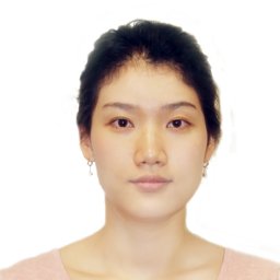 Yookyung Kim合作论文数University of Pittsburgh School of Nursing12
 Han Oh合作论文数12Vibhas S. Deshpande合作论文数11L Umapathy合作论文数Dept Med Imaging, Univ Arizona11
 Zhiyang Fu合作论文数United Imaging - North America11Sagar Mandava合作论文数Dept Med Imaging, Univ Arizona11
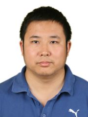 Feng Liu (刘峰)合作论文数College of Electronic Information and Optical Engineering, Nankai University11
 Jim Storer合作论文数Computer Science Department, Michtom School of Computer Science, Brandeis University11
 Amit Ashok合作论文数The University of Arizona11Brian Toner合作论文数Program Appl Math, Univ Arizona10
 Joan Serra-Sagristà合作论文数Department of Information and Communications Engineering, School of Engineering, Universitat Autònoma de Barcelona10
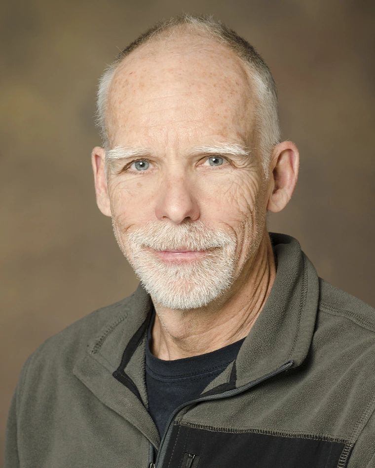 Arthur F. Gmitro合作论文数Department of Biomedical Engineering, College of Engineering, The University of Arizona10
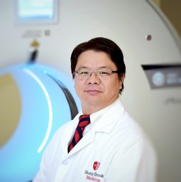 Chuan Huang合作论文数10
 Joseph C. Dagher合作论文数10Diego Martín de Andrés合作论文数9Zhitao Li合作论文数9
 Hariharan G. Lalgudi合作论文数Signal Processing and Coding Lab;Department of Electrical and Computing Engineering;University of Arizona;Signal Processing and Coding Lab, University of Arizona9Christian G. Graff合作论文数Center for Devices and Radiological Health, U. S. Food and Drug Administration9
 Eric W. Clarkson合作论文数Department of Radiology and Optical Sciences Center, The University of Arizona8
 Manojkumar Saranathan合作论文数Indian Institute of Science8Yuzhang Lin合作论文数Univ Arizona, Dept Elect & Comp Engn, Tucson, AZ 85721 USA7
 Lingling Pu合作论文数7Tp Trouard合作论文数6Bobby R. Hunt合作论文数Department of Electrical and Computer Engineering, The University of Arizona6
 Puneet Sharma合作论文数Siemens Healthineers6Zhiqiang Li合作论文数5
 Srinivasan Vedantham合作论文数UMass Memorial Medical Center, University of Massachusetts Medical School5Theodore P. Trouard合作论文数5Z. Li合作论文数4K Panchapakesan合作论文数4Mi Altbach合作论文数4Pj Sementilli合作论文数4benjamin p berman合作论文数4
 Abhishek Pandey合作论文数4David G. Sheppard合作论文数4
 Karthik Krishnan合作论文数Signal Processing and Coding Laboratory, University of Arizona;Signal Processing and Coding Laboratory, University of Arizona4
 Bane Vasić合作论文数Department of Electrical and Computer Engineering, College of Engineering, University of Arizona;Error Correction Laboratory, University of Arizona4
 Nathan A. Goodman合作论文数School of Electrical and Computer Engineering, The University of Oklahoma4Hüseyin Abut合作论文数4B A Winegar合作论文数3




