Bilateral posterior C1 lateral mass–C2 pedicle screw fixation is the standard surgical treatment for unstable traumatic odontoid fractures. However, unilateral fixation may be required when bilateral instrumentation cannot be safely completed because of anatomical constraints or intraoperative complications. This study compared the clinical and radiological outcomes of unilateral and bilateral posterior C1–C2 fixation in patients with traumatic odontoid fractures. This retrospective comparative cohort study included 43 adult patients who underwent posterior C1 lateral mass–C2 pedicle screw fixation for traumatic odontoid fractures between 2016 and 2024. Fourteen patients underwent unilateral fixation and 29 underwent bilateral fixation. The primary outcome was CT-confirmed fusion. Secondary outcomes included pain, neurological function, radiological parameters, operative variables, complications, and reoperation. Longitudinal radiological outcomes were evaluated using baseline-adjusted linear mixed-effects models where appropriate. CT-confirmed fusion was achieved in 92.9
Introduction:Lumbar microdiscectomy is an established treatment for symptomatic lumbar disc herniation; however, persistent pain and functional limitation may occur, particularly in patients with annular defects. Research question:Is adjunctive external bipolar thermal annuloplasty (EBTA) associated with improved outcomes after lumbar microdiscectomy in patients with Carragee Type I and III annular defects? Material and methods:This retrospective single-center matched cohort study included 60 patients with symptomatic single-level lumbar disc herniation and intraoperatively confirmed Carragee Type I or III annular defects. Thirty patients underwent microdiscectomy with adjunctive EBTA and were matched 1:1 with 30 patients treated with microdiscectomy alone based on age, sex, operated level, Carragee classification, baseline VAS-leg, VAS-back, and Oswestry Disability Index (ODI) scores. Outcomes were assessed preoperatively and at 6 and 12 months. Exploratory clinical relevance was assessed using MCID thresholds of ≥4.7 points for VAS-leg and >20 points for ODI. Results:Both groups improved significantly in VAS-leg, VAS-back, and ODI scores (p < 0.001). Compared with controls, the EBTA group had significantly lower VAS-leg, VAS-back, and ODI scores at both follow-ups. VAS-back decreased from 5.84 ± 1.17 to 1.20 ± 0.49 at 12 months in the EBTA group and from 5.96 ± 1.08 to 2.06 ± 0.72 in controls. EBTA was also associated with earlier return to work and higher satisfaction, without increased complications. Mean ODI improvement exceeded the MCID threshold in both groups, whereas VAS-leg exceeded the threshold at both follow-ups only in the EBTA group. Discussion and conclusion:Adjunctive EBTA was associated with greater improvements in leg pain, back pain, and disability without increased perioperative morbidity. These exploratory findings require confirmation in larger prospective studies.
This study aims to compare the risk of instrumentation failure between dynamic and rigid posterior spinal stabilization systems using propensity score matched cohorts, and to investigate the role of spinopelvic parameters and patient-related factors in construct longevity. A retrospective analysis was conducted on 952 patients who underwent thoracic, thoracolumbar, thoracolumbosacral, lumbar, or lumbosacral instrumentation between 2018 and 2024. Instrumentation failure (rod fracture, screw fracture, migration, pull-out, or loosening) was identified in a subgroup of 106 patients. Propensity score matching (1:1 nearest-neighbor, caliper 0.1) was applied using age, sex, osteoporosis, number of instrumented segments, and involvement of S1 as the lowest instrumented vertebra, yielding two matched cohorts of 196 dynamic and 196 rigid stabilized cases with improved baseline comparability. Survival differences were assessed by Kaplan-Meier and log-rank tests, and multivariable Cox regression was performed in both the full cohort and matched groups to identify potential risk factors. The overall failure rate was 11.1
Objectives: Retrospective comparative cohort study. This study aimed to compare the clinical and radiological outcomes of rigid, dynamic, and hybrid stabilization systems in the surgical management of degenerative lumbar spine disease, with a focus on adjacent segment disease (ASD), fusion-related complications, and postoperative recovery. Rigid fusion techniques provide immediate stability but may increase mechanical stress at adjacent segments, leading to ASD. Dynamic and hybrid constructs aim to preserve more physiological motion, potentially reducing fusion-related complications. Methods: A retrospective analysis was conducted on 86 patients who underwent posterior lumbar stabilization between January 2017 and December 2021. Patients were categorized into four groups based on the surgical technique: Rigid (titanium rod), Dynamic (PEEK rod), Rigid + TLIF (Transforaminal Lumbar Interbody Fusion), and Hybrid (Dynamic + TLIF). Clinical evaluation included the Visual Analog Scale (VAS) for back and leg pain. Radiological assessments comprised segmental and total range of motion (ROM), disc and foraminal height, fusion success, pseudoarthrosis, and the presence of ASD. Results: All surgical approaches resulted in significant postoperative improvement in back and leg pain (p < 0.001). The incidence of ASD was highest in the Rigid + TLIF group (45.5%, p < 0.001) and lowest in the Dynamic (4.8%) and Hybrid (4.2%) groups. Pseudoarthrosis was most frequently observed in the Rigid group (26.3%). The Hybrid group demonstrated favorable outcomes, with a high anterior fusion rate (91.7%) and low rates of ASD and pseudoarthrosis. Conclusions: All stabilization techniques were associated with significant postoperative clinical improvement. In this retrospective cohort, dynamic and hybrid constructs showed lower observed rates of early symptomatic ASD than the Rigid + TLIF construct, while TLIF-treated groups showed higher anterior fusion rates. Because treatment allocation was nonrandomized and adjustment for all potential confounders was not possible, these findings demonstrate associations rather than causal treatment effects. Larger prospective studies with longer follow-up are required to determine whether the stabilization strategy independently influences ASD, fusion, or pseudoarthrosis.
Background: Multisegmental lumbar degenerative disease (ms-LDD) is a common condition in older adults, often requiring surgical intervention. While rigid stabilization remains the gold standard, it is associated with complications such as adjacent segment disease (ASD), higher blood loss, and longer recovery times. The Dynesys dynamic stabilization system offers an alternative by preserving motion while stabilizing the spine. However, data comparing Dynesys with fusion in multisegmental cases are limited. Objective: This study evaluates the clinical and radiographic outcomes of Dynesys dynamic stabilization versus rigid stabilization in the treatment of ms-LDD. Methods: A retrospective analysis was conducted on 53 patients (mean age: 62.25 ± 15.37 years) who underwent either Dynesys dynamic stabilization (n = 27) or PLIF (n = 26) for ms-LDD involving at least seven motion segments. Clinical outcomes were assessed using the Visual Analog Scale (VAS) and Oswestry Disability Index (ODI), while radiological parameters such as lumbar lordosis (LL), sagittal vertical axis (SVA), and spinopelvic parameters (pelvic incidence, pelvic tilt and, sacral slope) were analyzed. A two-stage surgical approach was employed in the Dynesys group to enhance osseointegration, particularly in elderly osteoporotic patients. Results: Both groups showed significant improvements in VAS and ODI scores postoperatively (p < 0.001), with no significant differences between them. However, the Dynesys group demonstrated superior sagittal alignment correction, with a significant increase in LL (p < 0.002) and a significant decrease in SVA (p < 0.0015), whereas changes in the rigid stabilization group were not statistically significant. Additionally, the Dynesys group had fewer complications, including a lower incidence of ASD (0 vs. 6 cases). The two-stage technique facilitated improved screw osseointegration and reduced surgical risks in osteoporotic patients. Conclusions: Dynesys dynamic stabilization is an effective alternative to rigid stabilization in ms-LDD, offering comparable pain relief and functional improvement while preserving motion and reducing ASD risk. The two-stage approach enhances long-term stability, making it particularly suitable for elderly or osteoporotic patients. Further long-term studies are needed to confirm these findings.
Background and Objectives: Unilateral spinal stabilization has emerged as a less invasive alternative to bilateral fixation in the management of lateralized spinal pathologies. While both rigid and dynamic systems are utilized, comparative data regarding their clinical efficacy, radiological outcomes, and complication profiles—particularly in multilevel applications—remain limited. Materials and Methods: A retrospective, two-center analysis was conducted on 113 patients who underwent unilateral posterior spinal stabilization between 2019 and 2023. Patients were divided into unilateral rigid stabilization (URS, n = 41) and unilateral dynamic stabilization (UDS, n = 72) groups. Pathologies of the patients include disc herniations, foraminal and spinal stenosis, tumoral lesions and spondylolisthesis. Clinical outcomes were assessed using the Visual Analogue Scale (VAS) over a 24-month follow-up. Radiological parameters included fusion status, superior adjacent disc height, and foraminal height index. Complication rates, including adjacent segment degeneration (ASD), pseudoarthrosis, and screw loosening, were analyzed according to type-of-stabilization and construct length (two, three, or four levels). Results: Both URS and UDS groups demonstrated significant VAS improvement at final follow-up, with no significant differences between groups (p < 0.001). Fusion rates were significantly higher in the URS group (85.37% vs. 27.78%, p < 0.001), while pseudoarthrosis (39.02% vs. 16.62%, p = 0.081) were more frequent in URS. No cases of rod fracture or infection were observed. Complication rates, particularly ASD, increased with longer constructs (6.56%, 21.21%, vs. 31.58% p = 0.01), independent of stabilization type. Conclusions: Unilateral stabilization—whether rigid or dynamic—offers effective symptom relief with reduced surgical morbidity. However, dynamic systems may provide biomechanical advantages by preserving motion and minimizing adjacent segment stress. While rigid constructs yield higher fusion rates, they are associated with increased complications. These findings support the use of dynamic stabilization, particularly in multilevel constructs, and highlight the need for patient-specific surgical strategies to optimize outcomes and mitigate long-term complications.
Background & objectivesBasilar invagination (BI) represents a complex anomaly of the craniovertebral junction, characterized by the displacement of the odontoid process towards the foramen magnum. Current surgical interventions include anterior decompression and combined anterior-posterior decompression with posterior fusion. Traditional methods for odontoid resection encompass transoral, transnasal, and endonasal approaches. However, these techniques are fraught with significant risks. Furthermore, the restricted exposure provided by the endonasal corridor's anatomical limitations hampers surgical manipulation, prompting spine surgeons to seek alternative techniques. This report details a case of BI managed through an endoscopic posterolateral odontoidotomy, showcasing an innovative surgical approach. We aim to describe our experience in partially removing the odontoid via posterolateral approach with a novel endoscopic technique, preventing the need for additional approach and related complications.MethodsA 16-year-old male patient presented with complaints of imbalance and difficulty swallowing. Clinical examination revealed upper extremity muscle weakness, ataxic gait, and dysphagia. Upon the diagnosis of BI, a posterior occipito-cervical fusion was performed. However, six months postoperatively, the patient returned with exacerbated symptoms. During the subsequent surgical intervention, the odontoid body was resected using a posterolateral transmass endoscopic approach. Due to the patient's neck and shoulder anatomy, cranial angulation of the endoscope was restricted, necessitating the retention of the odontoid tip.ResultsPost-operative CT revealed that the tip was closer to the base and a subarachnoid space was formed. Follow-up CT after a year showed a complete migration of the tip to the base of C2 with a clear decompression of the brainstem.ConclusionOur findings demonstrate that partial or total resection of the odontoid process via a posterolateral approach is feasible using endoscopic techniques. The endoscopic posterolateral transmass odontoidotomy should be considered a viable alternative method and route for patients necessitating partial or total odontoidectomy.
Osteoporotic vertebral compression fractures (OVCFs) lead to posture restrictions, back muscle fatigue, difficulty walking, impaired lung function, an increased risk of disability, and severe pain. Collectively, these symptoms significantly diminish patients’ quality of life. While non-surgical management is often attempted, it may prove inadequate for cases involving walking difficulties and sagittal imbalance. In such instances, surgical interventions like vertebroplasty (VP) and kyphoplasty (KP) become necessary. To our knowledge, no study in the literature has directly compared VP and KP in terms of their effectiveness in improving sagittal balance. This study aims to compare the clinical and radiological outcomes of VP and KP, with a specific focus on their impact on global spinal alignment and sagittal balance. Seventy-six patients with OVCFs (mean age: 64.12 ± 11.85 years) underwent either kyphoplasty (59
 Tunc Oktenoglu合作论文数Neurosurgery Department, American Hospital124
 Mehdi Sasani合作论文数Neurosurgery Department, American Hospital108
 Mehmet Yigit Akgun合作论文数Department of Neurosurgery, Koc University Hospital34
 Ahmet Levent Aydin合作论文数Neurosurgery Department, VKV American Hospital34
 Kaner Tuncay合作论文数Dept Neurosurg, Pendik State Hosp33
 Ozkan Ates合作论文数Department of Neurosurgery, Inonu University School of Medicine33
 Deniz U. Erbulut合作论文数24
 M. Necmettin Pamir合作论文数Department of Neurosurgery, School of Medicine, Acibadem Mehmet Ali Aydınlar University22Mehdi Hekimoğlu合作论文数20
 Caner Gunerbuyuk合作论文数19
 Onur Yaman合作论文数19
 Murat Cosar合作论文数Faculty of Medicine, Canakkale 18 March University17Suzer Tuncer合作论文数16
 Salim Senturk合作论文数Department of Neurosurgery, Corum Education and Research Hospital16
 Basak Ahmet Tulgar合作论文数15M Sasani合作论文数12Hakan Bozkus合作论文数11
 Goktug Akyoldas合作论文数Department of Neurosurgery, Koc University Hospital11Canan Erzen合作论文数11Hadi Sasani合作论文数10
 Zafarparandeh Iman合作论文数10Gomleksiz Cengiz合作论文数School of Medicine, Erzincan University10Nazan Canbulat合作论文数9
 Omur Ercelen合作论文数9
 Sedat Dalbayrak合作论文数Department of Neurosurgery, Lütfi Kirdar Kartal Teaching and Research Hospital8Cerezci Onder合作论文数8
 Ataker Yaprak合作论文数8
 Vijay Goel合作论文数Engineering Center for Orthopaedic Research Excellence, Departments of Bioengineering and Orthopaedic Surgery, Colleges of Engineering and Medicine, The University of Toledo8
 A. Celal Iplikcioglu合作论文数Neurosurgery Department, Okmeydani Training Hospital8Ali Cetin Sarioglu合作论文数8G.Evren Keles合作论文数7A. C. SARIOGLU, M. Y. KAYNAR, M. HANCI合作论文数7
 Turgut Akgul合作论文数7
 Ismail Lazoglu合作论文数Department of Mechanical Engineering, Koc University7Mehmet Ali Tepebasili合作论文数6Ege Anil Ucar合作论文数6Hıdır ÖZER合作论文数6Ali C. Sarioglu合作论文数5MM Ozek合作论文数5
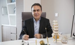 Canbay Suat合作论文数Department of Neurosurgery, Oncology Training and Research Hospital5
 Solmaz Bilgehan合作论文数Department of Neurosurgery, Istanbul Education and Research Hospital5
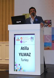 Atilla Yilmaz合作论文数Tayfur Ata Sökmen Medical Faculty, Mustafa Kemal University5
 Ulkun Unsal合作论文数5Senol Carilli合作论文数General Surgery Department, V.K.V. American Hospital5
 T. Ali Zirh合作论文数5T. Kaner合作论文数4Mehmet Kürşat Karadağ合作论文数4Idris Avci合作论文数4
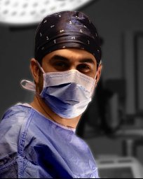 Muhammet Arif Ozbek合作论文数Neurosurgery Clinic, Haseki Training and Research Hospital4




