HIV-1 infection is enhanced by cell–cell adhesions between infected and uninfected T cells called virological synapses (VS). VS are initiated by the interactions of cell-surface HIV-1 envelope glycoprotein (Env) and CD4 on target cells and act as sites of viral assembly and viral transfer between cells. To study the process that recruits and retains HIV-1 Env at the VS, a replication-competent HIV-1 clone carrying an Env-sfGFP fusion protein was designed to enable live tracking of Env within infected cells. Combined use of surface pulse-labeling of Env and fluorescence recovery after photobleaching (FRAP) studies, enabled the visualization of the targeted accumulation and sustained recycling of Env between endocytic compartments (EC) and the VS. We observed dynamic exchange of Env at the VS, while the viral structural protein, Gag, was largely immobile at the VS. The disparate exchange rates of Gag and Env at the synapse support that the trafficking and/or retention of a majority of Env towards the VS is not maintained by entrapment by a Gag lattice or immobilization by binding to CD4 on the target cell. A FRAP study of an Env endocytosis mutant showed that recycling is not required for accumulation at the VS, but is required for the rapid exchange of Env at the VS. We conclude that the mechanism of Env accumulation at the VS and incorporation into nascent particles involves continuous internalization and targeted secretion rather than irreversible interactions with the budding virus, but that this recycling is largely dispensable for VS formation and viral transfer across the VS.
The availability of cost-effective, highly portable, and easy to use high-resolution live-cell imaging systems could present a significant technological break-through in challenging environments, such as high-level biosafety laboratories or sites where new viral outbreaks are suspected. We describe and demonstrate a cost-effective high-speed fluorescence microscope enabling the live tracking of virus particles across virological synapses that form between infected and uninfected T cells. The dynamics of HIV-1 proteins studied at the cellular level and the formation of virological synapses in living T cells reveals mechanisms by which cell-cell interactions facilitate infection between immune cells. Dual-color 3D fluorescence deconvolution microscopy of HIV-1 particles at frames rates of 100 frames per second allows us to follow the transfer of HIV-1 particles across the T cell virological synapse between living T cells. We also confirm the successful transfer of virus by imaging T cell samples fixed at specific time points during cell-cell virus transfer by super-resolution structured illumination microscopy.
Digital micromirror devices (DMDs) are spatial light modulators that employ the electro-mechanical movement of miniaturized mirrors to steer and thus modulate the light reflected off a mirror array. Their wide availability, low cost and high speed make them a popular choice both in consumer electronics such as video projectors, and scientific applications such as microscopy. High-end fluorescence microscopy systems typically employ laser light sources, which by their nature provide coherent excitation light. In super-resolution microscopy applications that use light modulation, most notably structured illumination microscopy (SIM), the coherent nature of the excitation light becomes a requirement to achieve optimal interference pattern contrast. The universal combination of DMDs and coherent light sources, especially when working with multiple different wavelengths, is unfortunately not straight forward. The substructure of the tilted micromirror array gives rise to a blazed grating, which has to be understood and which must be taken into account when designing a DMD-based illumination system. Here, we present a set of simulation frameworks that explore the use of DMDs in conjunction with coherent light sources, motivated by their application in SIM, but which are generalizable to other light patterning applications. This framework provides all the tools to explore and compute DMD-based diffraction effects and to simulate possible system alignment configurations computationally, which simplifies the system design process and provides guidance for setting up DMD-based microscopes. This article is part of the Theo Murphy meeting 'Super-resolution structured illumination microscopy (part 1)'.
Raw datasets of structured illumination microscopy measurements, accompanying the publication "Cost-effective live cell structured illumination microscopy with video-rate imaging"
ABSTRACT HIV-1 infection is enhanced by cell-cell adhesions between infected and uninfected T cells called virological synapses (VS). VS are initiated by the interactions of cell-surface HIV-1 envelope glycoprotein (Env) and CD4 on target cells and act as sites of viral assembly and viral transfer between cells. To study the process that recruits and retains HIV-1 Env at the VS, a replication-competent HIV-1 clone carrying an Env-sfGFP fusion protein was designed to enable live tracking of Env within infected cells. Using surface pulse-labeling of Env and fluorescence recovery after photobleaching (FRAP) studies, we observed targeted accumulation and sustained recycling of Env between the endocytic recycling compartment (ERC) and the VS. We observed dynamic exchange of Env at the VS while the viral structural protein, Gag, was largely immobile at the VS. The disparate exchange rates of Gag and Env at the synapse indicate that retention of Env is not likely to be maintained by entrapment into an immobile Gag lattice or through immobilizing interactions with CD4 on the target cell. A FRAP study of an Env endocytosis mutant showed that recycling is required for the rapid exchange of Env at the VS. We conclude that the mechanism of Env accumulation at the VS and incorporation into nascent particles involves continuous internalization and targeted secretion rather than irreversible interactions with the budding virus.
microscopy (SIM) allows for sub-diffraction imaging with low phototoxicity and high frame rates, making it a popular technique for live cell imaging. Current-generation instruments provide video-rate imaging speeds, and thus enable the observation of even fast dynamical processes with sub-diffraction resolution. In 2016, we released a free and open-source implementation of the SIM reconstruction algorithm [1]. Since then, the fairSIM project has been greatly extended in various ways: The reconstruction algorithm itself gained new filter options [2], the 3D data processing [3] implementation is well on its way, and microscope profiling tools such as optical transfer function generation have been added. We also pushed for the development of bespoke SIM microscopes, such as a multi-color version of the SLM-based fastSIM [4] system, a cost-effective DMD-based SIM and a modular, customizable and easily replicable SIM illumination system. All these microscope systems can now be linked to an on-the-fly reconstruction feature, which merges data acquisition and post-processing, and allows the user to obtain an immediate superresolution views of their samples. This is achieved by tight integration of the data acquisition process with a GPU-accelerated implementation of the reconstruction process. On our fastest data acquisition systems (based on [4]), we can achieve 25fps simultaneously on 3 color channels, and provide reconstructed super-resolution images within half a second after data acquisition. Here, we present an overview of the fairSIM project, its applications, features and capabilities, but also its current limitations and ongoing development. We hope to connect groups in need of bespoke, high speed SIM imaging with those developing these methods and algorithms.
We investigate the internal morphology of smart core-shell microgels by super-resolution fluorescence microscopy exploiting a combination of 3D single molecule localization and structured illumination microscopy utilizing freely diffusing fluorescent dyes. This approach does not require any direct chemical labeling and does not perturb the network structure of these colloidal gels. Hence, it allows us to study the morphology of the particles with very high precision. We found that the structure of the core-forming seed particles is drastically changed by the second synthesis step necessary for making the shell, resulting in a core region with highly increased dye localization density. The present work shows that super-resolution microscopy has great potential with respect to the study of soft colloidal systems.
Super-resolution (SR) fluorescence microscopy is typically carried out on research microscopes equipped with high-NA TIRF objectives and powerful laser light sources. Super-resolution optical fluctuation imaging (SOFI) is a fast SR technique capable of live-cell imaging, that is compatible with many wide-field microscope systems. However, especially when employing fluorescent proteins, a key part of the imaging system is a very sensitive and well calibrated camera sensor. The substantial costs of such systems preclude many research groups from employing SR imaging techniques. Here, we examine to what extent SOFI can be performed using a range of imaging hardware comprising different technologies and costs. In particular, we quantitatively compare the performance of an industry-grade CMOS camera to both state-of-the-art emCCD and sCMOS detectors, with SOFI-specific metrics. We show that SOFI data can be obtained using a cost-efficient industry-grade sensor, both on commercial and home-built microscope systems, though our analysis also readily exposes the merits of the per-pixel corrections performed in scientific cameras.
Arrhythmogenic right ventricular cardiomyopathy (ARVC) is a genetic heart disease characterized by ventricular arrhythmia, dilation of the right ventricle and fibro-fatty replacement of the myocardium [1]. ARVC is mainly caused by mutations in genes encoding desmosomal proteins. Desmosomes are cell-cell connections which are linked to the intermediate filament system contributing to the mechanical stabilization of cardiomyocytes [2]. The most prominent ARVC gene is PKP2, encoding plakophilin-2. Most ARVC associated PKP2 mutations cause premature termination codons leading to a putative truncation of the protein [3]. Plakophilin-2 is a linker protein of the Armadillo family and connects desmosomal cadherins with desmoplakin.
Injection techniques to deliver macromolecules to cells such as microinjection have been around for decades with applications ranging from probing whole organisms to the injection of fluorescent molecules into single cells. A similar technique that has raised recent interest is nanoinjection. The pipettes used here are much smaller and allow for the precise deposition of molecules into single cells via electrokinetics with minimal influence on the cells’ health. Unfortunately, the equipment utilized for nanoinjection originates from scanning ion conductance microscopy (SICM) and is therefore expensive and not portable, but usually fixed to a specific microscope setup. The level of precision that these systems achieve is much higher than what is needed for the more robust nanoinjection process. We present Mobile Nanoinjection (MoNa), a portable, cost-efficient and easy to build system for the injection of single cells. Sacrificing unnecessary sub-nanometer accuracy and low ion current noise levels, we were able to inject single living cells with high accuracy. We determined the noise of the MoNa system and investigated the injection conditions for 16 prominent fluorescent labels and fluorophores. Further, we performed proof of concepts by injection of ATTO655-Phalloidin and MitoTracker Deep Red to living human osteosarcoma (U2OS) cells and of living adult human inferior turbinate stem cells (ITSC’s) following neuronal differentiation with the MoNa system. We achieved significant cost reductions of the nanoinjection technology and gained full portability and compatibility to most optical microscopes.
We have carefully studied the blazed grating effect of DMDs by simulations, varying a range of parameters and compared the simulation results with experiments. This allowed us to identify settings which result in very high modulation contrast across all angles and phases required to generate 2-beam SIM pattern. The use of inexpensive industry-grade CMOS cameras as well as low-cost lasers enabled us to construct a cost-effective, high-speed SIM system. Reconstruction of the super-resolved SIM images is achieved on a recently demonstrated parallel-computing platform, which allowed us to visualize living cells with super-resolution at multiple reconstructed frames per second in real time. We demonstrate the versatility of this new platform by imaging cellular organelle dynamics based on live-cell fluorescent stains as well as with fluorescent protein stained samples.
Alice Sandmeyer, Mario Lachetta, Hauke Sandmeyer, Wolfgang Hübner, Thomas Huser, Marcel Müller 1 Biomolecular Photonics, Faculty of Physics, University of Bielefeld, Germany 2 Numerical Simulations and Field Theory on the Lattice, Faculty of Physics, University of Bielefeld, Germany 3 Laboratory for NanoBiology, Department of Chemistry, KU Leuven, Belgium E-Mail: asandmeyer@physik.uni-bielefeld.de
ABSTRACT Super-resolution (SR) fluorescence microscopy, especially at high speeds, is typically carried out on high-performance research microscopes. The substantial cost of such equipment, combined with the limited distribution of such instruments in imaging facilities, have complicated access to super-resolution imaging for research groups in the biosciences. In this work we promote the accessibility of Super-resolution Optical Fluctuation Imaging (SOFI) to the scientific community by demonstrating its flexibility in terms of the minimal required performance of the imaging system. We show that SOFI data can be acquired using a very cost-efficient industrial-grade detector on both a standard research microscope and an entirely home-built wide-field system. This enables more scientists to enter the field of live-cell super-resolution imaging, as it provides access to a robust and fast SR imaging modality at comparatively modest cost.
 Thomas R. Huser合作论文数Department of Physics, Faculty of Physics, University of Bielefeld/Department of Internal Medicine, University of California, Davis11
 Wolfgang Hubner合作论文数Biomol Photon, Univ Bielefeld9
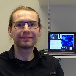 Marcel Muller合作论文数Department of Physics, University of Bielefeld8Mario Lachetta合作论文数5Hauke Sandmeyer合作论文数4Robin Van den Eynde合作论文数3
 Benjamin K Chen合作论文数Rockefeller University3
 Peter Dedecker合作论文数Department of Chemistry, Katholieke Universiteit Leuven3
 Stephan Bergmann合作论文数2
 Lili Wang合作论文数2Hongru Li合作论文数2
 Sam Duwe合作论文数Department of Chemistry, University of Leuven2
 Wim Vandenberg合作论文数Department of Chemistry, University of Leuven2
 Oliver Wrede合作论文数1Maxim Dirksen合作论文数1Andreas Markwirth合作论文数Biomol Photon, Bielefeld Univ1
 Andreas Brodehl合作论文数E. &H. Klessmann Institute for Cardiovascular Research &Ruhr-University Bochum;E. & H. Klessmann Institute for Cardiovascular Research & Development, Ruhr-University Bochum1
 Johannes Greiner合作论文数Molecular Neurobiology, University of Bielefeld1
 Thomas Hellweg合作论文数Department of Physical and Biophysical Chemistry, Faculty of Chemistry, Bielefeld University1Simonis Matthias合作论文数1
 Mönkemöller Viola合作论文数Department of Physics, University of Bielefeld1
 Rainer Heintzmann合作论文数Institut fur Physikalische Chemie, Friedrich Schiller Universitat Jena1Ian M. Dobbie合作论文数1
 Jan Schulte am Esch合作论文数1
 Hendrik Milting合作论文数Erich und Hanna Klessmann-Institut für Kardiovaskuläre Forschung und Entwicklung, Klinik der Ruhr Universitaet Bochum Klinik der Ruhr-Universitat Bochum1
 Simon Hennig合作论文数Physics Department, Bielefeld University1
 Lothar Schermelleh合作论文数Pembroke College, University of Oxford;Department of Biochemistry, University of Oxford1
 Barbara Kaltschmidt合作论文数Faculty of Biology, Bielefeld University1




