Chemical control of protein secretion using a small molecule approach provides a powerful tool to optimize tissue engineering strategies by regulating the spatial and temporal dimensions that are exposed to a specific protein. We placed fibroblast growth factor 2 (FGF-2) under conditional control of a small molecule and demonstrated greater than 50-fold regulation of FGF-2 release as well as tunability, reversibility, and functionality in vitro. We then applied conditional control of FGF-2 secretion to a cell-based, skeletal tissue engineering construct consisting of adipose stem cells (ASCs) on a biomimetic scaffold to promote bone formation in a murine critical-sized calvarial defect model. ASCs are an easily harvested and abundant source of postnatal multipotent cells and have previously been demonstrated to regenerate bone in critical-sized defects. These results suggest that chemically controlled FGF-2 secretion can significantly increase bone formation by ASCs in vivo. This study represents a novel approach toward refining protein delivery for tissue engineering applications.
This study investigated the delivery of bone morphogenetic protein (BMP)4-secreting muscle-derived stem cells (MDSC-B4) capable of inducing bone formation in mice using collagen gel (CG), fibrin sealant (FS), and gelatin sponge carriers. After implanting these various cell-loaded scaffolds intramuscularly or into critical-size skull defects, we measured the extent of heterotopic ossification and calvarial defect healing over a 6-week period via radiographic, radiomorphometric, histological, and micro-computed tomography analyses. As expected, in the absence of MDSC-B4, there was no ectopic ossification and only minimal calvarial regeneration using each type of scaffold. Although CG and gelatin sponges loaded with BMP4-secreting cells produced the most ectopic bone, FS constructs produced bone with comparably less mineralization. In the mouse calvaria, we observed MDSC-B4-loaded scaffolds able to promote bone defect healing to a variable degree, but there were differences between these implants in the volume, shape, and morphology of regenerated bone. MDSC-B4 delivery in a gelatin sponge produced hypertrophic bone, whereas delivery in a CG and FS healed the defect with bone that closely resembled the quantity and configuration of native calvarium. In summary, hydrogels are suitable carriers for osteocompetent MDSCs in promoting bone regeneration, especially at craniofacial injury sites.
Background Over a hundred years ago, Wolff originally observed that bone growth and remodeling are exquisitely sensitive to mechanical forces acting on the skeleton. Clinical studies have noted that the size and the strength of bone increase with weight bearing and muscular activity and decrease with bed rest and disuse. Although the processes of mechanotransduction and functional response of bone to mechanical strain have been extensively studied, the molecular signaling mechanisms that mediate the response of bone cells to mechanical stimulation remain unclear. Results Here, we identify a novel germline mutation at the mouse Bone morphogenetic protein 5 ( Bmp5 ) locus. Genetic analysis shows that the mutation occurs at a site encoding the proteolytic processing sequence of the BMP5 protein and blocks proper processing of BMP5. Anatomic studies reveal that this mutation affects the formation of multiple skeletal features including several muscle-induced skeletal sites in vivo . Biomechanical studies of osteoblasts from these anatomic sites show that the mutation inhibits the proper response of bone cells to mechanical stimulation. Conclusion The results from these genetic, biochemical, and biomechanical studies suggest that BMPs are required not only for skeletal patterning during embryonic development, but also for bone response and remodeling to mechanical stimulation at specific anatomic sites in the skeleton.
Introduction: Angiogenesis is closely associated with bone formation during normal bone development and is important for the bone formation elicited by BMP4. However, it remains unknown whether vascular endothelial growth factor (VEGF) also interacts with other BMPs, especially BMP2, in bone formation and bone healing.Materials and Methods: For this study, mouse muscle-derived stem cells were transduced to express BMP2, VEGF, or VEGF antagonist (sFlt1). We studied the angiogenic process during endochondral bone formation elicited by BMP2, a prototypical osteogenic BMP. Using radiographic and histologic analyses, we also evaluated the interaction between VEGF and BMP2 during bone formation and bone healing.Results: Our results indicate that BMP2-elicited bone formation comprises two phases of angiogenesis, with an early phase occurring before the appearance of hypertrophic cartilage, followed by a late phase coupled with the appearance of hypertrophic cartilage. Our finding that the administration of sFlt1, a specific antagonist of VEGF, significantly inhibited BMP2-induced bone formation and the associated angiogenesis indicates that endogenous VEGF activity is important for bone formation. Furthermore, we found that the delivery of exogenous VEGF enhanced BMP2-induced bone formation and bone healing by improving angiogenesis, which in turn led to accelerated cartilage resorption and enhanced mineralized bone formation. Our findings also indicate that the ratio between VEGF and BNP2 influences their synergistic interaction, with a higher proportion of VEGF leading to decreased synergism. Our study also revealed unique VEGF-BMP2 interactions that differ from the VEGF-BMP4 interactions that we have described previously.Conclusions: This study, along with previously published work, shows that VEGF interacts synergistically with both BMP4 and BMP2 but elicits substantially different effects with these two BMPs.
Objective : To examine the role of non-steroidal anti-inflammatory drugs (NSAIDs) in treating exercise-induced injuries. Data sources : Electronic databases MEDLINE, PreMEDLINE, and SPORTDiscus were searched for articles published between 1966 and 2004 using the keywords AND exercise AND skeletal muscle in combination. The bibliographies of the retrieved articles were examined to identify other relevant papers, which also were included in the review. Study selection : The 26 articles reviewed in this paper described studies that used the injury models most relevant to exercise-induced injuries, incorporated NSAIDs as the main therapy, included multiple modes of outcome measurement, and contained appropriate controls and follow-up. Data extraction : Data were analysed with a focus on the method of injury, NSAID dosing regimen, outcome measurement, control, and conclusion of study. The strengths and weaknesses of the studies were also examined. Data synthesis : The data on the role of NSAIDs in injuries are conflicting. Some studies suggested that NSAIDs may improve function acutely after injury, but delay regeneration later. Other studies did not show any negative effect of NSAIDs on healing. Most agreed that NSAIDs are most efficacious if given shortly after injury, and that prolonged NSAID administration provides little benefit to recovery. Conclusions : NSAIDs should be given shortly after exercise-induced injury to provide analgesia and reduce early inflammatory damages. There is no benefit to prolonged NSAID therapy. Future studies should focus on combining NSAIDs with other modalities, including growth factors and ex vivo gene therapy, to enhance regeneration and prevent tissue fibrosis.
Purpose of review: Skeletal muscle injuries constitute the single most common musculoskeletal complaint encountered by orthopaedic surgeons. In addition, various inherited skeletal muscle disorders manifest themselves through repeated muscle injuries. A more thorough and comprehensive understanding of the pathways by which skeletal muscle regeneration occurs should lead to the development of more effective treatment modalities for these ailments. Recent findings: The regeneration of skeletal muscle after injury is a highly coordinated and intricate process. Initially, an inflammatory response removes necrotic debris, thereby preparing the zone of injury for regeneration, and activates regenerating muscle precursors (satellite cells) to reconstitute normal muscle architecture. Many cytokines, chemokines, and proteins are upregulated and activate signaling transduction pathways to carry out this process; disruption of any single step appears to delay regeneration. Often, the ultimate fate for severely injured skeletal muscle is the formation of residual fibrous tissue, a process mediated in part by the protein transforming growth factor-β1; alteration of this pathway can modulate fibrous tissue formation and muscle regeneration. Recent findings suggest that skeletal muscle regeneration may be further enhanced at the molecular, cellular, and tissue levels. Summary: Researchers investigating skeletal muscle regeneration continue to make exciting discoveries that foster a better understanding of muscle tissue response to injury. Garnering a greater understanding of the process of muscle regeneration should broaden the therapeutic options for even the most severe injuries.
Chondrocalcinosis (CC) is a common cause of joint pain and arthritis that is caused by the deposition of calcium-containing crystals within articular cartilage. Although most cases are sporadic, rare familial forms have been linked to human chromosomes 8 (CCAL1) or 5p (CCAL2) (Baldwin et al. 1995; Hughes et al. 1995; Andrew et al. 1999). Here, we show that two previously described families with CCAL2 have mutations in the human homolog of the mouse progressive ankylosis gene (ANKH). One of the human mutations results in the substitution of a highly conserved amino acid residue within a predicted transmembrane segment. The other creates a new ATG start site that adds four additional residues to the ANKH protein. Both mutations segregate completely with disease status and are not found in control subjects. In addition, 1 of 95 U.K. patients with sporadic CC showed a deletion of a single codon in the ANKH gene. The same change was found in a sister who had bilateral knee replacement for osteoarthritis. Each of the three human mutations was reconstructed in a full-length ANK expression construct previously shown to regulate pyrophosphate levels in cultured cells in vitro. All three of the human mutations showed significantly more activity than a previously described nonsense mutation that causes severe hydroxyapatite mineral deposition and widespread joint ankylosis in mice. These results suggest that small sequence changes in ANKH are one cause of CC and joint disease in humans. Increased ANK activity may explain the different types of crystals commonly deposited in human CCAL2 families and mutant mice and may provide a useful pharmacological target for treating some forms of human CC.
Mutation at the mouse progressive ankylosis (ank) locus causes a generalized, progressive form of arthritis accompanied by mineral deposition, formation of bony outgrowths, and joint destruction. Here, we show that the ank locus encodes a multipass transmembrane protein (ANK) that is expressed in joints and other tissues and controls pyrophosphate levels in cultured cells. A highly conserved gene is present in humans and other vertebrates. These results identify ANK-mediated control of pyrophosphate levels as a possible mechanism regulating tissue calcification and susceptibility to arthritis in higher animals.
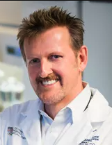 Johnny Huard合作论文数Center for Regenerative Sports Medicine, Steadman Philippon Research Institute;Department of Clinical and Biomedical Sciences, College of Veterinary Medicine, Colorado State University6
 Hairong Peng合作论文数4
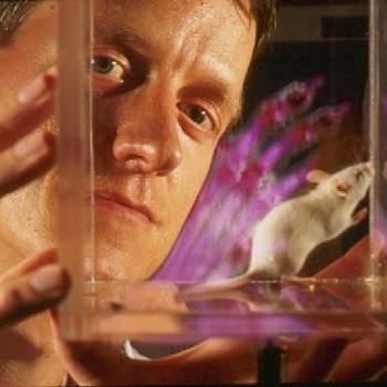 David Kingsley合作论文数Department of Developmental Biology, School of Medicine, Stanford University4
 Arvydas Usas合作论文数Department of Orthopaedic Surgery, University of Pittsburgh3Michelle D. Johnson合作论文数3
 Freddie Fu (傅浩強)合作论文数Department of Orthopaedic Surgery, School of Medicine, University of Pittsburgh2Anne Olshanski合作论文数2
 Gregory Cooper合作论文数Department of Biomedical Informatics, University of Pittsburgh2B Gearhart合作论文数1D. Loeuille合作论文数1A Reginato合作论文数1Matthew Kwan合作论文数1KA Gurley合作论文数1
 Paul C Marker合作论文数Pharmaceutical Sciences Division, School of Pharmacy, University of Wisconsin-Madison1G. Wright合作论文数1
 Sellmyer Mark A合作论文数Univ Penn, Radiol, Philadelphia, PA 19104 USA1Dixey Josh合作论文数1
 Amanda Hughes合作论文数Department of Epidemiology and Public Health, University College London1
 Andres Quintero合作论文数Universidad Nacional de Colombia, Universidad Nacional de Colombia-Sede Bogotá1
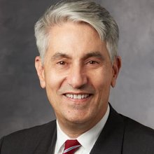 Michael T. Longaker合作论文数Department of Materials Science & Engineering, Stanford University1
 Michael Doherty合作论文数Division of Rheumatology, Orthopaedics and Dermatology, School of Medicine, University of Nottingham;Faculty of Medicine and Health Sciences, University of Nottingham1Rita Shiang合作论文数Department of Pediatrics, University of Texas Southwestern Medical Center;University of Utah School of Medicine, University of Iowa;Departments of Pediatrics and Center of Medical Genetics, The Johns Hopkins School of Medicine;Centre d'Etudes du Bouchet, University of Stellenbosch;and Department of Genetics, Stanford University School of Medicine1Charlene J. Williams合作论文数1Hany Bedair合作论文数1
 Adrian Pendleton合作论文数1
 Pierre Gillet合作论文数1
 Patrick Netter合作论文数Université de Lorraine1Rodney Mcgrath合作论文数1
 Tom Wandless合作论文数Department of Chemical and Systems Biology, Stanford University1Natalina Quarto合作论文数1




