Many membrane proteins are known to form higher-order oligomers, but the degree to which membrane regions could facilitate protein complex assembly remains largely unclear. Clusters of chemotaxis receptors are among the most prominent structures in the bacterial cell membrane, and they play important functions in processing of chemotactic signals. Although much work has been done to elucidate mechanisms of cluster formation, it almost exclusively focused on cytoplasmic interactions among receptors and other chemotaxis proteins, whereas involvement of membrane-mediated interactions was only hypothesized. Here we used imaging of constructs composed of only a fluorescent protein and the TM helices of Tar to demonstrate that interactions between the lipid bilayer and transmembrane (TM) helices of Escherichia coli chemoreceptors alone are sufficient to mediate clustering. We found that the ability to cluster depends on the sequence or length of the TM helices, implying that certain conformations of these helices facilitate clustering, whereas others do not. Notably, observed sequence specificity was apparently consistent with differences in clustering between native E. coli receptors, with the TM sequence of better-clustering high-abundance receptors being more efficient in promoting membrane-mediated complex formation. These results indicate that being more than just membrane anchors, TM helices could play an important role in the clustering and organization of membrane proteins in bacteria.
Most bacteria use transmembrane sensors to detect a wide range of environmental stimuli. A large class of such sensors are the chemotaxis receptors used by motile bacteria to follow environmental chemical gradients. In Escherichia coli, chemotaxis receptors are known to mediate highly sensitive responses to ligands, making them potentially useful for biosensory applications. However, with only four ligand-binding chemotaxis receptors, the natural ligand spectrum of E. coli is limited. The design of novel chemoreceptors to extend the sensing capabilities of E. coli is therefore a critical aspect of chemotaxis-based biosensor development. One path for novel sensor design is to harvest the large natural diversity of chemosensory functions found in bacteria by creating hybrids that have the signaling domain from E. coli chemotaxis receptors and sensory domains from other species. In this work, we demonstrate that the E. coli receptor Tar can be successfully combined with most typical sensory domains found in chemotaxis receptors and in evolutionary-related two-component histidine kinases. We show that such functional hybrids can be generated using several different fusion points. Our work further illustrates how hybrid receptors could be used to quantitatively characterize ligand specificity of chemotaxis receptors and histidine kinases using standardized assays in E. coli.
SummaryChemotaxis enables bacteria to navigate chemical gradients in their environment, accumulating toward high concentrations of attractants and avoiding high concentrations of repellents. Although finding nutrients is likely to be an important function of bacterial chemotaxis, not all characterized attractants are nutrients. Moreover, even for potential nutrients, the exact relation between the metabolic value of chemicals and their efficiency as chemoattractants has not been systematically explored. Here we compare the chemotactic response of amino acids with their use by bacteria for two well‐established models of chemotactic behavior, Escherichia coli and Bacillus subtilis. We demonstrate that in E. coli chemotaxis toward amino acids indeed strongly correlates with their utilization. However, no such correlation is observed for B. subtilis, suggesting that in this case, the amino acids are not followed because of their nutritional value but rather as environmental cues.
The signaling apparatus that controls bacterial chemotaxis is composed of a core complex containing chemoreceptors, the histidine autokinase CheA, and the coupling protein CheW. Site-specific spin labeling and pulsed dipolar ESR spectroscopy (PDS) have been applied to investigate the structure of a soluble ternary complex formed by Thermotoga maritima CheA (TmCheA), CheW, and receptor signaling domains. Thirty-five symmetric spin-label sites (SLSs) were engineered into the five domains of the CheA dimer and CheW to provide distance restraints within the CheA:CheW complex in the absence and presence of a soluble receptor that inhibits kinase activity (Tm14). Additional PDS restraints among spin-labeled CheA, CheW, and an engineered single-chain receptor labeled at six different sites allow docking of the receptor structure relative to the CheA:CheW complex. Disulfide cross-linking between selectively incorporated Cys residues finds two pairs of positions that provide further constraints within the ternary complex: one involving Tm14 and CheW and another involving Tm14 and CheA. The derived structure of the ternary complex indicates a primary site of interaction between CheW and Tm14 that agrees well with previous biochemical and genetic data for transmembrane chemoreceptors. The PDS distance distributions are most consistent with only one CheW directly engaging one dimeric Tm14. The CheA dimerization domain (P3) aligns roughly antiparallel to the receptor-conserved signaling tip but does not interact strongly with it. The angle of the receptor axis with respect to P3 and the CheW-binding P5 domains is bound by two limits differing by approximately 20 degrees . In one limit, Tm14 aligns roughly along P3 and may interact to some extent with the hinge region near the P3 hairpin loop. In the other limit, Tm14 tilts to interact with the P5 domain of the opposite subunit in an interface that mimics that observed with the P5 homologue CheW. The time domain ESR data can be simulated from the model only if orientational variability is introduced for the P5 and, especially, P3 domains. The Tm14 tip also binds beside one of the CheA kinase domains (P4); however, in both bound and unbound states, P4 samples a broad range of distributions that are only minimally affected by Tm14 binding. The CheA P1 domains that contain the substrate histidine are also broadly distributed in space under all conditions. In the context of the hexagonal lattice formed by trimeric transmembrane chemoreceptors, the PDS structure is best accommodated with the P3 domain in the center of a honeycomb edge.
Transmembrane chemoreceptors, also known as methyl-accepting chemotaxis proteins (MCPs), translate extracellular signals into intracellular responses in the bacterial chemotaxis system. MCP ligand binding domains control the activity of the CheA kinase, situated approximately 200 A away, across the cytoplasmic membrane. The 2.17 A resolution crystal structure of a Thermotoga maritima soluble receptor (Tm14) reveals distortions in its dimeric four-helix bundle that provide insight into the conformational states available to MCPs for propagating signals. A bulge in one helix generates asymmetry between subunits that displaces the kinase-interacting tip, which resides more than 100 A away. The maximum bundle distortion maps to the adaptation region of transmembrane MCPs where reversible methylation of acidic residues tunes receptor activity. Minor alterations in coiled-coil packing geometry translate the bulge distortion to a >25 A movement of the tip relative to the bundle stalks. The Tm14 structure discloses how alterations in local helical structure, which could be induced by changes in methylation state and/or by conformational signals from membrane proximal regions, can reposition a remote domain that interacts with the CheA kinase.
In bacterial chemotaxis, an assembly of transmembrane receptors, the CheA histidine kinase and the adaptor protein CheW processes environmental stimuli to regulate motility. The structure of a Thermotoga maritima receptor cytoplasmic domain defines CheA interaction regions and metal ion–coordinating charge centers that undergo chemical modification to tune receptor response. Dimeric CheA–CheW, defined by crystallography and pulsed ESR, positions two CheWs to form a cleft that is lined with residues important for receptor interactions and sized to clamp one receptor dimer. CheW residues involved in kinase activation map to interfaces that orient the CheW clamps. CheA regulatory domains associate in crystals through conserved hydrophobic surfaces. Such CheA self-contacts align the CheW receptor clamps for binding receptor tips. Linking layers of ternary complexes with close-packed receptors generates a lattice with reasonable component ratios, cooperative interactions among receptors and accessible sites for modification enzymes.
Signal transduction underlying bacterial chemotaxis involves excitatory phosphorylation and feedback control through deamidation and methylation of sensory receptors. The structure of a complex between the signal-terminating phosphatase, CheC, and the receptor-modifying deamidase, CheD, reveals how CheC mimics receptor substrates to inhibit CheD and how CheD stimulates CheC phosphatase activity. CheD resembles other cysteine deamidases from bacterial pathogens that inactivate host Rho-GTPases. CheD not only deamidates receptor glutamine residues contained within a conserved structural motif but also hydrolyzes glutamyl-methyl-esters at select regulatory positions. Substituting Gln into the receptor motif of CheC turns the inhibitor into a CheD substrate. Phospho-CheY, the intracellular signal and CheC target, stabilizes the CheC:CheD complex and reduces availability of CheD. A point mutation that dissociates CheC from CheD impairs chemotaxis in vivo. Thus, CheC incorporates an element of an upstream receptor to influence both its own effect on receptor output and that of its binding partner, CheD.
 Brian Crane合作论文数Department of Chemistry & Chemical Biology, The College of Arts & Sciences, Cornell University4
 A.M. Bilwes合作论文数4
 Victor Sourjik合作论文数Department of Systems and Synthetic Microbiology, Max Planck Institute for Terrestrial Microbiology;Philipps-Universitat Marburg3Jaya Bhatnagar合作论文数2
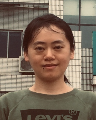 Yiling Yang (杨毅玲)合作论文数Institute of Hydrobiology, Chinese Academy of Sciences2Park SangYoun合作论文数2
 Jack Freed合作论文数Department of Chemistry and Chemical Biology, College of Arts & Sciences, Cornell University;National Biomedical Center for Advanced Electron Spin Resonance Technology, Cornell University2Peter P. Borbat合作论文数2Gabriela Gonzalez-Bonet合作论文数1
 Markus Wirtz合作论文数Heidelberg Univ, Ctr Organismal Studies, D-69120 Heidelberg, Germany1
 Gernot Poschet合作论文数Centre for Organismal Studies, Ruprecht-Karls-University Heidelberg1Höfler Carolin合作论文数1
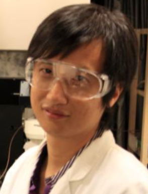 Fan Jin (金帆)合作论文数University of Science and Technology of China1
 Shuangyu Bi (毕双玉)合作论文数State Key Laboratory of Microbial Technology, Shandong University1
 George Winford Ordal合作论文数1
 Travis Muff合作论文数Department of Biochemistry, University of Illinois1
 Rüdiger Hell合作论文数Centre for Organismal Studies, Department of Molecular Biology of Plants, Heidelberger Institut fur Pflanzenwissenschaften, Universitat Heidelberg1Xingjuan Chao合作论文数1




