Human sterile alpha motif domain-containing 9 (hSAMD9L) is a large (~185 kDa) multi-domain interferon-stimulated antiviral effector with strong translation-inhibitory activity. Inherited heterozygous gain-of-function (GoF) mutations in SAMD9L directly associated with severe bone marrow failure syndromes. Using single-particle cryo-electron microscopy (cryo-EM), we determined the first structures of both full-length wild-type hSAMD9L and an N-terminal-truncated mutant at resolutions ranging from 2.8 to 3.7 Å. Both proteins exist in monomeric and dimeric states, providing clear evidence that the sterile alpha motif (SAM) and AlbA domains are not essential for dimerization. Our cryo-EM analysis reveals a tightly packed, closed architecture defined by interlocking multi-domains. We precisely mapped the extensive dimer interface mediated by Sir2-like and an oligonucleotide/oligosaccharide-binding (OB) domains. Biochemical analyses show that hSAMD9L binds double-stranded DNA in vitro but has no detectable NTP hydrolysis activity under our assay conditions. Accordingly, in our cryo-EM map we observed a clear density for a non-hydrolyzed NTP in the pocket, suggesting nucleotide binding without turnover like in STAND (Signal Transduction ATPases with Numerous Domains) proteins. Together, these results provide a structural framework for hSAMD9L and also providing key insights into its organization, domain packing and dimerization and offer a basis for understanding how GoF variants may alter hSAMD9L regulation thus impacting cell proliferation. ### Competing Interest Statement The authors have declared no competing interest.
Cyclic AMP (cAMP) compartmentalization at the plasma membrane employs export by ABCC4, yet the protein machinery that creates spatially restricted signaling has remained unclear. We show that robust PKA activation increases ABCC4 at the cell surface and assembles a PDZ-dependent macromolecular complex that constrains ABCC4 mobility and stabilizes the transporter. Using Avi-tag crosslinking/AP-MS and APEX proximity labeling, we map a PKA-induced ABCC4 neighborhood enriched for PDZ-domain scaffolds that couple ABCC4 to actin and cell junctions. SCRIB emerges as a previously unrecognized interactor whose depletion lowers surface ABCC4 and blunts ABCC4-dependent drug resistance. Deleting the ABCC4 PDZ motif accelerates lateral diffusion ~4-fold, halves protein half-life, and attenuates PKA-stimulated increases in surface ABCC4 and cAMP efflux, establishing a requirement for the PDZ motif in optimal export under PKA drive. Finally, the selective ABCC4 inhibitor Ceefourin-2 lowers ABCC4's melting temperature and dismantles the actin/junction-enriched neighborhood, revealing a non-classical small molecule inhibitory mechanism based on network disruption. These findings define a PKA-driven, PDZ-dependent ABCC4 protein neighborhood that stabilizes the transporter and optimizes cAMP efflux-an advance over prior work that localized ABCC4 but lacked a mechanistic blueprint for its membrane microdomain organization.
Stress granules (SGs) are dynamic RNA-protein assemblies that form in response to cellular stress and must be efficiently disassembled to restore normal cell function. Valosin-containing protein (VCP), an enzyme implicated in neurodegenerative diseases, is essential for SG disassembly, but whether and how this process is coordinated with SG assembly remains unclear. Here, we identify the VCP cofactor, Alveolar soft part sarcoma locus (ASPL) as a key regulator linking SG assembly and disassembly. ASPL promotes SG assembly by facilitating biomolecular condensation of Ras guanosine triphosphatase-activating protein-binding protein (G3BP) and stabilizing its interactions with other SG proteins. ASPL also facilitates phosphorylation and activation of VCP by UNC-51-like kinases 1 and 2 (ULK1/2), enabling G3BP extraction and efficient SG disassembly. Pathogenic VCP mutations that disrupt ASPL binding impair SG disassembly, a defect rescued by phosphomimetic mutations or ASPL depletion. Our findings suggest that disruptions in the ASPL-VCP interaction uncouple SG assembly and disassembly, representing a potential mechanism underlying VCP-associated neurodegenerative diseases.
Abstract ID 95276Poster Board 431The ATP-binding cassette transporter ABCC4 regulates cAMP membrane microdomain formation after adenylate cyclase activation but how ABCC4 localizes to these membrane microdomains are still unclear. Herein we demonstrate that the assembly and spatiotemporal organization of ABCC4’s protein neighborhood by PKA requires the PDZ motif of ABCC4 and PKA activation. To investigate the role of the highly conserved ABCC4 PDZ-motif, we performed live cell imaging using total internal reflectance fluorescence, TIRF, of ABCC4-GFP expression constructs with or without the PDZ motif. The expression constructs were introduced into HEK293 cells where endogenous ABCC4 had been deleted by CRISPR-Cas9 (ABCC4-null HEK293 cells). Deletion of the PDZ motif untethered ABCC4 promoting rapid movement. Treatment with the adenylate cyclase agonist, forskolin and the phosphodiesterase inhibitor, iso-butyl-methyl xanthine increased intracellular cAMP and promoted the membrane localization of ABCC4 harboring the PDZ motif, which was significantly reduced by treatment with a PKA specific peptide inhibitor, PKI-14-22. Density gradient ultracentrifugation revealed the formation of an ABCC4 PDZ motif–specific, large protein complex upon PKA activation. To elucidate how PKA ushers ABCC4 to form this membrane supercomplex, we identified proteins that closely interact with ABCC4 and proteins in ABCC4’s “neighborhood” network using Avi-tagged or APEX-tagged ABCC4 constructs. Cell lines were generated in ABCC4-null HEK293 cells by inserting Avi- and APEX-tagged ABCC4 constructs, and their deletion mutants lacking the PDZ motif. Each Avi-expression construct harbored a biotin ligase, thus enabling ABCC4 to be tagged by biotin. We used a crosslinker, DSP (dithiobis(succinimidyl propionate)) to capture weakly or transiently interacting proteins. Ascorbic acid peroxidase (APEX) - tagged to ABCC4 catalyzes the externally added biotin-phenol molecules in the presence of hydrogen peroxidase into biotin-phenol radicals which bind to tyrosine residues of proteins within ∼10 nm of ABCC4. The biotinylated proteins were isolated by affinity purification using avidin beads and mass spectrometry. Our results from the proteomic analysis indicated that ABCC4 interacts with numerous PDZ domain containing proteins (PDZ proteins), and the PKA-induced enrichment of actin filament-binding proteins in the vicinity of ABCC4 requires the PDZ motif. The assembly of the supercomplex was crucially dependent on the actin-binding protein Moesin (MSN), which also interacts with the PDZ motif. siRNA knockdown of MSN disrupted the ABCC4-SCRIB interaction within the PKA-driven supercomplex, as well as ABCC4 interaction with actin and filamin A. Furthermore, we demonstrated that Ceefourin 2, an ABCC4 specific inhibitor, reduced membrane levels of ABCC4, induced by PKA, by disrupting the actin cytoskeletal network around ABCC4. Altogether, these findings indicate that PKA activation promotes ABCC4 membrane localization by enhancing the assembly of a protein scaffold at its PDZ motif through PDZ proteins, MSN and actin cytoskeletal proteins.This work was supported by NIH and by ALSAC.
The regulation of hematopoietic stem cell (HSC) function has been extensively studied at genomic and transcriptional levels, but the roles of ubiquitin E3 ligase-associated factors in the hematopoietic system are not well understood. DCAFs (DDB1-CUL-Associated Factors) interact with Cullin-Ring Ligases to recruit proteins for ubiquitination, facilitate protein assemblies, redirect E3 ligase complexes, and recognize chromatin features. Our studies focus on elucidating the function of DCAF7, a poorly characterized protein and one of the few DCAFs (a family of more than 100 proteins) expressed in HSCs. Previous studies on DCAF7 were performed in cell lines, and its physiological role in vivo is unknown. DCAF7 has been shown to bind kinases and target substrates for degradation such as Menin and TFEB. Mutations in DCAF7 have been reported in myeloid leukemia in Down Syndrome and other hematological malignancies. Thus, we aimed to investigate DCAF7 function in hematopoiesis using a novel conditional knockout mouse line. Dcaf7-deficient mice (Dcaf7F/F Mx1-Cre+) showed an increased frequency of phenotypically defined long-term HSCs and multipotent progenitors, with a decrease in more committed progenitors. Similar results were observed in Dcaf7F/F Vav1-Cre+ mice. We assessed DCAF7 function in HSC regeneration upon myelotoxic stress by injecting mice with 5-fluorouracil (5-FU). KO mice showed delayed recovery and died earlier than WT mice upon serial injections. To investigate the effect of Dcaf7 loss on hematopoietic cell fitness and long-term self-renewal, we performed bone marrow transplants. In competitive transplants, KO cells (CD45.2) were outcompeted by CD45.1 cells with no differences in KO-derived lymphoid or myeloid cells. In non-competitive transplants, KO-recipient mice showed impaired hematopoietic reconstitution and hypocellular bone marrow, consistent with a bone marrow failure phenotype. Using single-cell RNA sequencing and single-cell time-lapse microscopy, we observed that loss of Dcaf7 accelerates HSC differentiation including their direct differentiation into megakaryocytes. KO cells show a dysregulated trajectory toward erythroid, basophil/mast cell, and dendritic progenitors, with reduced stemness in HSCs. Overall, these results indicate that lack of Dcaf7 induces HSC differentiation into the myeloid lineage, leading to stem cell exhaustion. Next, we investigated the molecular mechanism by which DCAF7 contributes to hematopoiesis. By performing IP-MS, we found that DCAF7 interacts with components of the E3 ligase-containing polycomb repressive complex 1.5 (PRC1.5), which is involved in gene activation unlike other PRC1 complexes. DCAF7 absence did not alter PRC1.5 protein levels. Glycerol-density sedimentation analysis showed that without DCAF7, all PRC1 subunits eluted in lower molecular-weight fractions compared to WT complexes, indicating DCAF7 promotes the assembly of large PRC1.5 complexes. Given that PRC1 binds chromatin and its configuration varies among cell types, we performed chromatin profiling in HSCs and progenitors. We analyzed the genome-wide distribution of RING1B (the core PRC1 component that ubiquitinates H2A), H2AK119ub, H3K27me3, and H3K4me3 using CUT&RUN. Our results revealed that the loss of DCAF7 leads to increased RING1B localization at transcriptionally active loci marked by H3K4me3. This increased binding was observed at genes involved in myeloid differentiation and was associated with increased mRNA expression in HSCs. Together, our study elucidates a new pathway for the regulation of PRC1 complex activity mediated by DCAF7 that controls HSC differentiation.
Bacteriophage T4 is a classic model system for studying the mechanisms of DNA processing. A key protein in T4 DNA processing is the gp32 single-stranded DNA-binding protein. gp32 has two key functions: it binds cooperatively to single-stranded DNA (ssDNA) to protect it from nucleases and remove regions of secondary structure, and it recruits proteins to initiate DNA processes including replication and repair. Dda is a T4 helicase recruited by gp32, and we purified and crystallized a gp32-Dda-ssDNA complex. The low-resolution structure revealed how the C-terminus of gp32 engages Dda. Analytical ultracentrifugation analyses were consistent with the crystal structure. An optimal Dda binding peptide from the gp32 C-terminus was identified using surface plasmon resonance. The crystal structure of the Dda-peptide complex was consistent with the corresponding interaction in the gp32-Dda-ssDNA structure. A Dda-dependent DNA unwinding assay supported the structural conclusions and confirmed that the bound gp32 sequesters the ssDNA generated by Dda. The structure of the gp32-Dda-ssDNA complex, together with the known structure of the gp32 body, reveals the entire ssDNA binding surface of gp32. gp32-Dda-ssDNA complexes in the crystal are connected by the N-terminal region of one gp32 binding to an adjacent gp32, and this provides key insights into this interaction.
AbstractSpecificity of the ubiquitin-proteasome system depends on E3 ligase-substrate interactions. Many such pairings depend on E3 ligases binding to peptide-like sequences - termed N- or C-degrons - at the termini of substrates. However, our knowledge of structural features distinguishing closely related C-degron substrate-E3 pairings is limited. Here, by systematically comparing ubiquitylation activities towards a suite of common model substrates, and defining interactions by biochemistry, crystallography, and cryo-EM, we reveal principles of C-degron recognition across the KLHDCX family of Cullin-RING ligases (CRLs). First, a motif common across these E3 ligases anchors a substrate’s C-terminus. However, distinct locations of this C-terminus anchor motif in different blades of the KLHDC2, KLHDC3, and KLHDC10 β-propellers establishes distinct relative positioning and molecular environments for substrate C-termini. Second, our structural data show KLHDC3 has a pre-formed pocket establishing preference for an Arg or Gln preceding a C-terminal Gly, whereas conformational malleability contributes to KLHDC10’s recognition of varying features adjacent to substrate C-termini. Finally, additional non-consensus interactions, mediated by C-degron binding grooves and/or by distal propeller surfaces and substrate globular domains, can substantially impact substrate binding and ubiquitylatability. Overall, the data reveal combinatorial mechanisms determining specificity and plasticity of substrate recognition by KLDCX-family C-degron E3 ligases.
E3 ligase recruitment of proteins containing terminal destabilizing motifs (degrons) is emerging as a major form of regulation. How those E3s discriminate bona fide substrates from other proteins with terminal degron-like sequences remains unclear. Here, we report that human KLHDC2, a CRL2 substrate receptor targeting C-terminal Gly-Gly degrons, is regulated through interconversion between two assemblies. In the self-inactivated homotetramer, KLHDC2's C-terminal Gly-Ser motif mimics a degron and engages the substrate-binding domain of another protomer. True substrates capture the monomeric CRL2KLHDC2, driving E3 activation by neddylation and subsequent substrate ubiquitylation. Non-substrates such as NEDD8 bind KLHDC2 with high affinity, but its slow on rate prevents productive association with CRL2KLHDC2. Without substrate, neddylated CRL2KLHDC2 assemblies are deactivated via distinct mechanisms: the monomer by deneddylation and the tetramer by auto-ubiquitylation. Thus, substrate specificity is amplified by KLHDC2 self-assembly acting like a molecular timer, where only bona fide substrates may bind before E3 ligase inactivation.
We recently demonstrated that a protein interaction between the PDZ motif of ABCC4 and a single PDZ‐domain‐containing protein, MPP1 had an important role in ABCC4 function in acute myeloid leukemia (Pitre et al. Nature Comm. 2017). To gain a better understanding into the role of the highly conserved ABCC4 PDZ‐motif, we performed live cell imaging using total internal reflectance fluorescence, TIRF, of ABCC4‐GFP expression constructs with or without the PDZ motif. The expression constructs were introduced into HEK293 cells where ABCC4 had been deleted by CRISPR‐Cas9 technology. ABCC4‐GFP was static at the membrane showing little movement, while deletion of the PDZ motif promoted rapid movement. Activation of PKA with forskolin and the phosphodiesterase inhibitor, iso‐butyl‐methyl xanthine increased intracellular cAMP and promoted membrane movement of ABCC4 harboring the PDZ motif. To get an unbiased understanding of the proteins interacting with ABCC4 both in cis‐ and trans‐, and under PKA activation conditions, we developed stable cell lines in the ABCC4‐null HEK293 cells. These stable cell lines harbored either an APEX2‐ABCC4 or an APEX2‐ABCC4 lacking a PDZ motif. APEX2‐derived ascorbate oxidase and biotin phenol, coupled with Avidin‐beads was used to interrogate the neighborhood of proteins within ~10 nm of ABCC4. We also developed stable cells harboring expression constructs for either Avi‐tagged ABCC4 or Avi‐ABCC4 without the PDZ motif to determine proteins that directly interacted with ABCC4 through the PDZ motif. Each Avi‐expression construct harbored a biotin ligase, thus enabling ABCC4 to be tagged by biotin and readily “pulled down” by avidin‐beads. The interacting proteins were identified by quantitative proteomics. Several PDZ specific interacting proteins were identified in these cell lines. By using analytical density gradient ultracentrifugation, we identified distinct PDZ specific ABCC4‐complexes that formed upon PKA activation. The actin‐binding cytoskeletal protein, Moesin (MSN) associated with ABCC4, requiring the PDZ‐motif. We used quantitative proteomics and APEX2 ABCC4 constructs to show that the neighborhood surrounding ABCC4 lacking the PDZ‐motif had fewer proteins, especially cytoskeletal proteins interacting with it. We propose that the ABCC4‐MSN interaction is important for tethering ABCC4 to the cytoskeletal actin network. Importantly, siRNA knockdown of MSN disrupted the association between ABCC4 and its interactors indicating the key role of MSN in the dynamic protein interactions occurring between ABCC4 and its interactors.
Ozz, a member of the SOCS-box family of proteins, is the substrate-binding component of CRL5Ozz, a muscle-specific Cullin-RING ubiquitin ligase complex composed of Elongin B/C, Cullin 5 and Rbx1. CRL5Ozz targets for proteasomal degradation selected pools of substrates, including sarcolemma-associated β-catenin, sarcomeric MyHCemb and Alix/PDCD6IP, which all interact with the actin cytoskeleton. Ubiquitination and degradation of these substrates are required for the remodeling of the contractile sarcomeric apparatus. However, how CRL5Ozz assembles into an active E3 complex and interacts with its substrates remain unexplored. Here, we applied a baculovirus-based expression system to produce large quantities of two subcomplexes, Ozz-EloBC and Cul5-Rbx1. We show that these subcomplexes mixed in a 1:1 ratio reconstitutes a five-components CRL5Ozz monomer and dimer, but that the reconstituted complex interacts with its substrates only as monomer. The in vitro assembled CRL5Ozz complex maintains the capacity to polyubiquitinate each of its substrates, indicating that the protein production method used in these studies is well-suited to generate large amounts of a functional CRL5Ozz. Our findings highlight a mode of assembly of the CRL5Ozz that differs in presence or absence of its cognate substrates and grant further structural studies.
Proteins that exhibit intrinsically disordered regions (IDRs) are prevalent in the human proteome and perform diverse biological functions, including signaling and regulation. Due to these important roles, misregulation of intrinsically disordered proteins (IDPs) is associated with myriad human diseases, including neurodegeneration and cancer. The inherent flexibility of IDPs limits the applicability of the traditional structure-based drug design paradigm; therefore, IDPs have long been considered "undruggable". Using NMR spectroscopy and other methods, we previously discovered small, drug-like molecules that bind specifically, albeit weakly, to dynamic clusters of aromatic residues within p27(Kip1) (p27), an archetypal disordered protein involved in cell cycle regulation. Here, using synthetic chemistry, NMR spectroscopy and other biophysical methods, we discovered elaborated analogs of our previously reported molecules with 30-fold increased affinity for p27 (apparent K-d = 57 +/- 19 lM). Strikingly, using analytical ultracentrifugation methods, we showed that the highest affinity compounds caused p27 to form soluble, disordered oligomers. Based on these observations, we propose that sequestration within soluble oligomers may represent a general strategy for therapeutically targeting disease-associated IDPs in the future. (C) 2021 Elsevier Ltd. All rights reserved.
Liquid-liquid phase separation underlies the membrane-less compartmentalization of cells. Intrinsically disordered low-complexity domains (LCDs) often mediate phase separation, but how their phase behavior is modulated by folded domains is incompletely understood. Here, we interrogate the interplay between folded and disordered domains of the RNA-binding protein hnRNPA1. The LCD of hnRNPA1 is sufficient for mediating phase separation in vitro. However, we show that the folded RRM domains and a folded solubility-tag modify the phase behavior, even in the absence of RNA. Notably, the presence of the folded domains reverses the salt dependence of the driving force for phase separation relative to the LCD alone. Small-angle X-ray scattering experiments and coarse-grained MD simulations show that the LCD interacts transiently with the RRMs and/or the solubility-tag in a salt-sensitive manner, providing a mechanistic explanation for the observed salt-dependent phase separation. These data point to two effects from the folded domains: (i) electrostatically-mediated interactions that compact hnRNPA1 and contribute to phase separation and (ii) increased solubility at higher ionic strengths mediated by the folded domains. The interplay between disordered and folded domains can modify the dependence of phase behavior on solution conditions and can obscure signatures of physicochemical interactions underlying phase separation.
Anaplastic lymphoma kinase (ALK) is a receptor tyrosine kinase (RTK) that regulates important functions in the central nervous system1,2. The ALK gene is a hotspot for chromosomal translocation events that result in several fusion proteins that cause a variety of human malignancies3. Somatic and germline gain-of-function mutations in ALK were identified in paediatric neuroblastoma4-7. ALK is composed of an extracellular region (ECR), a single transmembrane helix and an intracellular tyrosine kinase domain8,9. ALK is activated by the binding of ALKAL1 and ALKAL2 ligands10-14 to its ECR, but the lack of structural information for the ALK-ECR or for ALKAL ligands has limited our understanding of ALK activation. Here we used cryo-electron microscopy, nuclear magnetic resonance and X-ray crystallography to determine the atomic details of human ALK dimerization and activation by ALKAL1 and ALKAL2. Our data reveal a mechanism of RTK activation that allows dimerization by either dimeric (ALKAL2) or monomeric (ALKAL1) ligands. This mechanism is underpinned by an unusual architecture of the receptor-ligand complex. The ALK-ECR undergoes a pronounced ligand-induced rearrangement and adopts an orientation parallel to the membrane surface. This orientation is further stabilized by an interaction between the ligand and the membrane. Our findings highlight the diversity in RTK oligomerization and activation mechanisms.
Amyotrophic lateral sclerosis (ALS) is a lethal and incurable neurodegenerative disorder commonly associated with repeat expansion in the C9orf72 gene; termed C9-ALS. A major pathological feature of this is the accumulation of arginine-rich (R-rich) dipeptide repeat (DPR) polypeptides in neurons. R-rich DPRs interact with low complexity domains in proteins, accumulate in membraneless organelles (MLOs), alter the material properties of MLOs, and induce cell death. The molecular mechanisms underlying pathogenesis, though, remain unknown. R-rich DPRs infiltrate nucleoli, co-localize with nucleophosmin (NPM1), and alter NPM1 phase separation in vitro. NPM1 is crucial to the maintenance of nucleolar liquid-like properties through its ability to phase separate with proteins and nucleic acids. Elucidating the effects of DPR interactions with NPM1 on the liquid-like properties and overall architecture of nucleoli that ultimately lead to nucleolar dysfunction and cell death is critical to understanding C9-ALS. Here we employ poly(PR) as an archetypal R-rich DPR to (1) identify the interactions mediating phase separation with NPM1; (2) elucidate the mechanisms causing the DPR-dependent dissolution of in vitro NPM1/DPR droplets; and (3) confirm hypotheses stimulated by our biophysical results regarding the mechanisms of DPR-mediated nucleolar disruption in cells. Results from multiple, complementary biochemical and biophysical techniques show that (1) NPM1/DPR interactions are mediated by acidic tracts within the intrinsically disorder region of NPM1, (2) R-rich DPRs dissolve NPM1-containing droplets in vitro by sequestering NPM1 into large saturated complexes, and (3) exogenous poly(PR) induces NPM1 release from nucleoli, disrupting nucleolar organization and function. These results support the hypothesis that R-rich DPRs, mediate their toxic effects in part through saturation/sequestration of NPM1, perturbing NPM1 mediated phase separation in nucleoli, disrupting nucleolar function, and inducing cell death.
The cellular stress response has a vital role in regulating homeostasis by modulating cell survival and death. Stress granules are cytoplasmic compartments that enable cells to survive various stressors. Defects in the assembly and disassembly of stress granules are linked to neurodegenerative diseases, aberrant antiviral responses and cancer1–5. Inflammasomes are multi-protein heteromeric complexes that sense molecular patterns that are associated with damage or intracellular pathogens, and assemble into cytosolic compartments known as ASC specks to facilitate the activation of caspase-1. Activation of inflammasomes induces the secretion of interleukin (IL)-1β and IL-18 and drives cell fate towards pyroptosis—a form of programmed inflammatory cell death that has major roles in health and disease6–12. Although both stress granules and inflammasomes can be triggered by the sensing of cellular stress, they drive contrasting cell-fate decisions. The crosstalk between stress granules and inflammasomes and how this informs cell fate has not been well-studied. Here we show that the induction of stress granules specifically inhibits NLRP3 inflammasome activation, ASC speck formation and pyroptosis. The stress granule protein DDX3X interacts with NLRP3 to drive inflammasome activation. Assembly of stress granules leads to the sequestration of DDX3X, and thereby the inhibition of NLRP3 inflammasome activation. Stress granules and the NLRP3 inflammasome compete for DDX3X molecules to coordinate the activation of innate responses and subsequent cell-fate decisions under stress conditions. Induction of stress granules or loss of DDX3X in the myeloid compartment leads to a decrease in the production of inflammasome-dependent cytokines in vivo. Our findings suggest that macrophages use the availability of DDX3X to interpret stress signals and choose between pro-survival stress granules and pyroptotic ASC specks. Together, our data demonstrate the role of DDX3X in driving NLRP3 inflammasome and stress granule assembly, and suggest a rheostat-like mechanistic paradigm for regulating live-or-die cell-fate decisions under stress conditions. The RNA helicase DDX3X has a critical role in regulating both the induction of stress granules and the activation of the NLRP3 inflammasome in cells under stress conditions.
The nucleolus is a multi-layered, liquid-like membraneless organelle that mediates the multi-step process of ribosome biogenesis and also coordinates signaling responses to various cellular stresses. Initiation of ribosomal RNA (rRNA) transcription during interphase of the cell division cycle triggers phase separation with the nucleolar protein, Fibrillarin, forming the Dense Fibrillar Component region of the nucleolus wherein rRNA is sequentially modified and spliced. Mature rRNAs flux outwards from the DFC into the surrounding Granular Component (GC) region wherein they phase separate with another nucleolar protein, Nucleophosmin (NPM1). Utilizing an intrinsically disordered region (IDR) displaying alternating acidic and basic tracts, NPM1 independently phase separates with ribosomal proteins (rProteins), sequestering them within the GC and enabling their assembly with rRNA to form nascent ribosomal subunits. We seek to understand how the molecular interactions that independently drive phase separation of NPM1 with rRNA and rProteins also enable the molecular handoffs that underlie ribosomal subunit assembly. Our studies employ structural and biophysical methods that probe molecular interactions at the atomic length scale as well as single-molecule, scattering and imaging methods that reveal the structural and material properties of liquid-phase condensates on the micron length scale. Only by bridging these disparate length scales can we understand how phase separation contributes to the essential process of ribosome biogenesis. We will present our latest findings on the liquid phase organization of the nucleolus and how this mediates vectorial ribosomal subunit assembly.
Repeat expansion in the C9orf72 gene is the most common cause of the neurodegenerative disorder amyotrophic lateral sclerosis (C9-ALS) and is linked to the unconventional translation of five dipeptide-repeat polypeptides (DPRs). The two enriched in arginine, poly(GR) and poly(PR), infiltrate liquid-like nucleoli, co-localize with the nucleolar protein nucleophosmin (NPM1), and alter the phase separation behavior of NPM1 in vitro. Here, we show that poly(PR) DPRs bind tightly to a long acidic tract within the intrinsically disordered region of NPM1, altering its phase separation with nucleolar partners to the extreme of forming large, soluble complexes that cause droplet dissolution in vitro. In cells, poly(PR) DPRs disperse NPM1 from nucleoli and entrap rRNA in static condensates in a DPR-length-dependent manner. We propose that R-rich DPR toxicity involves disrupting the role of phase separation by NPM1 in organizing ribosomal proteins and RNAs within the nucleolus.
The Sonic Hedgehog (Hh) signaling pathway is inappropriately activated in multiple malignancies, including basal cell carcinoma and medulloblastoma (MB). MB is the most common malignant pediatric brain tumor, accounting for 20% of all childhood brain tumors. While frontline therapy of MB, which includes tumor resection, radiation, and targeted chemotherapy yields 70% survival rate amongst average‐risk patients, radiation results in devastating neurocognitive impairments, and targeted therapy can give rise to drug‐resistant tumors. As such, further advances in therapy of MB will require identification of factors that modulate Hh pathway. We discovered that ABCC4 was, along with other Hh pathway genes, specifically over‐expressed in Hh‐driven MB. High ABCC4 expression was associated with significantly worse overall survival in Hh‐driven MB patients. In Hh pathway cellular model systems, ligand‐dependent pathway activation or loss of negative regulators upregulated ABCC4 expression, suggesting ABCC4 has a positive regulatory role and that blocking expression may impair signaling. Ablation of Abcc4 suppressed both normal, ligand‐dependent signaling as well as aberrant, cancer‐like, ligand‐independent signaling. Furthermore, targeted Abcc4 ablation using CRISPR technology in a murine model of Hh‐MB tumor extended survival of Hh tumor‐bearing mice. This study reveals ABCC4 as a critical regulator of the Hh pathway that contributes to MB pathogenesis and present a new molecular target and opportunity to improve therapy against Hh‐driven tumors.Support or Funding InformationThis work was supported by ALSAC and NIH P30CA021765 Cancer Center Support GrantThis abstract is from the Experimental Biology 2019 Meeting. There is no full text article associated with this abstract published in The FASEB Journal.
 Richard Kriwacki合作论文数Department of Structural Biology, St. Jude Graduate School of Biomedical Sciences, St. Jude Children's Research Hospital20
 Brenda Schulman合作论文数Max Planck Institute of Biochemistry;Department of Chemistry, Technical University of Munich;St. Jude Children's Research Hospital10
 Diana M Mitrea合作论文数DeMix Solutions, LLC9
 Grace Royappa合作论文数Department of Structural Biology, St. Jude Children’s Research Hospital;Department of Structural Biology, St. Jude Children’s Research Hospital8
 Stephen H. White合作论文数Department of Physiology & Biophysics, School of Medicine, University of California, Irvine8
 Douglas Green合作论文数Comprehensive Cancer Center, St. Jude Children's Research Hospital;Department of Immunology, St. Jude Children's Research Hospital8
 Darcie J. Miller合作论文数8
 Tanja Mittag合作论文数Structural Biology Department, St Jude Children's Research Hospital7Mi Kyung Yun合作论文数6
 Pitre Aaron合作论文数Department of Cellular Biology and Anatomy, Louisiana State University Health Sciences Center6Schuetz John D合作论文数6
 Christopher Stanley合作论文数6
 Junmin Peng合作论文数Department of Structural Biology, St. Jude Children's Research Hospital5
 Aaron Phillips合作论文数Department of Physiology and Pharmacology, Cumming School of Medicine, University of Calgary;Department of Clinical Neurosciences, Cumming School of Medicine, University of Calgary;Hotchkiss Brain Institute, University of Calgary5Ashok A. Deniz合作论文数The Scripps Research Institute5
 J. Paul Taylor合作论文数Cell & Molecular Biology Department, St. Jude Children's Research Hospital;Graduate School of Biomedical Sciences, St. Jude Children's Research Hospital5Sivashankar G. Sivakolundu合作论文数4Jaclyn Cika合作论文数4Juwina Wijaya合作论文数Dept Chem & Biochem, Univ Calif Los Angeles4
 Anjaparavanda Naren合作论文数4M Brett Waddell合作论文数St Jude Childrens Res Hosp, Hartwell Ctr Bioinformat & Biotechnol, Memphis, TN 38105 USA4Yao Wang合作论文数4
 Melissa R Marzahn合作论文数4
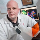 Clifford S Guy合作论文数4
 Priya R Banerjee合作论文数4
 Jihun Lee合作论文数4
 Ravi C. Kalathur合作论文数The New York Consortium on Membrane Protein Structure (NYCOMPS),, New York Structural Biology Center4John C Fisher合作论文数3Edmond R Watson合作论文数Max Planck Inst Biochem, Munich, Germany3Zhenmei Li合作论文数3Rebecca Crawford合作论文数3
 John Lynch合作论文数3Jerry Edward Chipuk合作论文数3Ban David合作论文数Department of NMR-based Structural Biology, Max Planck Institute for Biophysical Chemistry3Gose Tomoka合作论文数3
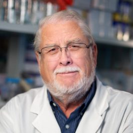 Charles O. Rock合作论文数Department of Host-Microbe Interactions, St. Jude Children's Research Hospital3
 Peter W. Schuck合作论文数National Institute of Biomedical Imaging and Bioengineering3
 Marada Suresh合作论文数Department of Biochemistry, St Jude Children's Research Hospital3
 Michele Tolbert合作论文数3
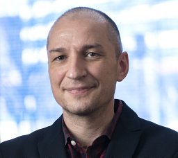 Tudor Moldoveanu合作论文数Engineering Network of Centres of Excellence, Queen's University3David M. Duda合作论文数3Cristina D. Guibao合作论文数3Igor Kurinov合作论文数3
 Stacey K. Ogden合作论文数Department of Biochemistry, St. Jude Children’s Research Hospital3Jie J. Zheng合作论文数3John Satumba合作论文数3Huaying Zhao合作论文数College of Chemical & Life Sciences, University of Maryland3Cheon-Gil Park合作论文数3Vishwajeeth R. Pagala合作论文数3




