Trogocytosis modulates immune responses, with still unclear underlying molecular mechanisms. Using leukemia mouse models, we found that lymphocytes perform trogocytosis at high rates with tumor cells. While performing trogocytosis, both Natural Killer (NK) and CD8 + T cells acquire the checkpoint receptor PD-1 from leukemia cells. In vitro and in vivo investigation revealed that PD-1 on the surface of NK cells, rather than being endogenously expressed, was derived entirely from leukemia cells in a SLAM receptor–dependent fashion. PD-1 acquired via trogocytosis actively suppressed NK cell antitumor immunity. PD-1 trogocytosis was corroborated in patients with clonal plasma cell disorders, where NK cells that stained for PD-1 also stained for tumor cell markers. Our results, in addition to shedding light on a previously unappreciated mechanism underlying the presence of PD-1 on NK and cytotoxic T cells, reveal the immunoregulatory effect of membrane transfer occurring when immune cells contact tumor cells.
Leucocytes often perform trogocytosis, the process by which cells acquire parts of the plasma membrane from interacting cells. Accumulating evidence indicates that trogocytosis modulates immune responses, but the underlying molecular mechanisms are unclear. Here, using two mouse models of leukemia, we found that cytotoxic lymphocytes perform trogocytosis at high rates with tumor cells. While performing trogocytosis, both Natural Killer and CD8 + T cells acquire the checkpoint receptor PD-1 from leukemia cells. In vitro and in vivo investigation revealed that PD-1 protein found on the surface of Natural Killer cells, rather than being endogenously expressed, was derived entirely from leukemia cells. Mechanistically, SLAM receptors were essential for PD-1 trogocytosis. PD-1 acquired via trogocytosis actively suppressed anti-tumor immunity, as revealed by the positive outcome of PD-1 blockade in PD-1-deficient mice. PD-1 trogocytosis was corroborated in patients with clonal plasma cell disorders, where Natural Killer cells that stained for PD-1 also stained for tumor cell markers. Our results, in addition to shedding light on a previously unappreciated mechanism underlying the presence of PD-1 on Natural Killer and cytotoxic T cells, reveal the immune-regulatory effect of membrane transfer occurring when immune cells contact tumor cells. Once sentence summary Natural Killer cells are inhibited by PD-1 acquired from the surface of tumor cells via trogocytosis.
While the COVID-19 pandemic is causing important loss of life, knowledge of the effects of the causative SARS-CoV-2 virus on human cells is currently limited. Investigating protein-protein interactions (PPIs) between viral and host proteins can provide a better understanding of the mechanisms exploited by the virus and enable the identification of potential drug targets. We therefore performed an in-depth computational analysis of the interactome of SARS-CoV-2 and human proteins in infected HEK 293 cells published by Gordon et al. (Nature2020, 583, 459-468) to reveal processes that are potentially affected by the virus and putative protein binding sites. Specifically, we performed a set of network-based functional and sequence motif enrichment analyses on SARS-CoV-2-interacting human proteins and on PPI networks generated by supplementing viral-host PPIs with known interactions. Using a novel implementation of our GoNet algorithm, we identified 329 Gene Ontology terms for which the SARS-CoV-2-interacting human proteins are significantly clustered in PPI networks. Furthermore, we present a novel protein sequence motif discovery approach, LESMoN-Pro, that identified 9 amino acid motifs for which the associated proteins are clustered in PPI networks. Together, these results provide insights into the processes and sequence motifs that are putatively implicated in SARS-CoV-2 infection and could lead to potential therapeutic targets.
NK cells are key effectors of cancer immunosurveillance and immunotherapy and yet, much is still unknown about how cancer evades NK cell responses. Recent studies showed that checkpoint receptors, including PD-1, inhibit NK cell functions, but the mechanisms underlying the expression of these receptors remains unknown. Here, using two mouse models of leukemia, we show that NK cells, rather than intrinsically expressing the protein, are decorated with exogenous PD-1 by acquisition of membrane fragments from tumor cells. PD-1 acquisition, both ex vivo and in vivo, was a feature not only of NK cells, but also of CD8+ T cells. PD-1 acquisition occurred with a mechanism consistent with trogocytosis and did not require engagement of PD-1-ligands on NK cells. In vivo results were corroborated in humans, where PD-1+ NK cells from multiple myeloma patients also stained for cancer cell markers. Our results, in addition to shedding light on a previously unappreciated mechanism underlying the presence of PD-1 on NK and T cells, reveal the immuno-regulatory effect of membrane transfer occurring when immune cells contact tumor cells.
Checkpoint blockade immunotherapy targeting the PD-1/PD-L1 inhibitory axis has produced remarkable results in the treatment of several types of cancer. Whereas cytotoxic T cells are known to provide important antitumor effects during checkpoint blockade, certain cancers with low MHC expression are responsive to therapy, suggesting that other immune cell types may also play a role. Here, we employed several mouse models of cancer to investigate the effect of PD-1/PD-L1 blockade on NK cells, a population of cytotoxic innate lymphocytes that also mediate antitumor immunity. We discovered that PD-1 and PD-L1 blockade elicited a strong NK cell response that was indispensable for the full therapeutic effect of immunotherapy. PD-1 was expressed on NK cells within transplantable, spontaneous, and genetically induced mouse tumor models, and PD-L1 expression in cancer cells resulted in reduced NK cell responses and generation of more aggressive tumors in vivo. PD-1 expression was more abundant on NK cells with an activated and more responsive phenotype and did not mark NK cells with an exhausted phenotype. These results demonstrate the importance of the PD-1/PD-L1 axis in inhibiting NK cell responses in vivo and reveal that NK cells, in addition to T cells, mediate the effect of PD-1/PD-L1 blockade immunotherapy.
 Ardolino Michele合作论文数Department of Molecular and Cell Biology and Cancer Research Laboratory, University of California at Berkeley4Jonathan J. Hodgins合作论文数3Olivia MacMillan合作论文数3Mohamed S Hasim合作论文数3
 Elisabetta Vulpis合作论文数Sapienza Univ Rome, Dept Mol Med, Rome, Italy3
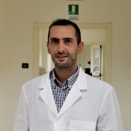 Giuseppe Sciume合作论文数Department of Molecular Medicine-Istituto Pasteur-Fondazione Cenci Bolognetti, Sapienza University of Rome3
 Angela Santoni合作论文数Department of Molecular Medicine, Sapienza University of Rome3Alessandra Zingoni合作论文数3
 Maria Teresa Petrucci合作论文数Dipartimento di Biotecnolgie Cellulari ed Ematologia, Università Sapienza di Roma3
 David Cook合作论文数2Kelly P Burke合作论文数2
 Marie Marotel合作论文数2
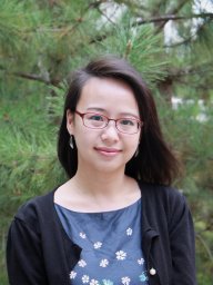 Han-Yu Shih合作论文数National Eye Institute, National Institutes of Health2
 Padraic Fallon合作论文数School of Medicine, Trinity College Dublin2
 Arlene Sharpe合作论文数Department of Immunology, Blavatnik Institute, Harvard Medical School, Harvard University2McCurdy Arleigh合作论文数2
 André Veillette合作论文数Department of Medicine, University of Montreal2Thornton W Thompson合作论文数1Sara Asif合作论文数1Troy N Trevino合作论文数1Nikhita Mathur合作论文数1Iryna Abramchuk合作论文数1Christopher J Nicolai合作论文数Univ Calif Berkeley, Dept Mol & Cell Biol, Div Immunol & Pathogenesis, Berkeley, CA 94720 USA1Rachel Nadeau合作论文数1Joy Hsu合作论文数1
 Haley E Randolph合作论文数Genet Genom & Syst Biol, Univ Chicago1
 David Raulet合作论文数Division of Immunology and Pathogenesis, Department of Molecular and Cell Biology, College of Letters & Science, University of California, Berkeley1
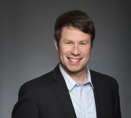 Lavallée-Adam Mathieu合作论文数Department of Chemical Physiology, The Scripps Research Institute1
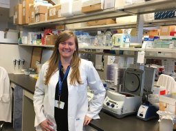 Bourgeois-Daigneault Marie-Claude合作论文数Département de Microbiologie et Immunologie, Université de Montréal1
 Azimi Camillia S合作论文数1
 Douglas A. Gray合作论文数Department of Microbiology and Immunology, University of Western Ontario1
 Lily Zhang合作论文数UCLA Center for Health Services and Society1Karen Jardine合作论文数1
 John Bell合作论文数Ottawa Hospital Research Institute1
 Alexandre Iannello合作论文数Ste-Justine Hospital Research Center, University of Montreal1
 Steffany A. L. Bennett合作论文数Department of Biochemistry, Microbiology and Immunology, University of Ottawa Neural Regeneration Laboratory1
 Michael McBurney合作论文数Cancer Therapeutics Program, Ottawa Hospital Research Institute;Departments of Medicine and Biochemistry, Microbiology and Immunology, University of Ottawa, Canada1




