Plerixafor (PLX) is FDA approved for use in autologous peripheral blood stem cell donors but not in allogeneic donors. This study was completed by members of the ASFA HPC Donor Subcommittee to examine the incidence and characteristics of poor mobilizers (PM) among matched related donors (MRD), as well as factors associated with PLX use in MRD. Risks of poor mobilization in MRD were older age, especially donors older than 60 years, lower baseline platelet counts, and heavier recipients. PLX use in PM was low but safe, tripling the success rate for collection. This study adds evidence to the body of literature to support use of PLX in allogeneic donors who are PM.
COVID‐19 convalescent plasma (CCP) was approved under emergency authorization to treat critically ill patients with COVID‐19 in the United States in 2020. We explored the demographics of donors contributing plasma for a hyperimmune, plasma‐derived therapy to evaluate factors that may be associated with anti‐SARS‐CoV‐2 antibody response variability and, subsequently, antibody titers.
Background: Plasma contains many important proteins of therapeutic interest including albumin, clotting factors, and antibodies. Source plasma (SP) is in great demand particularly due to a shortage of immunoglobulin. To better understand how to increase supply, we examined SP donor deferrals for the previous 3 years. Study design: This is a description of donor deferrals at 255 plasma donation centers in the United States for April 1, 2017 to March 31, 2020. Results: A total of 4 587 923 events were evaluated for the 3-year period 2017-2020. There were 873 227 deferrals analyzed for 2017-2018, 1 765 582 in 2018-2019, and 1 949 114 for 2019-2020. The most common deferral each year was for unacceptable blood pressure (BP) or pulse which comprised 27.9%, 28.2%, and 28.3% of deferrals in 2017-2018, 2018-2019, and 2019-2020, respectively. The second most common cause of deferral was for unacceptable hematocrit which comprised 14.1% of deferrals in 2017-2018, and 16.0% in 2018-2019 and 2019-2020. The majority of these deferred donors had low hematocrits and were predominately (similar to 80%) female. Deferral for unacceptable total protein comprised a smaller percentage (similar to 4%) of deferrals. Discussion: Most donor deferrals were due to unacceptable screening results, particularly high BP, elevated pulse, low protein, and low hematocrit. Although rates of deferrals in other categories have been slightly increasing over time, they comprise a small percentage. Donor education regarding healthy lifestyle choices may improve overall donor health, decrease deferrals, and increase SP supply.
There are three main components manufactured from whole blood: red blood cells (RBCs), plasma, and platelets. Plasma contains a multitude of different proteins, peptides, and biologic substances. Approximately 53 million liters of plasma was collected in the United States in 2019. Following collection, plasma is frozen and manufactured into plasma-derived medicinal products (PDMPs). During the manufacture process, several thousand plasma units are pooled for Cohn fractionation, which is based upon cold ethanol precipitation of proteins. The PDMPs are further prepared using ion exchange or affinity chromatography and additional steps to inactivate and remove infectious diseases such as viruses. Almost 20 different therapeutic plasma proteins are purified from plasma via these multi-step manufacturing processes. Interestingly, the demand for pharmaceutical plasma products, particularly intravenous immunoglobulin (IVIG) products, has been increasing. The manufacture and therapeutic role of blood derivatives particularly immunoglobulin therapy, Rh immunoglobulin (RhIG), COVID-19 convalescent plasma (CCP) and hyperimmune globulins, albumin, clotting factors, fibrin sealants, and platelet rich plasma will be described.Copyright © 2022 AME Publishing Company. All Rights Reserved.
Factor V Leiden (FVL) is a point mutation of factor V resulting in an elimination of cleavage site in factor V and factor Va. This genetic defect leads to an increased risk of thrombosis especially in homozygous or pseudo-homozygous FVL mutations. Many individuals with the mutation will never develop a venous thrombotic event (VTE). The decision about VTE risk reduction (both primary and secondary and prevention of recurrence) requires a great deal of clinical acumen, given that most of the people who carry the mutation will never have VTE.
Coagulation factor VIIa (FVIIa) consists of a γ-carboxyglutamic acid (GLA) domain, two epidermal growth factor-like (EGF) domains and a protease domain. FVIIa binds three Mg2+ ions and four Ca2+ ions in the GLA domain, one Ca2+ ion in the EGF1 domain and one Ca2+ ion in the protease domain. Further, FVIIa contains an Na+ site in the protease domain. Since Na+ and water share the same number of electrons, Na+ sites in proteins are difficult to distinguish from waters in X-ray structures. Here, to verify the Na+ site in FVIIa, the structure of the FVIIa-soluble tissue factor (TF) complex was solved at 1.8 Å resolution containing Mg2+, Ca2+ and Rb+ ions. In this structure, Rb+ replaced two Ca2+ sites in the GLA domain and occupied three non-metal sites in the protease domain. However, Rb+ was not detected at the expected Na+ site. In kinetic experiments, Na+ increased the amidolytic activity of FVIIa towards the synthetic substrate S-2288 (H-D-Ile-Pro-Arg-p-nitroanilide) by ∼20-fold; however, in the presence of Ca2+, Na+ had a negligible effect. Ca2+ increased the hydrolytic activity of FVIIa towards S-2288 by ∼60-fold in the absence of Na+ and by ∼82-fold in the presence of Na+. In molecular-dynamics simulations, Na+ stabilized the two Na+-binding loops (the 184-loop and 220-loop) and the TF-binding region spanning residues 163-180. Ca2+ stabilized the Ca2+-binding loop (the 70-loop) and Na+-binding loops but not the TF-binding region. Na+ and Ca2+ together stabilized both the Na+-binding and Ca2+-binding loops and the TF-binding region. Previously, Rb+ has been used to define the Na+ site in thrombin; however, it was unsuccessful in detecting the Na+ site in FVIIa. A conceivable explanation for this observation is provided.
Rivaroxaban (Xarelto; Johnson & Johnson Services, Inc) is a direct oral anticoagulant (DOAC) that works by directly inhibiting the active site of factor Xa (FXa). Rivaroxaban is metabolized and cleared via the kidney and liver. The results of various studies have shown that patients with severe renal impairment should receive reduced dosages of rivaroxaban or another anticoagulant due to impaired clearance. Although it is not required, monitoring rivaroxaban is useful in some conditions; however, the assays required for such monitoring are not readily available. Herein, we present a case of a 68-year-old Caucasian male patient who was receiving rivaroxaban (20 mg/day) for atrial flutter and had mild renal impairment. The patient was found to have increased effect of rivaroxaban due to further impairment of renal clearance caused by several renally cleared medications. This case highlights the importance of closely examining the renal function of and medication list for a patient before starting DOACs such as rivaroxaban.
BACKGROUND:Factor (F) IX/IXa inactivation by plasmin has been studied; however, whether plasmin converts FIXa to a fibrinolytic enhancer is not known. OBJECTIVE:Investigate plasmin proteolysis site(s) in FIXa that inactivates and transforms it into a fibrinolytic enhancer. METHODS:NH2 -terminal sequencing, mass spectrometry analysis, and functional assays. RESULTS:Plasmin in the presence of Ca2+ /phospholipid (PL) rapidly cleaved FIXaβ at Lys316↓Gly317 to yield FIXaγ followed by a slow cleavage at Lys413↓Leu414 to yield FIXaδ. FIXaγ/FIXaδ migrated indistinguishably from FIXaβ in nondenaturing gel system indicating that C-terminal residues 317-415/317-413 of heavy chain remain noncovalently associated with FIXaγ/FIXaδ. However, as compared with FIXaβ, FIXaγ or FIXaγ/FIXaδ (25-75 mixture, 8-hour/24-hour incubation analysis by mass spectrometry) was impaired ~ 10-fold in hydrolyzing synthetic substrate CBS 31.39 (CH3-SO2-D-Leu-Gly-Arg-pNA), ~ 30-fold (~ 5-fold higher Km , ~ 6-fold lower kcat ) in activating FX in a system containing Ca2+ /PL, and ~ 650-fold in a system containing Ca2+ /PL and FVIIIa. Further, FIXaγ or FIXaγ/FIXaδ bound FVIIIa with ~ 60-fold reduced affinity compared with FIXaβ. Additionally, in ligand blots, plasminogen or diisopropylfluorophosphate-inhibited plasmin (DIP-plasmin) bound FIXaγ and FIXaδ but not FIXaβ. This interaction was prevented by ε-aminocaproic acid or carboxypeptidase B treatment suggesting that plasminogen/DIP-plasmin binds to FIXaγ/FIXaδ through newly generated C-terminal Lys316 and Lys413. Importantly, FIXaγ/FIXaδ mixture but not FIXaγ enhanced tissue plasminogen activator (tPA)-mediated plasminogen activation in a concentration dependent manner. Similarly, FIXaγ/FIXaδ mixture but not FIXaγ enhanced tPA-induced clot lysis in FIX-depleted plasma. CONCLUSION:Plasmin cleavage at Lys316↓Gly317 abrogates FIXaβ coagulant activity, whereas additional cleavage at Lys413↓Leu414 converts it into a fibrinolytic enhancer.
Platelet-transfusion refractoriness (PTR) is common in patients with hematological malignancies. The etiology of immune PTR is typically human leukocyte antigen (HLA) antibodies (Abs) from pregnancy or previous transfusion. Herein, we report PTR in the setting of induction chemotherapy for acute myelogenous leukemia (AML) from Abs against CD36/glycoprotein (GP)IV. A 66-year-old African American woman presented with anemia and thrombocytopenia. She was found to have transfusion-dependent AML, and a 7 + 3 regimen (7 days of standard-dose cytarabine and 3 days of an anthracycline antibiotic or an anthracenedione, most often daunorubicin) was initiated. The patient developed profound thrombocytopenia, with platelet nadir of 0 by day 13. The results of HLA antibody screening were negative. However, the results of a screening test for platelet-specific antibodies screen showed Abs against cluster of differentiation (CD)36. The platelets of the patient lacked expression of CD36, and DNA analysis showed mutations in the CD36 gene. HLA Ab-mediated PTR is common in patients with hematological malignancies. However, once HLA Abs are excluded, other less-frequent Abs should be considered, particularly in patient populations of Asian, African, or Middle Eastern descent.
Objectives To provide an overview of the complexities associated with the human leukocyte antigen (HLA)-mediated platelet refractoriness. HLA antibody detection technologies and limitations associated with methodologies are discussed. Methods A case scenario and review of relevant literature describing platelet refractoriness are presented, followed by a discussion of HLA antibody testing. Results Following diagnosis of HLA-mediated refractoriness, a decision is made regarding the approach to obtain the appropriate platelets. The panel reactive antibodies (PRA) % of the patient, HLA typing, and limitations of the HLA testing should be taken into account when deciding which type of product would be the best option for a given patient. Conclusions Following confirmation and review of HLA antibody testing, platelets are ordered based upon the PRA% and approach employed, HLA-matched platelets, antigen restricted platelets, or cross-matched platelets. The platelets are transfused and a posttransfusion increment count is monitored to determine transfusion success.
OBJECTIVES:To provide an overview of the clot viscoelastic testing technology and to describe its utility in guiding blood product transfusions.METHODS:A case scenario will be discussed as well as interpretation of thromboelastography (TEG) tracings. In addition, literature examining the utility of viscoelastic testing in guiding patient management and blood product transfusions will be reviewed.RESULTS:TEG/rotational thromboelastometry (ROTEM) is useful in evaluating clot kinetics in trauma and acutely bleeding patients. TEG/ROTEM parameters are reflective of values measured using standard coagulation assays; however, TEG/ROTEM parameters are more rapidly available and more costly. TEG and ROTEM are used in three main settings: cardiac surgery, liver transplantation, and trauma to assess global hemostasis and administration of blood products.CONCLUSIONS:TEG/ROTEM can be helpful in guiding resuscitation and blood product transfusion. Several studies have demonstrated a reduction in transfusion of blood components with TEG/ROTEM; however, other studies have suggested that TEG/ROTEM is not clinically effective in guiding transfusion.
Background Activated coagulation factor IX (FIXa) consists of a gamma-carboxyglutamic acid domain, two epidermal growth factor-like (EGF) domains, and a C-terminal protease domain. Consensus sequence and biochemical data support the existence of a Na+-site in the FIXa protease domain. However, soaking experiments or crystals grown in high concentration of ammonium sulfate did not reveal a Na+-site in wild-type or mutant FIXa EGF2/protease domain structure. Objective Determine the structure of the FIXa EGF2/protease domain in the presence of Na+; perform molecular dynamics (MD) simulations to explore the role of Na+ in stabilizing FIXa structure. Methods Crystallography, MD simulations, and modeling heparin binding to FIXa. Results Crystal structure at 1.37-angstrom resolution revealed that Na+ is coordinated to carbonyl groups of residues 184A, 185, 221A, and 224 in the FIXa protease domain. The Na+-site in FIXa is similar to that of FXa and is linked to the Asp189 S1-site. In MD simulations, Na+ reduced fluctuations in residues 217-225 (Na+-loop) and 70-80 (Ca2+-loop), whereas Ca2+ reduced fluctuations only in residues of the Ca2+-loop. Ca2+ and Na+ together reduced fluctuations in residues of the Ca2+-loop and Na+-loop (residues 70-80, 183-194, and 217-225). Moreover, we observed four sulfate ions that make salt bridges with FIXa protease domain Arg/Lys residues, which have been implicated in heparin binding. Based upon locations of the sulfate ions, we modeled heparin binding to FIXa, which is similar to the heparin binding in thrombin. Conclusions The FIXa Na+-site in association with Ca2+ contributes to stabilization of the FIXa protease domain. The heparin binding mode in FIXa is similar to that in thrombin.
Importance Neuromyelitis optica/neuromyelitis optica spectrum disorder patients' response to therapeutic plasma exchange (TPE) is currently incompletely characterized. Objective Our study aims to understand the clinical status improvement of neuromyelitis optica/neuromyelitis optica spectrum disorder patients treated with TPE. Design, Setting, and Participants This is a multicenter retrospective study conducted between 1 January 2003 and 31 July 2017 at 13 US hospitals performing apheresis procedures. Subjects studied were diagnosed with neuromyelitis optica/neuromyelitis optica spectrum disorder who received TPE during presentation with acute disease. Main Outcomes and Measures The primary outcome was clinical status improvement in patients treated with TPE. Secondary measures were procedural and patient characteristics associated with response to treatment. Results We evaluated 114 patients from 13 institutions. There was a female predilection. The largest ethnic group affected was non-Hispanic Caucasian. The average age of diagnosis was 43.1 years. The average time to diagnosis was 3.1 years. On average, five procedures were performed during each treatment series. The most commonly performed plasma volume exchange was 1.0 to 1.25 using 5% albumin as replacement fluid. Most patients (52%) did not require an additional course of TPE and noted "mild" to "moderate" clinical status improvement. Maximal symptom improvement appeared by the fourth or fifth TPE treatment. Conclusion and Relevance TPE improved the clinical status of patients. Adults responded more favorably than children. Procedural characteristics, including number of TPEs, plasma volume exchanged, and replacement fluid used, were similar between institutions. TPE was well-tolerated and had a low severe adverse event profile.
The main clinical distinction between post-transfusion purpura (PTP) and idiopathic thrombocytopenic purpura (ITP) is the sudden development of severe thrombocytopenia in the days after transfusion. Herein, we report the case of a 53-year-old Caucasian woman who developed multiple myeloma (MM) after peripheral blood-stem-cell transplant (PBSCT), along with severe thrombocytopenia (with a nadir of 1 × 10⁹/L); she also experienced severe adverse events after each platelet transfusion, including the first one. These reactions were absent with any other transfused blood products. The results of an human leukocyte antigen (HLA) class-1 panel reactive antibody assay were 0%, and the results of a platelet-antibody screening assay were positive for HLA class-1 antibodies and glycoprotein (Gp)IIb/IIIa antibodies. Her platelet count reached 42 × 109 per L on day 50, after rituximab on day 22 and daratumumab on day 29. Her clinical scenario was most consistent with the course of PTP.
OBJECTIVES:Washing cellular blood products is accepted to ameliorate repeated severe allergic reactions but is associated with RBC hemolysis and suboptimal platelet function. We compared in vitro hemolysis and platelet function in blood components after washing with Plasma-Lyte A (PL-A) vs normal saline (NS).METHODS:RBC (n = 14) were washed/resuspended in NS or PL-A. Free hemoglobin and heme were determined at 0, 24, 48, and 72 hours. Platelet concentrates (PCs; n = 21) were washed with NS or PL-A and resuspended in same washing solution (n = 13) or ABO-identical plasma (n = 8). Platelet aggregation and spreading were evaluated.RESULTS:The 24-hour free hemoglobin and heme levels were higher in NS (P < .05). Improved platelet function was observed in PL-A-washed PCs (P < .001).DISCUSSION:PL-A showed less RBC hemolysis and better platelet function than NS. Whether such differences would occur in vivo is unknown.
Oxidation reduction potential (ORP) or Redox is the ratio of activity between oxidizers and reducers. Oxidative stress (OS) can cause cellular injury and death, and is important in the regulation of immune response to injury or disease. In the present study, we investigated changes in the redox system as a function of cardiopulmonary bypass (CPB) in pediatric patients. 664 plasma samples were collected from 162 pediatric patients having cardiac surgery of various CPB times. Lower ORP values at 12 h post-CPB were associated with poor survival rate (mean ± SD 167 ± 20 vs. 138 ± 19, p = 0.005) and higher rate of thrombotic complications (153 ± 21 vs. 168 ± 20, p < 0.008). Similarly, patients who developed infections had lower ORP values at 6 h (149 ± 19 vs. 160 ± 22, p = 0.02) and 12 h (156 ± 17 vs. 168 ± 21, p = 0.004) post-CPB. Patients that developed any post-operative complication also had lower 6 h (149 ± 17 vs. 161 ± 23, p = 0.002) and 12 h (157 ± 18 vs. 170 ± 21, p = 0.0007) post-CPB ORP values. Free hemoglobin and IL-6, IL-10, and CRP were not associated with ORP levels. However, higher haptoglobin levels preoperatively were protective against decreases in ORP. Decreased ORP is a marker for poor outcome and predictive of post-operative thrombosis, infection, and other complications in critically ill pediatric cardiac surgery patients. These results suggest that redox imbalance and OS may contribute to the risk of complications and poor outcome in pediatric CBP patients. Haptoglobin may be a marker for increased resilience to OS in this population.
The concept of “universal donor” group O red blood cells (RBC) and “universal donor” group AB plasma antedate the modern era of evidence-based medicine. The absence of gross hemolysis led to the assumption that the small amounts of incompatible anti-A and anti-B (in O RBC concentrates) and the absence of ABO antibodies (in AB plasma) are almost always benign and of no consequence for the recipient. These assumptions, never tested in observational studies, much less randomized trials, have been called into question in recent decades. Evidence that ABO compatible (but nonidentical) transfusions of blood components may be less effective and less safe than ABO identical has accumulated steadily over the past 2 decades. Although transfusions of ABO “compatible” blood components have long been considered safe to use, ABO identical transfusions have been associated with better outcomes in terms of morbidity and mortality in multiple studies.1–4 Clinical studies, bedside observations, randomized clinical trials, and in vitro studies strongly suggest that ABO identical, and not ABO “compatible,” transfusions are likely the safest practice (Table).1,8,10,13–18Table.: A Summarized List of Some of the ABO Identical and ABO Nonidentical StudiesMost blood components, regardless of the methods of collection, contain significant amounts of donor plasma carrying soluble A and/or B antigens (except group O), in addition to anti-A and/or anti-B (except group AB). The amount of plasma in each blood component varies from 200 to 300 mL in platelet or plasma units to 30–70 mL in RBC concentrates, to 10–20 mL in a unit of cryoprecipitate. In addition, cellular components (ie, RBCs and platelets) express ABO antigens on their membranes. The recipient frequently possesses cell surface and soluble ABO antigens in considerable amounts (except group O) and anti-A/B (except group AB). The transfused anti-A and anti-B are rapidly bound to circulating soluble A and B antigens and to cell surface A and B antigens. Likewise, the transfused soluble antigen(s) also binds to circulating anti-A/B forming soluble high-molecular weight immune complexes.7 These complexes circulate for hours to days after nonidentical transfusions and can bind to RBCs, causing hemolysis, and to platelets, resulting in their activation and premature removal from the circulation by monocytes and macrophages via their FC receptors.7,19 The formation of anti-A and/or anti-B immune complexes may initiate inflammation, tissue injury, and/or modulate cellular immunity and potentially predispose to infection.8 Depending on their avidity, transfused anti-A and/or anti-B present in ABO nonidentical components can also bind to the tissue-bound A and B antigens, resulting in RBC hemolysis, platelet activation, and alteration of the normal hemostasis.20 In addition, similar to human leukocyte antigen antigens, A and B antigens are widely expressed on the endothelium, thus transfused anti-A/B are likely capable of causing direct injury to organs. The acute rejection of ABO-incompatible organs and bone marrows is well documented in literature.9,21,22 ABO NONIDENTICAL RBC TRANSFUSIONS The presence of anti-A and anti-B in ABO nonidentical RBC has been associated with higher morbidity and mortality in many subsets of patients.13,14,16 Most recently, in a large multihospital registry (Transfusion Registry for Utilization, Surveillance, and Tracking), the association between exposures to at least 1 unit of ABO nonidentical RBC increased in-hospital mortality.13 Pai et al13 reported that of all nongroup O patients (n = 18,843) admitted between 2002 and 2011, group A patients who received group O RBCs had significantly increased risk of in-hospital death (relative risk, 1.79; 95% confidence interval, 1.20–2.67; P = .005). This remarkable and important finding raises for the first time the issue of whether group O RBCs are truly “universal donor,” at least for group A recipients. Possible contributing factors are the higher titer, avidity, and biologic activity of anti-A, compared with anti-B, and/or the formation of the circulating immune complexes, or the induction of iatrogenic “autoimmune like process.”7 ABO NONIDENTICAL PLATELET TRANSFUSIONS The importance of ABO compatibility in platelet transfusion, in particular, has long been a subject of debate. Although it is not usual practice to transfuse ABO nonidentical plasma (usually “universal” donor group AB plasma), with the exception of trauma patients of unknown ABO blood group, platelets are often transfused across ABO barriers due to the difficulty in inventory management of a component with a short shelf-life. Approximately 10%–40% of all platelet transfusions in the United States are ABO nonidentical.14,15 Greater morbidity and mortality have been reported in several observational studies of ABO nonidentical platelet transfusion.1,8,10,16–18 Early randomized trials, although small, clearly demonstrated that use of ABO nonidentical platelets leads to dramatic increases in platelet transfusion refractoriness, a potentially lethal outcome.5,6 Intentional transfusion of ABO-mismatched platelets increased refractoriness by 5-fold and increased sensitization to human leukocyte antigen as well. The use of unselected ABO blood group platelets doubled the refractoriness rate compared with use of ABO identical platelets. This evidence was largely ignored because of the long standing conviction that ABO was not clinically important, and due to difficulties in managing blood bank inventories to provide only ABO identical platelets. Severe, even fatal hemolytic transfusion reactions have been reported after transfusion of ABO nonidentical (compatible and incompatible) platelets.16–18 Transfusion of ABO nonidentical platelets in transfusion-dependent patients with hematologic diseases revealed earlier platelet refractoriness than in patients receiving ABO identical platelets.5 Consistently higher platelet increments were also observed in a systematic review assessing outcomes of ABO identical versus ABO nonidentical platelet transfusion; however, no consistent benefit in clinical outcomes was considered proven.1 After transfusion of ABO nonidentical platelets in bone marrow transplant patients, Heal et al10 hypothesized an association with increased mortality due to bleeding, multiorgan failure, and sepsis, seen in immune complex diseases such as lupus and rheumatoid arthritis. Multivariate analysis of 1-hour platelet recovery in pediatric patients (N = 400) showed superiority of efficacy of ABO identical versus ABO major-mismatched platelet transfusions (odds ratio, 3.97; 95% confidence interval, 1.52–10.39; P = .005).11 Using flow cytometry and fluorescence microscopy, Julmy et al11 demonstrated a rapid clearance of group A platelets from the circulation of group O or B recipients. However, major-mismatched transfusions of group A platelets that expose A2 antigen were as efficient as ABO identical transfusions (P = .90), indicating a remarkable effect of the circulating anti-A on platelet recovery. In the multicenter Platelet Dose “PLADO” study, where 1102 pediatric and adult hematology-oncology patients with hypoproliferative thrombocytopenia were randomized to receive different doses of prophylactic platelet transfusions (for platelet count of <10 × 109/L), Kaufman et al23 found no correlation between platelet source and ABO matching with overall transfusion reaction risks. However, the absence of a substantial control group of patients receiving solely ABO identical platelets makes it impossible to ascertain the role of ABO matching of platelet transfusions in these patients. In a post hoc analysis of the same study (PLADO study), including 1272 hematology-oncology patients who received 6031 prophylactic platelet transfusions, Triulzi et al12 reported that randomized dose strategy, platelet source, ABO compatibility, and duration of storage have no impact on prevention of clinical bleeding. However, authors noted that platelet increments were generally higher after transfusions of apheresis platelets, ABO identical platelets, and platelets stored 3 vs 4–5 days.12 The major limitation of these data is that few patients received solely ABO identical platelets, thus there is no substantive control group for comparison. ABO NONIDENTICAL PLASMA TRANSFUSIONS Inaba et al2 compared the rate of mortality and complications (acute respiratory distress syndrome, sepsis, renal failure, and liver failure) in a retrospective study of trauma patients (N = 284) exposed to ABO nonidentical (but compatible) plasma who were matched 1:1 with patients receiving solely ABO identical plasma. Although mortality was not different between the groups (P = .66), the overall complication rate was significantly higher for ABO nonidentical plasma recipients (P = .002), in particular for acute respiratory distress syndrome and sepsis. A dose-dependent increase in complication rates was detected as the transfused volume of ABO nonidentical plasma increased.2 In a retrospective, nationwide cohort study, Shanwell et al3 evaluated the mortality rate at 14 days posttransfusion in Swedish recipients (N = 86,082) exposed to plasma between 1990 and 2002. Higher mortality rates were found among patients receiving ABO nonidentical (but compatible) plasma. This risk was found to be dose dependent. The excess risk was most strongly associated with transfusion of 5 or more AB plasma units to group O recipients. This suggests a likely mechanism for mortality caused by circulating ABO immune complexes made up of donor soluble A and B antigens and recipient anti-A and anti-B3. In summary, there is evidence that anti-A/B, along with soluble A/B antigens present in the donor and/or recipient plasmas, may cause substantial dose-dependent harm after ABO nonidentical transfusions (Table). Unless transfusion is urgent and lifesaving, we propose that ABO identical transfusions should be the standard of care for all relevant blood components when feasible. Although adherence to a strict ABO identical transfusion policy, for platelets in particular, may lead to difficulties in maintaining adequate inventory and increase wastage, patient safety should outweigh these logistic challenges. It may be necessary to overproduce platelets from whole blood (a relatively inexpensive blood component), so that adequate inventories of ABO identical platelets are available. Of note, in a survey of North American laboratories that was performed almost a decade ago, <22% of participating laboratories (N = 3156) reported the existence of a transfusion policy of only ABO identical plasma and platelet products.15 While we have introduced an ABO identical policy successfully with minimal effects on outdating,24 this is made possible by the use of washed group O RBCs and platelets, the proximity of our regional blood center, and our large inventory/transfusion volume. It is still difficult to provide ABO identical platelets to group B and AB patients in emergency situations, particularly patients of group AB, as washed platelets are not available at a moment’s notice. The availability of platelet additive solutions that remove 95% of plasma will likely help solve this remaining issue. The availability of closed system washed platelets and RBCs that can be stored for perhaps 2–5 days would make it possible to supply minimally toxic RBCs and platelets at distances from regional blood centers and to smaller hospitals. It has been suggested that components be titered to exclude higher titers of anti-A and anti-B. Unfortunately there is no evidence base for the efficacy and safety of this practice. In any case, unless we are prepared to titer antigen in donor plasma and antibody in recipients, this approach is not feasible to mitigate the formation of ABO immune complexes based on recipient antibody and donor soluble antigen. An alternate approach might be to remove the supernatant from donor RBCs and platelets before transfusion when isoagglutinins that react with the recipient’s A/B antigens are present. None of these approaches have been rigorously evaluated as yet. There is an urgent need for randomized trials and other definitive studies to confirm or refute the hypothesis that there really are no truly safe “universal donor” transfusions. These long established practices, based on “expert opinion,” reflect historical and traditional concepts which may no longer be tenable in the modern era. ABO immune complexes and direct binding of anti-A to group A RBCs may contribute to low level hemolysis, among other biologic phenomena. Low level hemolysis may be deleterious to patients in the same manner that it is in sickle cell disease and paroxysmal nocturnal hemoglobinuria, where it contributes to inflammation, vasculopathy, thrombosis, and infection. Further research into these possibilities will help transfusion services determine if and how we move forward from the likely outdated concept of “ABO compatible” transfusions and “universal donor” blood components. A number of approaches can be taken to assess the benefits and risks of an ABO identical transfusion policy. A randomized trial comparing current practice with ABO identical only (using washed or plasma-reduced platelets and RBCs when ABO identical are not available) is 1 design that is both feasible and unlikely to dramatically affect inventory practice. Another possibility would be to compare group O universal donor RBCs with washed or plasma reduced, or low-titer group O RBCs, in elective transfusions, because these are standard practice in many centers. Finally, an implementation of a clinical trial with before and after analysis of current practice versus ABO identical only (using washed, plasma reduced, or low-titer group O platelets/RBCs when necessary) would be feasible. The terminology “ABO compatible” is potentially confusing, only relevant for RBCs, likely incorrect, and should be discarded and replaced by the more accurate terms “ABO identical” and “ABO nonidentical.” Similarly, the concept of “universal donors,” although time honored, is no longer one which we can be confident is clinically sound. The terms provide the illusion of safety to health care providers, but not the reality of safety for the recipient. DISCLOSURES Name: Majed A. Refaai, MD. Contribution: This author helped collect and analyze data, and draft and revise the manuscript. Name: Christine Cahill, MS, RN. Contribution: This author helped collect the data, and review and edit the manuscript. Name: Debra Masel, MT, (ASCP) SBB. Contribution: This author helped revise the manuscript and approve the final version. Name: Amy E. Schmidt, MD, PhD. Contribution: This author helped revise the manuscript and approve the final version. Name: Joanna M. Heal, MD. Contribution: This author helped revise the manuscript and approve the final version. Name: Scott A. Kirkley, MD. Contribution: This author helped revise the manuscript and approve the final version. Name: Neil Blumberg, MD. Contribution: This author helped revise the manuscript and approve the final version. This manuscript was handled by: Marisa B. Marques, MD.
 S. Paul Bajaj合作论文数Molecular Biology Institute, University of California, Los Angeles33
 Majed Refaai合作论文数30
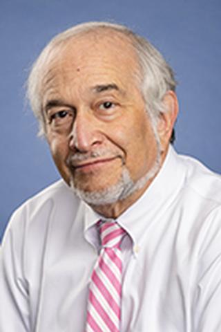 Neil Blumberg合作论文数School of Medicine and Dentistry, University of Rochester Medical Center;Department of Pathology and Laboratory Medicine, University of Rochester Medical Center22
 Scott A. Kirkley合作论文数10
 Christine Cahill合作论文数8Henrichs Kelly F合作论文数8Vadivel Kanagasabai合作论文数UCLA/Orthopaedic Hospital, University of California, Los Angeles8Kaillathe Padmanabhan合作论文数8Debra Masel合作论文数6
 Tanmay Sahai合作论文数5
 Sayeh Agah合作论文数5
 Walter Kisiel合作论文数School of Medicine, UNM5Joanna M. Heal合作论文数5Hitendra S. Chand合作论文数5Godwin I Ogueli合作论文数4Hannah L. McRae合作论文数4
 Duilio Cascio合作论文数Department of Biological Chemistry, University of California, Los Angeles4
 David Gailani合作论文数Vanderbilt Institute for Infection, Immunology and Inflammation, Vanderbilt University4
 Myra Coppage合作论文数4Haewon C. Kim合作论文数4
 Jill Cholette合作论文数4Anthony P. Pietropaoli合作论文数4Madhu S. Bajaj合作论文数4
 Laura L. Cooling合作论文数Department of Pathology, Medical School, University of Michigan-Ann Arbor3Simon Toby L合作论文数3
 Sreejesh Shanker合作论文数Baylor Coll Med, Dept Biochem & Mol Biol, Houston, TX 77030 USA3
 Richard P. Phipps合作论文数University of Rochester Medical Center3Sherry L. Spinelli合作论文数3
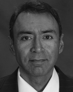 Ashwani Sharma合作论文数Space Vehicles Directorate, Kirtland AFB Air Force Research Laboratory3
 Joseph E. Schwartz合作论文数Stony Brook University3Angela Busacco合作论文数2Grace Conley合作论文数2Emily Gore合作论文数2Leonor Fernando合作论文数2Chelsea Milito合作论文数2Carrie A. Chastain合作论文数2yanyun wu合作论文数2Andrew Johnson合作论文数2Yogesh Kumar合作论文数2Peter Vogel合作论文数2
 Chisa Yamada合作论文数Department of Pathology, Medical School, University of Michigan-Ann Arbor2
 Kimberly B Bjugstad合作论文数Dept Med, Univ Colorado2Adamski Jill合作论文数2Roth Nathan J合作论文数2
 Dunbar Nancy M合作论文数2Underwood M C合作论文数2
 Jay S Raval合作论文数Department of Pathology, University of Pittsburgh;The Institute for Transfusion Medicine;The Institute for Transfusion Medicine, University of Pittsburgh2
 Patricia Janet Sime合作论文数School of Medicine and Dentistry, University of Rochester Medical Center2
 Sriram Krishnaswamy合作论文数Perelman School of Medicine, University of Pennsylvania2




