Optical transparency is a critical quality attribute of corneal lenticules intended for refractive correction, yet current evaluation methods rely primarily on qualitative visual inspection. In this study, we present a benchtop transparency measurement device that provides quantitative, spatially resolved transparency maps of corneal tissue samples, including lenticules measured directly within their clinical packaging. The system operates in transmission mode and quantifies transparency by analyzing contrast degradation of a high-precision grid pattern imaged through the sample. Device performance was validated using water-milk liquid phantoms with graded turbidity, enabling definition of quantitative thresholds corresponding to low, medium, and high transparency levels. These thresholds showed strong agreement with independent visual assessments and prior literature. The device demonstrated high repeatability, with a coefficient of variation below 1.1%. The system was applied to evaluate human corneal lenticules generated for presbyopia correction. Transparency measurements showed no dependence on stromal depth of origin. Additionally, paired measurements acquired before and after secondary electron-beam (e-beam) sterilization demonstrated that lenticules maintained high optical transparency following sterilization. Control measurements confirmed that minor transparency changes observed post-sterilization were attributable primarily to the storage medium rather than the tissue itself. This transparency-mapping approach enables objective, quantitative quality control of corneal lenticules and represents a significant improvement over traditional visual inspection methods, supporting reliable assessment of implant optical quality prior to clinical use.
AbstractTheory and observation show that glacier-flow regimes characterized by high basal slip enhance the projection of topographic detail to the surface, motivating this investigation into the efficacy of using glacier surges to improve bed estimation. Here we adapt a Bayesian inversion scheme and apply it to real and synthetic data as a proof of concept. Synthetic tests show a reduction in mean RMSE between true and inferred beds by more than half, and an increase in the mean correlation coefficient of ~0.5, when data from slip- versus deformation-dominated regimes are used. Multi-epoch inversions, which partition slip- and deformation-dominated regimes, are shown to outperform inversions that average over these flow regimes thereby squandering information. Tests with real data from a surging glacier in Yukon, Canada, corroborate these results, while highlighting the challenges of limited or inconsistent data. With the growing torrent of satellite-based observations, fast-flow events such as glacier surges offer potential to improve bed estimation for some of the world's most dynamic glaciers.
Optical Coherence Tomography (OCT) is a rapidly growing imaging modality in biomedical optics. OCT can perform high-resolution, cross-sectional imaging of the microstructure of biological tissues by measuring the coherent spectrum from the backscattered light. OCT systems with broad spectral bandwidths are often constructed using free-space optics to avoid dispersion by fibre optic components. This paper presents a fibre-based OCT system at a centre wavelength of 1300 nm with an axial resolution of 3.8 µm in air, surpassing any previously reported values to the best of our knowledge. Despite the challenges in transporting a broadband spectrum using fibre-optics, the system investigation was motivated by the ever-increasing demand for commercialization of high-resolution OCT systems and simplification of construction. We also evaluate and demonstrate the direct measurement method for axial resolution using an air wedge. Imaging of biomedical and other samples is demonstrated using a high numerical aperture sample lens and compared with images from a commercial OCT system. We discuss the effect of the improved structural visibility by achieving image voxels closer to an isometric shape with a high NA sample lens.
Abstract Glacier surges are periodic episodes of mass redistribution characterized by dramatic increases in ice flow velocity and, sometimes, terminus advance. We use optical satellite imagery to document five previously unexamined surge events of Sít’ Kusá (Turner Glacier) in the St. Elias Mountains of Alaska from 1983 to 2013. Surge events had an average recurrence interval of ~5 years, making it the shortest known regular recurrence interval in the world. Surge events appear to initiate in the winter, with speeds reaching up to ~25 m d−1. The surges propagate down-glacier over ~2 years, resulting in maximum thinning of ~100 m in the reservoir zone and comparable thickening at the terminus. Collectively, the rapid recurrence interval, winter initiation and down-glacier propagation suggest Sít’ Kusá's surges are driven by periodic changes in subglacial hydrology and glacier sliding. Elevation change observations from the northern tributary show a kinematic disconnect above and below an icefall located 23 km from the terminus. We suggest the kinematic disconnect inhibits drawdown from the accumulation zone above the icefall, which leads to a steady flux of ice into the reservoir zone, and contributes to the glacier's exceptionally short recurrence interval.
Corneal cross-linking (CXL) has grown from an interesting concept to a practical clinical treatment for corneal ectatic disease globally in the past three decades. In both understanding the principle of how CXL proceeds and monitoring the clinical procedure, detection of structural changes during cornea CXL plays a significant role. This paper demonstrates a novel over-sampling nano-sensitive optical coherence tomography (osnsOCT) method, which is potential to detect nanoscale structural changes in various tissues, to simultaneously measure the structural variations during the corneal CXL treatment.
In biomedical optics, Optical Coherence Tomography (OCT) is an emerging optical imaging modality during the last three decades. OCT can perform high resolution, cross-section imaging of the internal microstructure in biological tissues by measuring echoes of backscattered light. We have developed a broadband, high-resolution spectral domain OCT system whose central wavelength is 1300 nm with bandwidth ~400 nm. Theoretical axial resolution of the system is ~3.71 μm and experimentally we get ~5 μm in air. Furthermore, we will apply this system for nano-sensitive detection and visualization in varieties of biological tissues.
Corneal cross-linking (CXL) using ultraviolet-A (UVA) irradiation with a riboflavin photosensitizer has grown from an interesting concept to a practical clinical treatment for corneal ectatic diseases globally, such as keratoconus. To characterize the corneal structural changes, existing methods such as X-ray microscopy, transmission electron microscopy, histology and optical coherence tomography (OCT) have been used. However, these methods have various drawbacks such as invasive detection, the impossibility for in vivo measurement, or limited resolution and sensitivity to structural alterations. Here, we report the application of oversampling nanosensitive OCT for probing the corneal structural alterations. The results indicate that the spatial period increases slightly after 30 minutes riboflavin instillation but decreases significantly after 30 minutes UVA irradiation following the Dresden protocol. The proposed noninvasive method can be implemented using existing OCT systems, without any additional components, for detecting nanoscale changes with the potential to assist diagnostic assessment during CXL treatment, and possibly to be a real-time monitoring tool in clinics.
Multi-layer patterning schemes involve the use of Silicon containing Anti-Reflective Coating (SiARC) films for their anti-reflective properties. Patterning transfer completion requires complete and selective removal of SiARC which is very difficult due to its high silicon content (>40%). Typically, SiARC removal is accomplished through a non-selective etch during the pattern transfer process using fluorine containing plasmas, or an ex-situ wet etch process using hydrofluoric acid is employed to remove the residual SiARC, post pattern transfer. Using a non-selective etch may result in profile distortion or wiggling, due to distortion of the underlying organic layer. The drawbacks of using wet etch process for SiARC removal are increased overall processing time and the need for additional equipment. Many applications may involve patterning of active structures in a poly-Si layer with an underlying oxide stopping layer. In such applications, SiARC removal selective to oxide using a wet process may prove futile. Removing SiARC selectively to SiO2 using a dry etch process is also challenging, due to similarity in the nature of chemical bonds (Si - O) in the two materials. In this work, we present highly selective etching of SiARC, in a plasma driven by a surface wave radial line slot antenna. The first step in the process involves an in-situ modification of the SiARC layer in O-2 plasma followed by selective etching in a NF3/H-2 plasma. Surface treatment in O-2 plasma resulted in enhanced etching of the SiARC layer. For the right processing conditions, in-situ NF3/H-2 dry etch process demonstrated selectivity values greater than 15:1 with respect to SiO2. The etching chemistry, however, was sensitive to NF3:H-2 gas ratio. For dilute NF3 in H-2, no SiARC etching was observed. Presumably, this is due to the deposition of ammonium fluorosilicate layer that occurs for dilute NF3/H-2 plasmas[1]. Additionally, challenges involved in selective SiARC removal (selective to SiO2, organic and Si layers) post pattern transfer, in a multi-layer structure will be discussed. [1] A. J. Sidhwa et al., "Reactive ion etching of crystalline silicon using NF3 diluted with H-2," J. Vac. Sci. Technol. A, vol. 11, no. 4, pp. 1156-1160, 1993.
 Martin J Leahy合作论文数University of Limerick5
 Yi Zhou (周翊)合作论文数Institute of Optics and Electronics, Chinese Academy of Sciences4
 Rajib Dey合作论文数JADAVPUR UNIVERSITY4
 Michael Mrochen合作论文数Department of Ophthalmology, University of Zurich4Sergey Alexandrov合作论文数Optical + Biomedical Engineering Laboratory;Department of Electrical and Electronic Engineering;University of Western Australia;Optical + Biomedical Engineering Laboratory, University of Western Australia4
 Ellyn M. Enderlin合作论文数Cryosphere Geophys & Remote Sensing CryoGARS Lab, Boise State Univ3
 Nandan Das合作论文数3
 Kai Neuhaus合作论文数School of Physics, National University of Ireland3
 Arthur B. Cummings合作论文数2boss daniel合作论文数2maria galligan合作论文数2
 William Kochtitzky合作论文数Department of Geography, Environment and Geomatics, University of Ottawa2
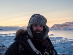 Robert McNabb合作论文数School of Geography and Environmental Sciences, Ulster University2
 Diana Bogusevschi合作论文数Res. Inst. for Networks & Commun. Eng. (RINCE),, Dublin City Univ.;c;Res. Inst. for Networks & Commun. Eng. (RINCE),, Dublin City Univ.2
 Pavel Zakharov合作论文数Solianis Monitoring AG2
 Karl J Kreutz合作论文数Woods Hole Oceanographic Institute2Ryan McAuley合作论文数1Peter Biolsi合作论文数1
 Douglas Brinkerhoff合作论文数Department of Computer Science, University of Montana1
 Ashkan Eliasy合作论文数Sch Engn, Univ Liverpool1
 Shyam Sridhar合作论文数1Curatolo Andrea合作论文数School of Electrical, Electronic and Computer Engineering, The University of Western Australia1Sergey Voronin合作论文数1
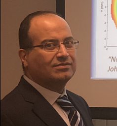 Abass Ahmed合作论文数1
 Ahmed Elsheikh合作论文数School of Engineering, University of Liverpool1Birkenfeld Judith合作论文数1
 Gwenn E. Flowers合作论文数Department of Earth and Ocean Sciences;University of British Columbia;Department of Earth and Ocean Sciences, University of British Columbia1
 Hester Jiskoot合作论文数Department of Geography;University of Lethbridge;Department of Geography, University of Lethbridge1
 Etienne Berthier合作论文数Laboratoire d'Etudes en Géophysique et Océanographie Spatiales, Centre National de la Recherche Scientifique1
 Alok Ranjan合作论文数Department of Information Technology, ABV - Indian Institute of Information Technology and Management1
 Timothy C. Bartholomaus合作论文数1
 Susana Marcos Celestino合作论文数Instituto De Optica, Consejo Superior De Investigaciones Cientificas1




