Epilepsy is characterized by widespread structural brain alterations extending beyond the epileptic zone, involving both cortical and subcortical regions. Importantly, the clinical manifestation of epilepsy, including seizure types, psychiatric comorbidities, and treatment responses, has been shown to differ between sexes. However, sex differences in structural alterations in epilepsy have been seldomly reported in neuroimaging studies, partly due to limited sample sizes and single-center designs. Here, we systematically investigated sex differences in common epilepsies and their related clinical variables using structural neuroimaging biomarkers in an international multi-center cohort of 1,253 epilepsy patients and 1,077 healthy controls. We studied cortical thickness and subcortical volume in two types of epilepsy: temporal lobe epilepsy (TLE) and genetic generalized epilepsy (GGE). Both male and female patients with TLE showed widespread cortical and subcortical thinning compared with controls. In GGE, when compared separately to controls, male patients showed only subtle structural alterations, whereas female patients exhibited more widespread structural alterations. Sex-stratified analyses revealed some variation in the extent and distribution of cortical thickness and subcortical volume alterations between male and female patients in both epilepsy cohorts. Yet, we did not find significant sex-by-diagnosis interaction effects in TLE and GGE. Similarly, no significant interaction effects were observed between sex and age of onset or disease duration in either patient group. Overall, although we observed some differences in regional cortical thickness and subcortical volume between male and female patients with epilepsy, we did not find significant sex-by-diagnosis interactions. Our findings indicate that sex differences in behavioral and clinical outcomes of epilepsy may involve biological or functional processes that require further investigation.
Diagnostic MRI evaluation of temporal lobe epilepsy (TLE) depends on the subjective visual interpretation of MRI images. These interpretations could be enhanced by quantitative artificial intelligence (AI) support tools. Humans often make sequential and conditional decisions during their radiological interpretations, such as whether an abnormality is present and, if present, characterizing the abnormality. It is not known whether it is superior to train AI to treat every decision separately in a similar step-wise manner or to train a model holistically on all decisions simultaneously. Here, we analysed three large epilepsy MRI datasets [n = 3676, 2320 people with epilepsy and 1356 healthy controls (HC)] to perform two tasks: (i) establish the presence of a TLE pattern on MRI and (ii) determine TLE pattern lateralization. We compared Step-wise models that independently classify TLE versus HC and lateralize patients as left TLE (L-TLE) or right TLE (R-TLE), against a simultaneous model trained to distinguish all three classes in a single step. To do this, 3D volumetric T1-weighted images were input into an EfficientNetV2 model multiple times to ensure reproducibility of results. Class prediction, model classification confidence and saliency maps were output for interpretability. Step-wise models outperformed the Simultaneous model on both tasks (both Ps < 0.001), with an average ∼2.8% accuracy increase for discriminating HC from TLE and an average 12.7% accuracy increase for distinguishing L-TLE from R-TLE. For both the Step-wise and Simultaneous models, important features discriminating TLE from HC included the known TLE limbic pattern involving the hippocampus, parahippocampal cortical regions, cingulate cortex and lateral temporal regions. However, there was less concordance between the Step-wise and Simultaneous models for the L-TLE versus R-TLE task (all Fisher's Zs > 10.5, Ps < 0.001); the Step-wise model focused less on subcortical regions such as the thalamus and hippocampus and focused more on distributed cortical pathology. Across the two Step-wise models, 95.1% of TLE patients had accurate classifications in either HC versus TLE and/or L-TLE versus R-TLE tasks. These results included 69.6% of patients being both correctly labelled as TLE and lateralized, 13.9% being correctly labelled TLE but lateralized incorrectly and 11.6% being lateralized correctly but not detected as TLE. These findings provide evidence that diagnostic tasks with simpler, Step-wise AI models may enhance diagnostic performance and interpretability in clinical workflows. Future AI clinical support tools can leverage this step-wise approach in the early identification of TLE-related structural patterns, supporting timely diagnosis and treatment decisions.
BACKGROUND:This study examined how slow- and fast-acting sodium-channel-blocker (SCB) antiseizure medications (ASMs) are associated with differences in normal and interictal resting-state EEG in people with focal epilepsy (FEs) compared with healthy controls (HCs). METHODS:The study included 44 FEs receiving Slow-SCB ASMs, 44 receiving Fast-SCB ASMs, and 54 HCs. Resting-state, eyes-closed EEG was recorded and preprocessed to remove artifacts. Power spectral density (PSD), band powers, and neuronal complexity (Katz fractal dimension, Shannon entropy, and Poincaré indexes) were computed. Group differences were statistically assessed, with p-values corrected for multiple comparisons using the Benjamini-Hochberg false discovery rate (FDR). RESULTS:Significant group differences were observed across spectral and complexity measures. The Fast-SCB group exhibited widespread reductions in PSD compare to both HCs and Slow-SCB, particularly in theta, alpha, and beta bands (p < 0.05), with marked decreases in band power (e.g., beta: 5.67 ± 0.68 vs. 10.08 ± 1.15 μV2/Hz in HCs; p < 0.01). In contrast, the Slow-SCB group showed more selective reductions, primarily in gamma bands (gamma: 1.09 ± 0.10 vs. 1.89 ± 0.20 μV2/Hz in HCs; p < 0.01). Topographic analyses indicated global alterations in Fast-SCB versus spatially restricted changes in Slow-SCB. Complexity measures were significantly lower in Fast-SCB compared with both groups in alpha and beta bands (p < 0.05), while both patient groups demonstrated comparable reductions in gamma relative to HCs. CONCLUSION:These findings provide insights into how different SCB-class medications are associated with distinct EEG activity patterns across frequency bands, and suggest that neuronal complexity may serve as a potential biomarker for distinguishing FEs from HCs.
BACKGROUND: In epilepsy, seizure freedom is widely regarded as the primary therapeutic endpoint. However, equating recovery with seizure cessation alone may overlook relevant psychological, social, and existential dimensions. The concept of the “burden of normality” describes the disequilibrium that can arise when individuals are expected to resume ordinary life after prolonged illness. We propose framing seizure freedom as the beginning of a transitional phase—analogous to developmental transitions—characterized by identity renegotiation and adjustment to new roles and expectations. METHODS: This paper stems from a multidisciplinary meeting held in Rome (June 2025) involving neurologists, a neuropsychologist, a medical anthropologist, and a patient representative. Through multidisciplinary discussion, participants explored the implications of seizure freedom across biomedical and psychosocial domains. Key themes were identified through qualitative synthesis and translated into practice-oriented considerations. RESULTS: Seizure freedom was conceptualized not as the end of illness experience but as the onset of a complex recovery phase. Key themes emerged, including: [1] recovery as a multidimensional process extending beyond symptom control; [2] psychosocial challenges, including identity disruption, emotional ambivalence, and relational strain; [3] persistent biological and neuropsychiatric burdens despite seizure control; and [4] gaps in current care models, which rarely address post-remission needs. CONCLUSIONS: Seizure freedom should be reframed as the start of a transitional recovery phase. Holistic care strategies and broader outcome measures are needed to support sustained reintegration and long-term well-being.
Background:Peri-ictal magnetic resonance imaging (MRI) brain abnormalities are increasingly recognized, yet their temporal evolution and imaging features in children remain incompletely characterized. This scoping review aims to map and synthesize two decades of evidence on MRI-based seizure-related changes in pediatric patients, providing an overview of the existing literature, reported imaging findings, diagnostic challenges, and knowledge gaps. Methods:PubMed and MEDLINE databases were searched (January 2000 to September 2025) for studies evaluating MRI changes in focal seizures (FS), prolonged febrile seizures (PFS), or status epilepticus (SE) in children aged 0-18 years. Studies reporting diffusion weighted images/apparent diffusion coefficient maps (DWI/ADC), T2/fluid attenuated inversion recovery (FLAIR), arterial spin labelling (ASL), functional MRI (fMRI), or diffusion tensor imaging (DTI) changes were considered. Results:Twenty-seven studies were included, most focusing on children with PFS and SE, whereas focal seizures were underreported, particularly extratemporal ones. Acute MRI findings (<1-week) most commonly showed hippocampal or limbic swelling with T2/FLAIR hyperintensity and diffusion restriction, consistent with edema, particularly following prolonged seizures. ASL reported cases of early focal hyperperfusion concordant with the seizure focus, often persisting longer than diffusion abnormalities. Subacute changes (days 4-10) included evolving edema and post-ictal hypoperfusion, while chronic findings included hippocampal atrophy, mesial temporal sclerosis (MTS), and persistent white matter abnormalities on DTI. Thalamic, basal ganglia, and subcortical white matter involvement have been hypothesized to reflect excitotoxic or inflammatory propagation beyond the seizure onset zone. Early DWI/ADC changes correlated with seizure duration, and when involving the hippocampus predicted MTS and long-term cognitive sequelae in some studies. Conclusions:Peri-ictal MRI abnormalities in children are most frequently described in PFS and SE, where recurring patterns of acute edema and later structural changes have been reported. However, existing evidence is heterogeneous and weighted toward early-life and acute symptomatic populations, limiting generalizability. Associations between early imaging changes and longer-term outcomes remain inconsistent. Advanced imaging techniques, largely underreported, may provide additional insights into seizure-related network alterations, although their prognostic relevance remains uncertain. Overall, larger, seizure-type specific longitudinal studies are needed before definitive conclusions can be made.
Introduction Brain structural differences consistent with an older-appearing brain have been reported in people with epilepsy, but the extent to which these differences reflect clinical characteristics vs broader socioeconomic context is unclear. We investigated whether country-level socioeconomic factors are associated with neuroanatomical differences in adults with epilepsy using MRI-based age prediction, along with epilepsy subtype, sex, and clinical factors. Methods Structural MRI and clinical data were collected from 26 epilepsy centres across 12 countries in the Americas, Australia, Europe, Asia and Africa. MRI-based age estimates were estimated using a previously developed prediction model trained on 29,175 healthy subjects. Brain predicted age difference (BrainPAD) was calculated as the difference between MRI-predicted brain age and chronological age. National gross domestic product (GDP) per capita and income inequality (Gini index) were obtained from the World Bank. Associations between BrainPAD and epilepsy subtype (temporal lobe epilepsy, extratemporal epilepsy, and genetic generalised epilepsy), national socioeconomic context (GDP per capita and Gini index), age and sex were assessed using regression models. Results We analyzed 2,109 individuals with epilepsy and 1,041 healthy non-epilepsy controls (57% female; median age = 35; range 17-83). BrainPAD was higher in epilepsy than controls (β 4.2 years, SE 0.4; t=10.6), with increases ranging from 2.5 to 6 years across subtypes. Male sex was associated with 1 year higher BrainPAD relative to females (SE 0.33, t=3.12). There were no main effects of GDP or Gini index; however, significant interactions between were observed. The effect of epilepsy on BrainPAD was greater in countries with lower GDP per capita (t=-2.74) and higher income inequality (t=2.72). Conclusions Clinical factors and socioeconomic context both influence brain structural ageing in epilepsy. These findings highlight the importance of geographic and economic diversity in neuroimaging research and underscore the relevance of global socioeconomic context when interpreting brain health measures.
In this multicentre, retrospective, real-world study, we evaluated 254 people with epilepsy initiating adjunctive perampanel in routine clinical practice. Treatment retention was high at 6 and 12 months (90.9% and 81.9%). In observed cases, ≥50% seizure reduction was achieved by 80.5% and 78.8%, while seizure freedom was reached by 28.1% and 25.5%, respectively. Conservative baseline-denominator estimates yielded responder rates of 73.2% and 64.6%, with seizure freedom in 25.6% and 20.9%; sustained seizure freedom occurred in 14.6% (37/254). Adverse events were reported in 10.4% at 6 months and 4.3% at 12 months, mostly irritability-related, leading to discontinuation in 14 patients. Median total drug load increased from 1.20 at baseline to 2.00 at 6 months and 1.92 at 12 months (p < 0.001), whereas the load of concomitant antiseizure medications remained largely stable, with modest reductions observed particularly among sodium channel blockers and SV2A ligands. Pittsburgh Sleep Quality Index and Epworth Sleepiness Scale scores improved significantly at both follow-ups, whereas Quality of Life in Epilepsy Inventory-31 total score remained stable, despite some domain-level improvements. Adjusted models indicated higher odds of response in temporal lobe epilepsy (OR 3.27; p = 0.006), and lower odds in patients with neuropsychiatric comorbidities, longer epilepsy duration, or female sex. Neuropsychiatric comorbidity was also strongly associated with adverse events (OR 6.01, p < 0.001). These results support sustained real-world effectiveness and manageable tolerability of perampanel across heterogeneous centres and patient subgroups, highlighting the importance of monitoring comorbidities and concomitant therapy adjustments, and suggesting potential benefits beyond seizure control.
Perampanel (PER) is a selective non-competitive antagonist of the AMPA receptor. While its efficacy in epilepsy very limited studies have focused on its impact on sleep quality, thus the aim of this study was to assess PER impact on sleep among patients with refractory epilepsy. Patients were recruited at the Epilepsy Center of the University of Messina, Italy according to the ILAE criteria and received PER as an add-on therapy. Sleep quality (PSQI), daytime sleepiness (ESS), and quality of life (QOLIE-31) were assessed at baseline and follow-up. Efficacy was evaluated based on seizure frequency reduction and classified as worsened, unchanged, < 50
OBJECTIVES:The gold standard for distinguishing epileptic seizures (ES) from non-epileptic events is video-EEG monitoring. In some cases, video alone might suffice, leading to increased utilization of home videos, to support the diagnosis. This study aimed to assess the feasibility of such practice and its accuracy compared to video-EEG, to identify key signs and symptoms of functional seizure (FS) and to establish if self-reported questionnaires would improve diagnostic accuracy. METHODS:All consecutive patients ≥14 years presenting to six Italian epilepsy centers with either recurrent paroxysmal events of uncertain nature or with confirmed ES were enrolled. Subjects had to record home videos of the events and to respond to ad-hoc questionnaires. De-identified data were randomly assigned to pairs of evaluators blinded to the gold standard diagnosis, one epileptologist and one neurologist, to predict the correct diagnosis in two steps: Step 1 (home video alone) and Step 2 (patient's and witness' questionnaires). RESULTS:Ninety-four videos (48 ES; 45 FS; 1 other), obtained from 36 patients, were independently evaluated by the 16 reviewers, providing a total of 188 assessments. Diagnostic accuracy for the whole group was 55.3% among epileptologists and 48.9% among neurologists (p = 0.6892) but was significantly higher in the FS subgroup (71.1%) compared to ES (41.7%) (p = 0.0043). SIGNIFICANCE:Diagnostic accuracy was moderate, without significant differences between epileptologists and neurologists, while it was higher for FS compared to ES. The addition of questionnaires did not improve accuracy. Eye closure for FS and abrupt ending for ES emerged as the only diagnostic signs. Thus, homemade videos have a role in differentiating these disorders. PLAIN LANGUAGE SUMMARY:This study aimed to assess if home videos can distinguish epileptic seizures from functional seizures. Patients over 14 years from six Italian centers recorded videos of their episodes and answered questionnaires. Data were evaluated by epileptologists and neurologists. Results showed moderate diagnostic accuracy, with higher accuracy for functional seizures compared to epileptic seizures. Key diagnostic signs included eye closure for functional seizures and abrupt endings for epileptic seizures. Questionnaires did not improve accuracy. In conclusion, home videos can help differentiate these events.
Lacosamide (LCS) is a third-generation antiseizure medication (ASM) approved for focal-onset seizures and generalized epilepsy. Although the branded formulation, Vimpat®, has shown efficacy and safety, the introduction of generic versions, such as Stutan®, raises concerns about clinical equivalence, especially considering the potential for therapeutic fluctuations that could result in breakthrough seizures or adverse events. This study aimed to compare the real-world efficacy, safety and tolerability of branded lacosamide (Vimpat®) versus its generic counterpart (Stutan®) in patients with focal or generalized epilepsy. A multicenter, retrospective, observational study was conducted at two epilepsy centers in Southern Italy. Sixty adult patients were included and divided into two groups: Group A (n = 30) received branded LCS and Group B (n = 30) received the generic formulation. Data were collected at treatment initiation (T0) and the first follow-up (T1), including seizure frequency, adverse events and dose adjustments. The primary outcome was the responder rate (≥ 50
Evidence directly comparing newer antiseizure medications (ASMs) is limited but crucial for guiding treatment decisions. This study compared the real-world effectiveness and tolerability of brivaracetam (BRV), lacosamide (LCM) and perampanel (PER) as add-on therapy in adults with epilepsy, applying a causal–inference extension of the COMPARE study. The aim of this approach was to overcome the limitations of standard multivariable analyses, better approximate causal effects, and reinforce the credibility of the results. Data were retrospectively collected in the Italian multicentre COMPARE study. To emulate a randomized setting, we estimated multinomial propensity scores and applied stabilized inverse probability weights. The primary analysis used a log-logistic accelerated failure time model to estimate time-to-treatment discontinuation, adjusting for adverse events (AEs), clinical response and follow-up duration. Secondary analyses evaluated changes in total and concomitant drug load and tolerability over time. Among the 850 subjects included in this analysis (259, 240 and 351 receiving LCM, BRV and PER, respectively; 53.4
INTRODUCTION:Drug-resistant focal epilepsy, as defined by the International League Against Epilepsy (ILAE), is characterized by the failure to achieve seizure control despite the use of at least two appropriately chosen and adequately dosed antiseizure medications (ASMs). This condition affects approximately 30% of patients and represents a significant clinical challenge. Cenobamate, a novel ASM with a unique dual mechanism of action-enhancing inhibitory GABAergic currents and attenuating persistent sodium currents-has emerged as a promising therapeutic option for drug-resistant focal epilepsy. METHODS:This expert consensus document was developed using a structured controversy methodology, integrating real-world experience and a narrative review of the literature focusing on the role of cenobamate in the management of drug-resistant focal epilepsy. This manuscript synthesizes current evidence on cenobamate and provides clinical recommendations for its integration into epileptological practice. RESULTS:The expert panel findings support the early use of cenobamate following the failure of two ASMs, emphasizing its efficacy in achieving substantial seizure reduction and increasing the likelihood of seizure freedom. Early prescription of cenobamate may offer a valuable therapeutic opportunity for patients with refractory focal epilepsy, potentially reducing seizure-related complications and improving quality of life. Identified challenges include limited long-term safety data in specific populations and regional disparities in drug access. CONCLUSION:Cenobamate represents a significant advancement in the treatment of drug-resistant focal epilepsy. Its early adoption in clinical practice has the potential to enhance patient outcomes. The expert panel provides recommendations that underscore individualized treatment planning, close monitoring during titration, and advocacy for improved accessibility. Continued research and policy initiatives are essential to fully realize the therapeutic potential of cenobamate.
Background. The corticobulbar (CB) tract links the motor cortex with orofacial muscles and is closely associated with motivational arousal and reward-related processing. In Parkinson’s disease (PD), where dopaminergic dysfunction is a core feature, alterations in corticobulbar excitability may provide insights into both motor and motivational deficits. Objective. This study investigated whether motor evoked potentials (MEPs) recorded from the submental muscle (SbM) and extensor carpi radialis (ECR) differentiate PD patients from matched healthy controls, and whether SbM excitability is modulated by acute dopaminergic stimulation. Methods. Twenty-two participants (10 PD patients, 12 healthy controls) were tested using transcranial magnetic stimulation (TMS). Patients underwent stimulation in both OFF (≥12 h levodopa withdrawal) and ON (1 h after levodopa) conditions. MEP amplitude, onset latency, and elicitation rate were extracted. Linear and generalized linear mixed models tested between- and within-group effects. Results. At baseline, patients showed a higher proportion of elicited MEPs in ECR compared to controls (p = 0.045), but no differences for SbM. Within patients, levodopa intake was associated with longer SbM onset latency (p = 0.012), reduced SbM elicitation rate (p = 0.034), and a robust increase in ECR amplitude (p < 0.001). Conclusions. These findings suggest a dissociation between control (ECR) and target (SbM) excitability, SbM results differently sensitive to dopaminergic state, supporting its potential linkage to broader dopaminergic network, comprising corticobulbar and motivational function that are disfunctional in PD.Keywords: corticobulbar tract; submental EMG; Parkinson’s disease; transcranial magnetic stimulation; motor evoked potentials; motivational arousal.
IMPORTANCE: Women with idiopathic generalized epilepsy (IGE) face challenges in treatment due to limited options that are both effective and safe. OBJECTIVE: To evaluate the effectiveness and safety of substitution monotherapy vs add-on therapy as second-line options for women who might become pregnant with IGE after failure of first-line antiseizure medications (ASMs) other than valproic acid. DESIGN, SETTING, AND PARTICIPANTS: Multicenter retrospective comparative effectiveness cohort study at 18 primary, secondary, and tertiary adult and children epilepsy centers across 4 countries, analyzing data from 1995 to 2023. Participants were women aged 10 to 50 years diagnosed with IGE who were prescribed a second line of ASM. MAIN OUTCOMES AND MEASURES: Treatment failure (TF), defined as the replacement or addition of a second ASM due to ineffectiveness, was compared between patients receiving ASM add-on or substitution monotherapy using inverse probability of treatment weighting (IPTW)-adjusted Cox proportional hazards regression. Exploratory analyses were also conducted to assess the effectiveness of individual ASMs and various ASM combinations. RESULTS: This study included 249 women with a median (IQR) age of 18.0 (15.5-22.0) years. Among them, 146 (58.6%) received an add-on regimen, and 103 (41.4%) received substitution monotherapy. During follow-up, TF occurred in 48 patients (32.9%) receiving add-on therapy and 36 (35.0%) using substitution monotherapy, with no significant differences between groups (IPTW-adjusted hazard ratio [HR], 0.89; 95% CI, 0.53-1.51; P = .69). ASM discontinuation due to ineffectiveness or adverse effects occurred in 36 patients (24.7%) receiving add-on therapy and 29 (28.2%) receiving substitution monotherapy, showing no significant differences (IPTW-adjusted HR, 0.97; 95% CI, 0.57-1.65; P = .92). Rates of ASM discontinuation due to adverse effects only were low in both groups, occurring in 13 patients (9.0%) receiving add-on therapy and 9 (8.7%) receiving a substitution monotherapy. Among add-on regimens other than valproic acid, the combination of levetiracetam and lamotrigine demonstrated a lower risk of TF compared with other combinations with levetiracetam plus other ASM (adjusted HR, 2.41; 95% CI, 1.12-5.17; P = .02) and lamotrigine plus other ASM (adjusted HR, 4.03; 95% CI, 1.73-9.39; P = .001). However, valproic acid remained the most effective second-line ASM when considering individual agents. CONCLUSIONS AND RELEVANCE: In this comparative effectiveness study of second-line treatment strategies for women with IGE, no significant differences were observed between substitution monotherapy and add-on therapy.
BACKGROUND AND OBJECTIVES:Temporal lobe epilepsy (TLE) is commonly associated with mesiotemporal pathology and widespread alterations of gray and white matter structures. Evidence supports a progressive condition, although the temporal evolution of TLE is poorly defined. In this ENIGMA-Epilepsy study, we aim to investigate structural alterations in gray and white matter across the adult lifespan in patients with TLE by charting both gray and white matter changes and explore the covariance of age-related alterations in both compartments. METHODS:Mega-analysis of parcellated T1-weighted and diffusion MRI data across 18 international sites for patients with TLE was compared against healthy controls. We combined median-age split groupwise comparisons with cross-sectional sliding age-window analyses to explore gray (cortical thickness, subcortical volume) and white matter microstructure (fractional anisotropy, mean diffusivity) age-related changes. Five-year range age windows were constructed from mean z scores of all patients. Covariance analyses examined the coupled correlations of gray and white matter lifespan curves for each region. RESULTS:We studied 769 patients with TLE and 885 healthy controls across an age range of 17-73 years. Robust (pFDR < 0.05) gray matter thickness/volume decline (d < -0.20) was seen across a broad cortico-subcortical territory, extending beyond the mesiotemporal lobe throughout the adult lifespan in patients with TLE. White matter changes were also widespread across multiple fiber tracts with peak effects in temporolimbic fibers in fractional anisotropy (d < -0.3, pFDR < 0.05) and mean diffusivity measures (d > 0.3, pFDR < 0.05). Changes spanned the adult time window and effects exceeded typical aging-related processes in patients at the level of cortical thickness, subcortical volume, and diffusion measures, particularly in patients older than 55 years. Covariance analyses revealed strong associations across multiple white matter tracts, subcortical structures, and cortical regions within and beyond the temporolimbic system. DISCUSSION:This study highlights that patients with TLE exhibit more pronounced and widespread gray and white matter atrophy across the lifespan. The cross-sectional nature of our study limits definitive conclusions on whether the atrophy shown is progressive but emphasizes the importance of prompt diagnosis and intervention in patients. Collectively, our results motivate future longitudinal studies to clarify consequences of drug-resistant epilepsy.
To live with epilepsy is to inhabit a space of perpetual uncertainty-between seizures, between wellness and risk, between life and the threat of sudden death. This editorial reflects on two recent contributions to Epilepsy & Behaviour addressing SUDEP risk communication and the redefinition of healing in epilepsy. Drawing on anthropological and phenomenological perspectives, the article explores how epilepsy disrupts categories of time, agency, and identity, and how biomedical approaches often fail to grasp the moral and relational dimensions of such disruption. Concepts such as the "burden of normality," liminality, and narrative healing are revisited, highlighting the inadequacy of a strictly pathological model of care. The editorial calls for a more symbolically aware and dialogical medical practice-one that acknowledges ambiguity, witnesses suffering, and remains present even in the absence of certainty. In doing so, it argues that true healing is not the eradication of risk, but the creation of a life that is liveable in spite of it.
The purpose of the second meeting of the Epilepsy Study Group of the Italian Society of Neurology, held in Sicily, Italy, and titled "Episicily" was on a new holistic approach to people with epilepsy. The most important points that the experts at the conference addressed and considered significant were the modern clinical approach of a physician dedicated to epilepsy that requests multidisciplinary collaboration and a dynamic vision throughout new diagnostic procedures. Then, the new antiseizures medications and their peculiarities, the role of neuroinflammation and finally the bidirectional relationship with sleep disorders.
Background: The corticobulbar (CB) tract connects the primary motor cortex to oral and facial effectors and may contribute to affective-motivational processes through its interactions with dopaminergic circuits. Prior studies have shown that excitability in the tongue motor cortex (tM1), as well as surface electromyographic (sEMG) activity of submental muscles (SbM), is modulated by hedonic and aversive stimuli. These findings suggest a potential role for the CB system as a physiological interface between motivational states and motor expression. Objectives: This protocol explores whether CB excitability can serve as an indirect marker of motivational arousal in both healthy individuals and patients with Parkinson's disease (PD), a condition marked by dopaminergic dysfunction. Methods: Study 1 protocol consists in a virtual reality-based shopping task where healthy adults (n = 100) are asked to do different food shipping based on motivational-affective value, while their SbM EMG activity is recorded. Study 2 assesses CB excitability via SbM motor evoked potentials (MEPs) in PD patients (n = 15) before and after a 3-month period of dopaminergic therapy (levodopa), and across ON/OFF medication states. The study design includes a control group (n = 15) tested twice, after a 3-month period, without any drug or placebo administration. Behavioral and self-report measures related to motivation and reward sensitivity are also included in both studies. Conclusion: This protocol combines advanced neurophysiological techniques with innovative experimental paradigms to investigate CB tract cortical excitability linkage with reward-related processing. By integrating neurophysiological, behavioral, and pharmacological measures, this protocol aims to clarify whether CB excitability reflects motivational and dopaminergic states. Findings may contribute to identifying non-invasive biomarkers of motivational functioning in both clinical and normative populations.
 Antonio Gambardella合作论文数Institute of Neurology, University Magna Graecia181
 Umberto Aguglia合作论文数Regional Epilepsy Centre, Hospital of Reggio Calabria67
 Aldo Quattrone合作论文数Università di Catanzaro59
 Edoardo Ferlazzo合作论文数IRCCS Centro Neurolesi “Bonino-Pulejo”,57
 Laura Mumoli合作论文数50Francesco Fortunato合作论文数40
 Pasquale Striano合作论文数Center of Experimental and Translational Myology, Dipartimento Di Neuroscienze, Riabilitazione, Oftalmologia, Genetica E Scienze Materno-Infantili, Università Degli Studi Di Genova;IRCCS 'G. Gaslini'39
 Maria Eugenia Caligiuri合作论文数Institute of Molecular Bioimaging and Physiology, National Research Council (IBFM-CNR),33
 Sara Gasparini合作论文数Institute of Neurology, Magna Græcia University30
 Giancarlo Di Gennaro合作论文数Epilepsy Surgery Unit, IRCCS “NEUROMED”,28
 Renzo Guerrini合作论文数Dipartimento Di Neuroscienze, Università Degli Studi Di Firenze27
 Fernando Cendes合作论文数Department of Neurology, State University of Campinas25
 Vittoria Cianci合作论文数24
 Stefano Meletti合作论文数22Carlo Di Bonaventura合作论文数Department of Neurological Sciences, La Sapienza University22
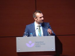 Emilio Russo合作论文数Faculty of Medicine and Surgery, University of Catanzaro20
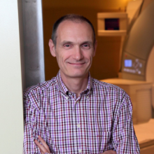 Andrea Bernasconi合作论文数Department of Neurology and Neurosurgery, Faculty of Medicine and Health Sciences, McGill University19
 Anna Elisabetta Vaudano合作论文数NOCSE Hospital, University of Modena and Reggio Emilia19Vivash Lucy合作论文数Royal Melbourne Hospital, University of Melbourne19Silvana Franceschetti合作论文数19
 Orrin Devinsky合作论文数Department of Neurology, Grossman School of Medicine, New York University;Department of Psychiatry, Grossman School of Medicine, New York University19
 Clarissa Yasuda合作论文数Neuroimaging Lab, Univ Estadual Campinas18Sanjay Sisodiya合作论文数Department of Clinical and Experimental Epilepsy, Queen Square Institute of Neurology, Faculty of Brain Sciences, University College London18
 Antonio Cerasa合作论文数Italian National Research Council18
 Iolanda Martino合作论文数17
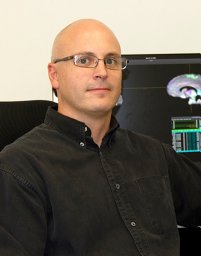 Luis Concha合作论文数17Graeme D. Jackson合作论文数17
 Paul M. Thompson合作论文数Imaging Genetics Center, Lab of Neuro Imaging, Institute for Neuroimaging and Informatics, Keck School of Medicine, University of Southern California17
 Reetta Kalviainen合作论文数University of Eastern Finland17
 Ida Manna合作论文数Institute of Neurological Sciences, National Research Council16Tortora Domenico合作论文数Department of Radiology, University Hospital of Chieti;Chieti;Department of Radiology, University Hospital of Chieti15Mario Mascalchi合作论文数15Norman Delanty合作论文数15
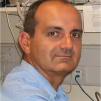 Hamandi Khalid合作论文数School of Psychology, Cardiff University15Laura Canafoglia合作论文数15Chantal Depondt合作论文数Université Libre de Bruxelles;Department of Neurology;Hôpital Erasme;Department of Neurology, Université Libre de Bruxelles15
 Neda Bernasconi合作论文数15
 Mariasavina Severino合作论文数14
 Colin Doherty合作论文数14Ilaria Sammarra合作论文数14
 Raviteja Kotikalapudi合作论文数Department of Neurology, University Hospital Essen14
 Carrie Mcdonald合作论文数Applied Sciences Division, Department of Radiation Medicine and Applied Sciences, School of Medicine, University of California, San Diego14
 Boris Bernhardt合作论文数Department of Neurology and Neurosurgery, Faculty of Medicine and Health Sciences, McGill University;Multimodal Imaging and Connectome Analysis Lab, McConnell Brain Imaging Centre, Montreal Neurological Institute, McGill University14
 Matteo Lenge合作论文数Dept. of Inf. Eng., Univ. of Florence;c;Dept. of Inf. Eng., Univ. of Florence14
 Bernd Weber合作论文数14
 Raul Rodriguez-Cruces合作论文数Instituto de Neurobiología, Universidad Nacional Autónoma de México14
 Terry O’Brien合作论文数Department of Medicine, Faculty of Medicine, Dentistry and Health Sciences, Monash University;Department of Neuroscience, School of Translational Medicine, Monash University14
 John Duncan合作论文数Department of Clinical and Experimental Epilepsy, University College London14
 Pascal GP Martin合作论文数Laboratoire de Pharmacologie et Toxicologie, Institut National de la Recherche Agronomique14




