The aim of this study was to produce a composite of microporous β-TCP filled with alginate-gelatin crosslinked hydrogel, clindamycin and bone morphogenetic protein (BMP-2) to prolong the drug-release behaviour for up to 28 days. The most promising alginate-di-aldehyde(ADA)-gelatin gel for drug release from microcapsules was used to fill microporous β-TCP ceramics under directional flow in a special loading chamber. Dual release of clindamycin and BMP-2 was measured on days 1, 2, 3, 6, 9, 14, 21 and 28 by high performance liquid chromatography (HPLC) and enzyme-linked immunosorbent assay (ELISA). After release, the microbial efficacy of the clindamycin was checked and the biocompatibility of the composite was tested in cell culture. Clindamycin and the model substance FITC-protein A were released from microcapsules over 28 days. The clindamycin burst release was 43 ± 1%. For the loaded ceramics, a clindamycin release above the minimal inhibitory concentration (MIC) until day 9 and a burst release of 90.56 ± 2.96% were detected. BMP-2 was released from the loaded ceramics in low concentrations over 28 days. The release of active substances from β-TCP and hydrogel have already been extensively studied. Directional flow loading is a special procedure in which the ceramic could act as a stabilizer in the bone and, as a biodegradable system, enables a single-stage surgical procedure. Whether ADA-gelatin gel is suitable for this procedure as a more biodegradable alternative to pure alginate or whether a dual release is possible in this composite has not yet been investigated. Graphical Abstract
The aim of this work was to establish an organ model for staphylococcal infection of human bone samples and to investigate the influence and efficacy of a microporous β-tricalcium phosphate ceramic (β-TCP, RMS Foundation) loaded with hydrogels (alginate, alginate-di-aldehyde (ADA)-gelatin) and clindamycin on infected human bone tissue over a period of 28 days. For this purpose, human tibia plateaus, collected during total knee replacement surgery, were used as a source of bone material. Samples were infected with S. aureus ATCC29213 and treated with differently loaded β-TCP composites (alginate +/- clindamycin, ADA-gelatin +/- clindamycin, unloaded). The loading of the composites was carried out by means of a flow chamber. The infection was observed for 28 days, quantifying bacteria in the medium and the osseus material on day 1, 7, 14, 21 and 28. All samples were histologically processed for bone vitality evaluation. Bone infection could be consistently performed within the organ model. In addition, a strong reduction in bacterial counts was recorded in the groups treated with ADA-gelatin + clindamycin and alginate + clindamycin, while the bacterial count in the control groups remained constant. No significant differences between groups could be observed in the number of lacunae filled with osteocytes suggesting no differences in bone vitality among groups. In an ex-vivo human bone infection model, over a period of 28 days bacterial growth could be reduced by treatment with ADA-Gel + CLI and ALG + CLI -releasing β-TCP composites. This could be relevant for its clinical use. Further work will be necessary to improve the loading of β-TCP and the bone infection organ model itself. STATEMENT OF SIGNIFICANCE: The common treatment of bone infections is debridement and systemic administration of antibiotics. In some cases, antibiotic-containing carriers are already used, but these must be removed again. Our work is intended to show another treatment option. The scaffold we have developed, made of a calcium phosphate ceramic and a hydrogel as the active substance carrier, can, in addition to releasing the active substance, also assume a load-bearing function of the bone and is biodegradable. In addition, the model we developed can also be used for the analysis and treatment of bone infections other than those of the musculoskeletal system. More importantly, it can also serve as a substitute for previously used animal experiments.
This work aimed to determine the influence of two hydrogels (alginate, alginate-di-aldehyde (ADA)/gelatin) on the mechanical strength of microporous ceramics, which have been loaded with these hydrogels. For this purpose, the compressive strength was determined using a Zwick Z005 universal testing machine. In addition, the degradation behavior according to ISO EN 10993-14 in TRIS buffer pH 5.0 and pH 7.4 over 60 days was determined, and its effects on the compressive strength were investigated. The loading was carried out by means of a flow-chamber. The weight of the samples (manufacturer: Robert Mathys Foundation (RMS) and Curasan) in TRIS solutions pH 5 and pH 7 increased within 4 h (mean 48 ± 32 mg) and then remained constant over the experimental period of 60 days. The determination surface roughness showed a decrease in the value for the ceramics incubated in TRIS compared to the untreated ceramics. In addition, an increase in protein concentration in solution was determined for ADA gelatin-loaded ceramics. The macroporous Curasan ceramic exhibited a maximum failure load of 29 ± 9.0 N, whereas the value for the microporous RMS ceramic was 931 ± 223 N. Filling the RMS ceramic with ADA gelatin increased the maximum failure load to 1114 ± 300 N. The Curasan ceramics were too fragile for loading. The maximum failure load decreased for the RMS ceramics to 686.55 ± 170 N by incubation in TRIS pH 7.4 and 651 ± 287 N at pH 5.0.
Orthopaedic and trauma research is a gateway to better health and mobility, reflecting the ever-increasing and complex burden of musculoskeletal diseases and injuries in Germany, Europe and worldwide. Basic science in orthopaedics and traumatology addresses the complete organism down to the molecule among an entire life of musculoskeletal mobility. Reflecting the complex and intertwined underlying mechanisms, cooperative research in this field has discovered important mechanisms on the molecular, cellular and organ levels, which subsequently led to innovative diagnostic and therapeutic strategies that reduced individual suffering as well as the burden on the society. However, research efforts are considerably threatened by economical pressures on clinicians and scientists, growing obstacles for urgently needed translational animal research, and insufficient funding. Although sophisticated science is feasible and realized in ever more individual research groups, a main goal of the multidisciplinary members of the Basic Science Section of the German Society for Orthopaedics and Trauma Surgery is to generate overarching structures and networks to answer to the growing clinical needs. The future of basic science in orthopaedics and traumatology can only be managed by an even more intensified exchange between basic scientists and clinicians while fuelling enthusiasm of talented junior scientists and clinicians. Prioritized future projects will master a broad range of opportunities from artificial intelligence, gene- and nano-technologies to large-scale, multi-centre clinical studies. Like Prometheus in the ancient Greek myth, transferring the elucidating knowledge from basic science to the real (clinical) world will reduce the individual suffering from orthopaedic diseases and trauma as well as their socio-economic impact.
Introduction The use of scaffolds in tissue engineering is becoming increasingly important as solutions need to be found to preserve human tissues such as bone or cartilage. Various factors, including cells, biomaterials, cell and tissue culture conditions, play a crucial role in tissue engineering. The in vivo environment of the cells exerts complex stimuli on the cells, thereby directly influencing cell behavior, including proliferation and differentiation. Therefore, to create suitable replacement or regeneration procedures for human tissues, the conditions of the cells’ natural environment should be well mimicked. Therefore, current research is trying to develop 3-dimensional scaffolds (scaffolds) that can elicit appropriate cellular responses and thus help the body regenerate or replace tissues. In this work, scaffolds were printed from the biomaterial polycaprolactone (PCL) on a 3D bioplotter. Biocompatibility testing was used to determine whether the printed scaffolds were suitable for use in tissue engineering. Material and Methods An Envisiontec 3D bioplotter was used to fabricate the scaffolds. For better cell-scaffold interaction, the printed polycaprolactone scaffolds were coated with type-I collagen. Three different cell types were then cultured on the scaffolds and various tests were used to investigate the biocompatibility of the scaffolds. Results Reproducible scaffolds could be printed from polycaprolactone. In addition, a coating process with collagen was developed, which significantly improved the cell-scaffold interaction. Biocompatibility tests showed that the PCL-collagen scaffolds are suitable for use with cells. The cells adhered to the surface of the scaffolds and as a result extensive cell growth was observed on the scaffolds. The inner part of the scaffolds, however, remained largely uninhabited. In the cytotoxicity studies, it was found that toxicity below 20% was present in some experimental runs. The determination of the compressive strength by means of the universal testing machine Z005 by ZWICK according to DIN EN ISO 604 of the scaffolds resulted in a value of 68.49 ± 0.47 MPa.
In the present work, an ex vivo organ model using human bone (explant) was developed for the evaluation of the initial osseointegration behavior of implant materials. The model was tested with additive manufactured Ti6Al4V test substrates with different 3D geometries. Explants were obtained from patients who underwent total knee replacement surgery. The tibial plateaus were used within 24 h after surgery to harvest bone cylinders (BC) from the anterior side using hollow burrs. The BCs were brought into contact with the test substrate and inserted into an agarose mold, then covered with cell culture media and subjected to the external load of 500 g. Incubation was performed for 28 days. After 28d the test substrate was removed for further analysis. Cells grown out BC onto substrate were immunostained with DAPI and with an antibody against Collagen-I and alkaline phosphatase (ALP) for visualization and cell counting. We show that cells stayed alive for up to 28d in our organ model. The geometry of test substrates influences the number of cells grown onto substrate from BCs. The model presented here can be used for testing implant materials as an alternative for in vitro tests and animal models.
The objective of this study was to vary the wall thicknesses and pore sizes of inversely printed 3D molded bodies. Wall thicknesses were varied from 1500 to 2000 to 2500 µm. The pores had sizes of 500, 750 and 1000 µm. The sacrificial structures were fabricated from polylactide (PLA) using fused deposition modeling (FDM). To obtain the final bioceramic scaffolds, a water-based slurry was filled into the PLA molds. The PLA sacrificial molds were burned out at approximately 450 °C for 4 h. Subsequently, the samples were sintered at 1250 °C for at least 4 h. The scaffolds were mechanically characterized (native and after incubation in simulated body fluid (SBF) for 28 days). In addition, the biocompatibility was assessed by live/dead staining. The scaffolds with a strand spacing of 500 µm showed the highest compressive strength; there was no significant difference in compressive strength regardless of pore size. The specimens with 1000 µm pore size showed a significant dependence on strand width. Thus, the specimens (1000 µm pores) with 2500 µm wall thickness showed the highest compressive strength of 5.97 + 0.89 MPa. While the 1000(1500) showed a value of 2.90 + 0.67 MPa and the 1000(2000) of 3.49 + 1.16 MPa. As expected for beta-Tricalciumphosphate (β-TCP), very good biocompatibility was observed with increasing cell numbers over the experimental period.
Background Horizontal instability following acute acromioclavicular joint (ACJ) reconstruction still occurs with a high prevalence. Although the human acromioclavicular ligament complex (ACLC) represents the major horizontal ACJ stabilizer, experimental studies on healing characteristics are lacking. Therefore, the purpose of this histological study was to investigate the healing potential of the ACLC following acute anatomical reconstruction Methods In this prospective clinical-experimental study, 28 ACLC biopsies were performed in patients with complete ACJ dislocations (Rockwood type 4 or 5) during acute hook plate stabilization (IG: implantation group; n = 14) and hook plate removal (EG: explantation group; n = 14). Histological analyses included Giemsa staining, polarized light microscopy and immunostaining against CD68, αSMA and collagen type I and type III. Histomorphological evaluation entailed cell counts, collagen expression score, ligament tissue maturity index (LTMI) and descriptive analysis of specific ligamentous structures. Statistics consisted of nonparametric Mann–Whitney U tests and a level of significance of P < .05. Results Total cell counts (cells/mm 2 1491 ± 296 vs. 635 ± 430; P < 0.001) and collagen III expression (3.22 ± 0.22 vs. 1.78 ± 0.41; P < 0.001) were higher in EG compared to IG. Inversely αSMA + (11 ± 9 vs. 179 ± 186; P < 0.001) and CD68 + cell counts (56 ± 20 vs. 100 ± 57; P 0.009) were significantly lower in the EG. The EG revealed a comparable reorientation of ligamentous structures. Consistently, ACLC samples of the EG (21.6 ± 2.4) displayed a high total but differently composed LTMI score (IG: 24.5 ± 1.2; P < 0.001). Conclusions This experimental study proved the ligamentous healing potential of the human ACLC following acute anatomical reconstruction. Histomorphologically, the ACLC reliably showed a ligamentous state of healing at a mean of about 12 weeks after surgery. However, processes of ligamentous remodeling were still evident. These experimental findings support recent clinical data showing superior horizontal ACJ stability with additional AC stabilization in the context of acute ACJ reconstruction. Though, prospective clinical and biomechanical studies are warranted to evaluate influencing factors on ACLC healing and potential impacts of acute ACLC repair on clinical outcome. Study type Controlled Laboratory Study
Soft tissues are commonly fiber-reinforced hydrogel composite structures, distinguishable from hard tissues by their low mineral and high water content. In this work, we proposed the development of 3D printed hydrogel constructs of the biopolymers chitosan (CHI) and cellulose nanofibers (CNFs), both without any chemical modification, which processing did not incorporate any chemical crosslinking. The unique mechanical properties of native cellulose nanofibers offer new strategies for the design of environmentally friendly high mechanical performance composites. In the here proposed 3D printed bioinspired CNF-filled CHI hydrogel biomaterials, the chitosan serves as a biocompatible matrix promoting cell growth with balanced hydrophilic properties, while the CNFs provide mechanical reinforcement to the CHI-based hydrogel. By means of extrusion-based printing (EBB), the design and development of 3D functional hydrogel scaffolds was achieved by using low concentrations of chitosan (2.0–3.0% (w/v)) and cellulose nanofibers (0.2–0.4% (w/v)). CHI/CNF printed hydrogels with good mechanical performance (Young’s modulus 3.0 MPa, stress at break 1.5 MPa, and strain at break 75%), anisotropic microstructure and suitable biological response, were achieved. The CHI/CNF composition and processing parameters were optimized in terms of 3D printability, resolution, and quality of the constructs (microstructure and mechanical properties), resulting in good cell viability. This work allows expanding the library of the so far used biopolymer compositions for 3D printing of mechanically performant hydrogel constructs, purely based in the natural polymers chitosan and cellulose, offering new perspectives in the engineering of mechanically demanding hydrogel tissues like intervertebral disc (IVD), cartilage, meniscus, among others.
Purpose Symptomatic horizontal instability is clinically relevant following acute acromioclavicular joint dislocations. However, the intrinsic healing response is poorly understood. The present study sought to investigate time-dependent healing responses of the human acromioclavicular ligament following acute traumatic rupture. Methods Biopsies of the acromioclavicular ligament were obtained from patients undergoing surgical treatment for acute acromioclavicular joint dislocations. Specimens were stratified by time between trauma and surgery: group 1, 0–7 days ( n = 5); group 2, 8–14 days ( n = 6); and group 3, 15–21 days ( n = 4). Time-dependent changes in cellularity, collagen (type 1 and 3) concentration, and histomorphological appearance were evaluated for the rupture and intact zone of the acromioclavicular ligament. Results Group 1 was characterized by cellular activation and early inflammatory response. The rupture zone exhibited a significantly higher count of CD68-positive cells than the intact zone (15.2 vs 7.4; P ≤ 0.05). Consistently, synovialization of the rupture end was observed. Within the second week, the rupture zone was subject to proliferation showing more fibroblast-like cells than the intact zone (66.8 vs 43.8; P ≤ 0.05) and a peak of collagen type 3 expression (group 1: 2.2 ± 0.38, group 2: 3.2 ± 0.18, group 3: 2.8 ± 0.57; P ≤ 0.05). Signs of consolidation and early remodeling were seen in the third week. Conclusions The acromioclavicular ligament exhibits early and dynamic healing responses following acute traumatic rupture. Our histological findings suggest that surgical treatment of acute ACJ dislocations should be performed as early as possible within a timeframe of 1 week after trauma to exploit the utmost biological healing potential. Prospective clinical studies are warranted to investigate whether early surgical treatment of ACJ dislocations translates into clinical benefits.
Background: Computed tomography (CT) scan is the standard for assessment of femoral torsion. This observational study was conducted to evaluate the comparability of the EOS radiation dose scanning system (EOS imaging, Paris, France) and the CT scan in patients with suspected torsional malalignment of the femur. Methods: Patients with suspected torsional malalignment of the femur were included in a study for surgical planning. The primary endpoint was to compare the 3-dimensional radiological (EOS) imaging system with the CT scan to determine femoral anteversion (AV) angle. Three independent raters performed measurements. Comparability of CT scan and EOS values was assessed by Pearson correlation, t test, interobserver reliability, and intraobserver reliability (Cronbach alpha). Results: About 34 femora were examined. Interobserver reliability/intraobserver reliability was 0.911 of 0.955 for EOS and 0.934 of 0.934 for CT scan. EOS system revealed an AV angle of 12.2 degrees +/- 10.0 degrees (-15.0 degrees to 32.0 degrees). CT examinations showed an AV angle of 12.6 degrees +/- 9.2 degrees (-3.2 degrees to 35.6 degrees). About 11 hips featured physiological AV, 14 hips showed decreased AV (<10 degrees) or retroversion (<0 degrees), and 9 hips showed increased AV (>20 degrees). Overall, a strong Pearson correlation of tau = 0.855 and a highly significant correlation in the t test for both methods was seen. In patients with decreased AV, retroversion, or increased AV, Pearson correlation only resulted in a moderate/low correlation of tau = 0.495 and tau = 0.292. The t test showed no significant correlation at malrotation. Conclusion: In torsional malalignment, EOS does not have correlation with CT measurements. In contrast to CT scan, EOS allows femoral torsion measurement independent of legs' positioning. (C) 2020 The Authors. Published by Elsevier Inc.
Recently, silicate materials have received attention as materials with promising applications in the bioceramics field. A recent study aimed to investigate the effect of forsterite (Mg2SiO4) addition to biogenic hydroxyapatite, Ca10(PO4)6(OH)2, on the phase formation, physical and mechanical properties, and biocompatibility of the produced composites. Different proportions of forsterite, 10 to 40 mass%, were added to hydroxyapatite obtained from fish bones to prepare the target composites. Various techniques, such as X-ray diffraction analysis (XRD), scanning electronic microscope (SEM), transmission electronic microscope (TEM), mechanical strength measurements and in vitro studies, were carried out to evaluate the composite properties. The results indicate that the addition of 20 to 40 mass% forsterite led to the transformation of forsterite into protoenstatite and the formation of Mg-rich whitlockite at the expense of hydroxyapatite. It is concluded that 20 mass% forsterite is the optimum addition amount to enhance the physical and mechanical properties of the produced composites. The cell culture tests and in vitro studies agree with the abovementioned results.
Just like menisci, articular cartilage is exposed to constant and varying stresses. Injuries to the meniscus are associated with the development of gonarthritis. Both the articular cartilage and the menisci are subject to structural changes under gonarthritis. The aim of this study was to investigate biomechanical alterations in articular cartilage and the menisci under gonarthritis by applying an indentation method. The study assessed 11 menisci from body donors as controls and 21 menisci from patients with severe gonarthritis. For the simultaneous examination of the articular cartilage and the menisci, we only tested the joint surfaces of the tibial plateau covered by the corresponding menisci. Over the posterior horn of the meniscus, the maximum applied load—the highest load registered by the load cell—of the arthritic samples of 0.02 ± 0.02 N was significantly greater (p = 0.04) than the maximum applied load of the arthritis-free samples of 0.01 ± 0.01 N. The instantaneous modulus (IM) at the center of the arthritic cartilage covered by the meniscus with 3.5 ± 2.02 MPa was significantly smaller than the IM of the arthritis-free samples with 5.17 ± 1.88 MPa (p = 0.04). No significant difference was found in the thickness of the meniscus-covered articular cartilage between the arthritic and arthritis-free samples. Significant correlations between the articular cartilage and the corresponding menisci were not observed at any point. In this study, the biomechanical changes associated with gonarthritis affected the posterior horn of the meniscus and the mid region of the meniscus-covered articular cartilage. The assessment of cartilage thickness as a structural characteristic of osteoarthritis may be misleading with regard to the interpretation of articular cartilage’s biomechanical properties.
Studies on osteoarthritis of the knee have examined isolated aspects of the meniscus biomechanically and histologically, but not the difference between instantaneous modulus (IM) in healthy and diseased samples. The aim of this study is to investigate the difference in the biomechanical behavior and proteoglycan content between arthritic and arthritis-free menisci. In addition, the relationship between the biomechanical behavior and proteoglycan content should be investigated. A novel indentation-based method was used, which allows the mapping of the entire meniscus, without damaging it for histological examinations. A total of 26 arthritic and 14 arthritis-free samples were examined in the present study. A Mach-1 Model V500css test machine was used for biomechanical testing. A position grid was placed over each sample allowing all measurements (indentation test and thickness measurements) to be taken at the same position. All sections were then graded for Safranin O staining intensity for proteoglycan content. The maximum applied load of our arthritic samples above the posterior horn was statistically significantly higher (p = 0.01) at 0.02 ± 0.02 N than the maximum applied load of the arthritis-free samples at 0.01 ± 0.01 N. The proteoglycan content of the meniscus, evaluated by the Safranin O score, correlated statistically significantly with the maximum applied load over the entire meniscus (p = 0.04, 95% CI: 0.06–0.71). The present study showed that in the final stage of gonarthritis, the attenuation behavior of the meniscus was significantly lower compared to the arthritis-free knee. The mapping of IM and histological examination of the meniscus showed a direct correlation between changes in proteoglycan content and altered mechanical properties of the meniscus in gonarthritis.
Symptomatic intervertebral disc (IVD) degeneration accounts for significant socioeconomic burden. Recently, the expression of the tissue renin-angiotensin system (tRAS) in rat and bovine IVD was demonstrated. The major effector of tRAS is angiotensin II (AngII), which participates in proinflammatory pathways. The present study investigated the expression of tRAS in human IVDs, and the correlation between tRAS, inflammation and IVD degeneration. Human IVD tissue was collected during spine surgery and distributed according to principal diagnosis. Gene expression of tRAS components, proinflammatory and catabolic markers in the IVD tissue was assessed. Hydroxyproline (OHP) and glycosaminoglycan (GAG) content in the IVD tissue were determined. Tissue distribution of tRAS components was investigated by immunohistochemistry. Gene expression of tRAS components such as angiotensin-converting enzyme (ACE), Ang II receptor type 2 (AGTR2), angiotensinogen (AGT) and cathepsin D (CTSD) was confirmed in human IVDs. IVD samples that expressed tRAS components (n = 21) revealed significantly higher expression levels of interleukin 6 (IL-6), tumour necrosis factor α (TNF-α), a disintegrin and metalloproteinase with thrombospondin motifs (ADAMTS) 4 and 5 compared to tRAS-negative samples (n = 37). Within tRAS-positive samples, AGT, matrix-metalloproteinases 13 and 3, IL-1, IL-6 and IL-8 were more highly expressed in traumatic compared to degenerated IVDs. Total GAG/DNA content of non-tRAS expressing IVD tissue was significantly higher compared to tRAS positive tissue. Immunohistochemistry confirmed the presence of AngII in the human IVD. The present study identified the existence of tRAS in the human IVD and suggested a correlation between tRAS expression, inflammation and ultimately IVD degeneration.
Background Arthrofibrosis is a painful and restraining complication that occurs after about 10% of total knee arthroplasty and cruciate ligament surgery. The pathogenesis of arthrofibrosis has not yet been fully understood. Stress signals stimulate immune cells, and fibroblast differentiates into myofibroblast, which produce a large amount of collagen. Xylosyltransferases also appear to be involved in these pathways. They catalyze proteoglycan biosynthesis, which is involved in tissue remodeling and myofibroblast differentiation. The aim of this study was to investigate the relationship between the disease arthrofibrosis and the expression of the two isoforms of xylosyltransferases I and II. Methods Tissue samples from 14 patients with arthrofibrosis were compared with tissue samples from seven healthy controls. The xylosyltransferases were detected by immunohistochemistry. The tissues were divided into four different areas of interest: vessels, synovialis, cell-poor and cell-rich fibrosis, or cell-poor and cell-rich areas in the control group. A quantification of the results was performed by modification of the immunoreactive score according to Remmele and Stegner. Results Xylosyltransferase I was expressed in the various tissue types at varying rates. Xylosyltransferase I expression was considerably and significantly stronger than that of xylosyltransferase II. The following sequences of xylosyltransferase I and xylosyltransferase II expression were determined as follows: vessels >> cell-rich fibrosis > cell-poor fibrosis > synovialis. A positive correlation between the number of positive fibroblasts and the immunoreactive scoring system (IRS) was documented. Conclusions The significant positive correlation of xylosyltransferase -I expression with increasing number of fibroblasts demonstrates a high myofibroblast differentiation rate, which implies a gradual event as the pathogenesis of arthrofibrosis.
Fragestellung Genau wie Menisken ist der Gelenkknorpel ständig wechselnden Belastungen ausgesetzt. Sind sie durch einen Unfall, eine Erkrankung oder degenerative Prozesse geschädigt, steigt das Risiko an Gonarthrose zu erkranken um ein Vielfaches. Sowohl der Gelenkknorpel als auch die Menisken unterliegen bei Gonarthrose strukturellen Veränderungen. Ziel dieser Studie ist es, die biomechanischen Veränderungen des Gelenkknorpels und der Menisken bei Gonarthrose zu untersuchen.
Connective tissue displays a large compositional and structural complexity that involves multiple length scales. In particular, on the molecular and the nanometer level, the elementary processes that determine the biomechanics of collagen fibrils in connective tissues are still poorly understood. Here, we use atomic force microscopy (AFM) to determine the three-dimensional (3D) depth profiles of the local nanomechanical properties of collagen fibrils and their embedding interfibrillar matrix in native (unfixed), hydrated Achilles tendon of sheep and chickens. AFM imaging in air with controlled humidity preserves the tissue's water content, allowing the assembly of collagen fibrils to be imaged in high resolution beneath an approximately 5-10 nm thick layer of the fluid components of the interfibrillar matrix. We collect pointwise force-distance (FD) data and amplitude- phase-distance (APD) data, from which we construct 3D depth profiles of the local tip-sample interaction forces. The 3D images reveal the nanomechanical morphology of unfixed, hydrated collagen fibrils in native tendon with a 0.1 nm depth resolution and a 10 nm lateral resolution. We observe a diversity in the nanomechanical properties among individual collagen fibrils in their adhesive and in their repulsive, viscoelastic mechanical response as well as among the contact points between adjacent collagen fibrils. This sheds new light on the role of interfibrillar bonds and the mechanical properties of the interfibrillar matrix in the biomechanics of tendon.
Objective The aim of the present paper is to analyze mid‐term and long‐term alterations of human anterior cruciate ligament (ACL) grafts during the remodeling process with special regards to cellularity, α‐smooth muscle protein (αSMP) expression, and crimp length in comparison to the native ACL. Methods A total of 34 patients were included (23 male and 11 female). Biopsies of 13 semitendinosus tendon and 14 patellar tendon autografts were obtained during surgical revision secondary to an ACL reconstruction. According to the interval between the index procedure and sample collection, the patients were divided into four groups: 4–12 months, 13–60 months, 61–108 months, and >108 months. Seven samples of native ruptured ACL tissue obtained during surgical intervention served as control. All biopsies were taken from the intraligamentous part of the ACL or the graft. Histomorphological and immunohistochemical analyses were conducted after samples were stained using hematoxylin–eosin, Giemsa, and αSMP enzyme‐labeled antibodies. The total cell density, the numbers of fibroblasts and fibrocytes, the fibroblast/fibrocyte ratio, the number of αSMP+ cell nuclei, and the percentage of αSMP+ cells per fibroblast as well as the crimp lengths were determined using light microscopy. Results In the early phase of remodeling, the grafts featured extensively high total cell counts (1021.2 ± 327.8, P = 0.001), with high numbers of fibroblasts (841.4 ± 245.2, P = 0.002), fibrocytes (174.5 ± 113.0, P = 0.04), and αSMP+ cells (78.3 ± 95.0, P = 0.02) compared to controls (390.1 ± 141.7, 304.5 ± 160.8, 65.6 ± 31.4 and 2.3 ± 2.6, respectively). Thereafter, the numbers of all cell entities decreased. After more than 108 months, the percentage of αSMP+ cells per fibroblast reached physiological values (ratio 1.3 ± 1.0, P = 0.41; control 0.8 ± 0.8), while the total cell count (834.3 ± 183.7, P = 0.001) as well as the numbers of fibroblasts (663.5 ± 192.6, P = 0.006) and fibrocytes (134.1 ± 73.0, P = 0.049) remained significantly high. The fibroblast/fibrocyte ratio showed no significant alterations over the course of time compared to the controls. The collagen crimp lengths were elongated by tendency in the early phase (28.8 ± 12.9 mm, P = 0.15; control 20.7 ± 2.2 mm) and significantly shortened over time, with the lowest values in the long term (14.8 ± 2.0 mm, P = 0.001). The comparison of biopsies from semitendinosus tendon and patellar tendon autografts revealed no significant differences for any of the histomorphological parameters investigated. Conclusion This study reveals distinctive mid‐term and long‐term immunomorphological alterations during human ACL graft remodeling. These data clearly indicate that the remodeling is a process that continues for 9 years or more. Furthermore, it seems to be a process of adaptation rather than full restoration. Even in the long run, several biological properties of the native ACL are not completely reestablished.
 Hermann O. Mayr合作论文数40Südkamp Norbert P合作论文数36
 Michael Seidenstuecker合作论文数22
 Hermann O Mayr合作论文数17
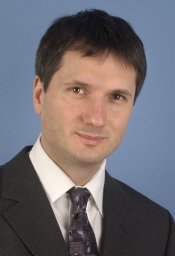 Hagen Schmal合作论文数Department of Orthopedic Surgery, University of Freiburg Medical Center15
 Robert Hube合作论文数13Amelie Stoehr合作论文数12Georg Berger合作论文数12Sergio H Latorre合作论文数10
 Philipp Niemeyer合作论文数10Renate Gildenhaar合作论文数10
 Marc Bohner合作论文数RMS Foundation/Acta Materialia Inc./SNSF Swiss National Science Foundation9
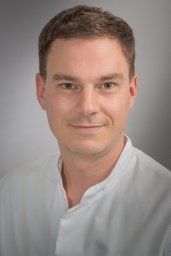 Feucht Matthias J合作论文数Department of Orthopaedic Sports Medicine, Technical University Munich7
 Rainer Gadow合作论文数Institute for Manufacturing Technologies of Ceramic Components and Composites, University of Stuttgart7Pia Schilling合作论文数6
 Martin Zens合作论文数6
 Peter Woias合作论文数Laboratory for the Design of Microsystems, Department of Microsystems Engineering, Faculty of Engineering, University of Freiburg6Joerg Brandt合作论文数6
 Eva Johanna Kubosch合作论文数5Doreen Nöbel合作论文数5Werner Hein合作论文数5Robert Magerle合作论文数5R. Von Eisenhart-Rothe合作论文数Asklepios Orthopädische Klinik Lindenlohe5
 Wolf Christian Prall合作论文数4Norbert P. Suedkamp合作论文数4Maik Reinhold合作论文数3T Krüger合作论文数3Lucas Ritschl合作论文数3H Reichel合作论文数3Steffen Esslinger合作论文数3Zeh Alexander合作论文数3
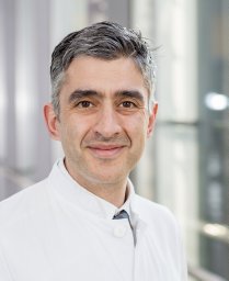 Kaywan Izadpanah合作论文数Department of Orthopeadic andTrauma Surgery, University Hospital Freiburg3senta senger合作论文数3Mayr Herrmann O合作论文数3
 Max D Kauther合作论文数3Kühle Jan合作论文数3
 Mauro Alini合作论文数AO Research Institute Davos3Czurratis P合作论文数3Andreas Birke合作论文数3
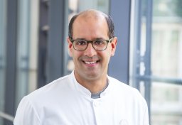 Lukas Konstantinidis合作论文数Department of Orthopaedics and Trauma Surgery, University Medical Centre3H.-J. Hein合作论文数3
 Sibylle Grad合作论文数AO Research Institute Davos;Department of Health Sciences and Technology, ETH Zürich3
 Gian Salzmann合作论文数Schulthess Clinic3
 Andreas Killinger合作论文数3
 Florian Haasters合作论文数3U. Schietsch合作论文数3




