Spinocerebellar ataxia type 27B (SCA27B), caused by an intronic GAA repeat expansion in the FGF14 gene, has recently emerged as a major cause of late-onset cerebellar ataxia (LOCA). Its prevalence and clinical profile in Central and Eastern Europe remain largely unknown. To determine the frequency and phenotypic characteristics of FGF14 GAA·TTC repeat expansions, a large cohort of Polish patients with undiagnosed adult-onset cerebellar ataxia was investigated. We retrospectively analyzed 701 patients (age of onset ≥ 25 years) with adult-onset cerebellar ataxia of unknown etiology, previously tested negative for SCA1, SCA2, SCA3, SCA8, and RFC1 expansions. GAA·TTC repeat lengths were assessed using long-range and repeat-primed PCR. Expansions ≥ 250 repeats were classified as pathogenic. Clinical and MRI data were evaluated where available. A control group of 66 neurologically healthy individuals was also screened. Pathogenic FGF14 expansions (≥ 250 repeats) were identified in 4.4
Hereditary spastic paraplegias (HSPs) comprise a heterogeneous group of rare, neurodegenerative disorders. The most prominent HSP features: spastic paraparesis, mild somatosensory deficits and bladder dysfunction may be accompanied by additional symptoms i.e.: neuropathy, epilepsy, dementia. We aimed to determine subclinical involvement of nonmotor or sensory brain structures in hereditary spastic paraplegias type 3 A (SPG3A) and type 4 (SPG4). Visual evoked potentials (VEPs), brainstem evoked potentials (BAEPs) and electroencephalography (EEG) were performed in 28 SPG4 and 9 SPG3A patients. Disease severity was evaluated with Spastic Paraplegia Rating Scale. The EEG examination revealed abnormalities in 9 SPG4 patients (35
Cerebellar ataxia with neuropathy and bilateral vestibular areflexia syndrome (CANVAS) is a late-onset, autosomal recessive neurodegenerative disorder characterised by the triad of cerebellar ataxia, bilateral vestibular areflexia, and sensory neuropathy. First identified more than 30 years ago, its clinical phenotype has since expanded to include chronic cough, dysautonomia, and pain, with isolated neuronopathy reported in some cases. The discovery of biallelic AAGGG repeat expansions in intron 2 of the RFC1 gene in 2019 established the genetic basis for CANVAS, with the pathogenic expansions disrupting gene function via secondary structures such as G-quadruplexes. Despite this breakthrough, the precise pathophysiological mechanisms behind CANVAS remain elusive, necessitating further research into the molecular, clinical, and genetic aspects of this disease. This review consolidates the current understanding of CANVAS, encompassing the expanding spectrum of RFC1-related disorders, clinical manifestations, molecular underpinnings, and epidemiology, while exploring future directions for diagnostics and therapeutic advancements.
BACKGROUND:Hereditary spastic paraplegias (HSPs) are a group of genetically heterogeneous, progressive disorders with a broad spectrum of clinical presentation. Among autosomal dominant inherited forms of HSP, the most common are SPG4, SPG3 and SPG31. MATERIAL AND METHODS:Our aim was the clinical characterisation of a large group of patients with SPG4, SPG3 and SPG31. Neurological assessments as well as neuroimaging, electrophysiological, neuropsychological and ophthalmological examinations were performed to characterise in detail the clinical picture in HSP patients. RESULTS:In our study group of 179 individuals from 60 families, three forms of AD-HSP: SPG4, SPG3 and SPG31 were diagnosed in 125, 36 and 18 patients, respectively. 48 of the probands had a positive family history of autosomal dominant hereditary disorder, while the other 12 probands represented apparently isolated cases. The pure form of HSP (pHSP) was found in a large majority (138 symptomatic patients), whereas a distinctly complex phenotype (cHSP) was found only in 32 individuals. The number of patients with cHSP became greater after including the results of electroneurography. CONCLUSIONS:The primarily pure form of HSP is present in patients with SPG4, SPG3 and SPG31. Nevertheless, the presence of additional symptoms provides evidence that in AD-HSP not only the cortico-spinal tract is involved. The most common additional symptom is polyneuropathy. We suggest that all patients with a genetic diagnosis of SPG4 and SPG3 should have electrophysiological testing performed, and then periodically repeated, for the purpose of controlling the involvement of peripheral nerves. We found a considerable variability, including in terms of age at onset and the progression rate of disability, mainly in SPG4.
Cerebellar ataxia with neuropathy and vestibular areflexia syndrome (CANVAS) is a recessively inherited neurodegenerative ataxic disorder, which has been associated with intronic biallelic repeat expansions in the RFC1 gene. Our objective was to assess retrospectively the prevalence of CANVAS in Polish population. We screened 2523 Polish patients in whom other repeat expansions were excluded. To determine the repeat expansions in the RFC1 gene in patients, we performed RFC1-flanking PCR and repeat primed PCR (RP-PCR) and to measure the size of the expansion we used Southern blotting and optical genome mapping to compare the results. We have observed the biallelic pathogenic motif/unit AAGGG expansions in 4.6% and expansions of non-pathogenic motifs AAAAG, AAAGG in 25% patients of our studied population. This is the first large-scale cohort study that confirms the relatively frequent occurrence of the CANVAS in Polish population. To increase the current diagnostics of late-onset ataxias within an unexplained molecular background, we suggest involving the RFC1 repeat expansions analysis to the routine diagnostic workflow.
Purpose:Extracellular vesicles are the subject of many studies in various medical specialties. Their role in neurodegenerative diseases is increasing and they worth introducing in more detail. Methods:This review was performed following an electronic search of the database PubMed/Medline and Web of Science for English-language articles between 2010 and 2024 in the fields of medicine, molecular biology, and biochemistry. Keywords searches included combinations of the following terms: "extracellular vesicles" OR "exosomes" AND "neurodeg*" AND "microRNA" OR "miRNA" AND "AD" OR "PD" OR "ALS" OR "HD". Articles had to be original work or reviews. Results:The classification of extracellular vesicles is based on their size or origin. Their content is of key importance in communication between cells and can be treated as a physiological determinant of the normal or pathological condition of a body. The cargo transported in the extracellular space and over longer distances in various body fluids is diversified and may be nucleic acids (DNA, RNA, miRNA) as well as proteins and lipids, and, in the case of apoptotic bodies also a cell's organelles. Exosomes are the most thoroughly studied extracellular vesicles and the most often considered for therapeutic applications. Vesicles carrying biological substances in the body perform three basic functions: participation in a pathological mechanism, a biomarker role that also has diagnostic and prognostic functions, and a role in therapeutic activities. In the case of neurodegenerative diseases, it appears that extracellular vesicles can transport misfolded proteins, initiating pathological processes in previously normal cells. Conclusions:The transport of various substances enclosed in vesicles seems to be very promising in therapeutic prospects in various diseases, and the possibility of their crossing the blood-brain barrier particularly indicates diseases of the central nervous system. Despite many years of research on extracellular vesicles in the context of neurodegenerative diseases, their practical use is currently limited to studies on animal and cellular models, and their practical application in clinical trials in neurodegenerative diseases is to date extremely rare.
Since 1991, several genetic disorders caused by unstable trinucleotide repeats (TNRs) have been identified, collectively referred to as triplet repeat diseases (TREDs). They share a common mutation mechanism: the expansion of repeats (dynamic mutations) due to the propensity of repeated sequences to form unusual DNA structures during replication. TREDs are characterized as neurodegenerative diseases or complex syndromes with significant neurological components. Spinocerebellar ataxia type 17 (SCA17) falls into the former category and is caused by the expansion of mixed CAA/CAG repeats in the TBP gene. To date, a five-unit organization of this region [(CAG)3 (CAA)3] [(CAG)n] [CAA CAG CAA] [(CAG)n] [CAA CAG], with expansion in the second [(CAG)n] unit being the most common, has been proposed. In this study, we propose an alternative organization scheme for the repeats. A search of the PubMed database was conducted to identify articles reporting both the number and composition of GAC/CAA repeats in TBP alleles. Nineteen reports were selected. The sequences of all identified CAG/CAA repeats in the TBP locus, including 67 cases (probands and b relatives), were analyzed in terms of their repetition structure and stability in inheritance, if possible. Based on the analysis of three units [(CAG)3 (CAA)2] [CAA (CAG)n CAA CAG] [CAA (CAG)n CAA CAG], the organization of repeats is proposed. Detailed analysis of the CAG/CAA repeat structure, not just the number of repeats, in TBP-expanded alleles should be performed, as it may have a prognostic value in the prediction of stability/instability during transmission and the possible anticipation of the disease.
Neuromuscular disorders (NMDs) include a wide range of diseases affecting the peripheral nervous system. The genetic diagnoses are increasingly obtained with using the next generation sequencing (NGS). We applied the custom-design targeted NGS panel including 89 genes, together with genotyping and multiplex ligation-dependent probe amplification (MLPA) to identify a genetic spectrum of NMDs in 52 Polish patients. As a result, the genetic diagnosis was determined by NGS panel in 29 patients so its diagnostic utility is estimated at 55.8%. The most pathogenic variants were found in CLCN1, followed by CAPN3, SCN4A, and SGCA genes. Genotyping of myotonic dystrophy type 1 and 2 (DM1 and DM2) as a secondary approach has been performed. The co-occurrence of CAPN3 and CNBP mutations in one patient as well as DYSF and CNBP mutations in another suggests possibly more complex inheritance as well as expression of a phenotype. In 7 individuals with single nucleotide variant found in NGS testing, the MLPA of the CAPN3 gene was performed detecting the deletion encompassing exons 2-8 in the CAPN3 gene in one patient, confirming recessive limb-girdle muscular dystrophy type 1 (LGMDR1). Thirty patients obtained a genetic diagnosis (57.7%) after using NGS testing, genotyping and MLPA analysis. The study allowed for the identification of 27 known and 4 novel pathogenic/likely pathogenic variants and variants of uncertain significance (VUS) associated with NMDs.In conclusion, the diagnostic approach with diverse molecular techniques enables to broaden the mutational spectrum and maximizes the diagnostic yield. Furthermore, the co-occurrence of DM2 and LGMD has been detected in 2 individuals.
Purpose:Muscular dystrophy is a group of heterogeneous diseases causing progressive muscle weakness and atrophy. Many types have been defined, including Duchenne/Becker, myotonic, limb-girdle, congenital, and facioscapulohumeral muscular dystrophies. This study aims to present the first patient with both a homozygous CAPN3 mutation and a CCTG expansion in the CNBP gene, which suggests the co-occurrence of two diseases in a single patient. Case description:Homozygous pathogenic variant c.550delA (p.Thr184ArgfsTer36) in the CAPN3 gene, as well as a heterozygous expansion of a CCTG repeat of the CNBP gene, were identified in a single patient. Segregation analysis showed both maternal and paternal heterozygous carriers for CAPN3 mutation, and a maternally inherited CNBP expansion. Comment:In general, the co-occurrence of two diseases in a single patient is considered as uncommon, although possible, and therefore it should be taken into consideration in the populations with a relatively high prevalence of myotonic dystrophy type 2.
The aim of this study was to determine the time between the first detection of postural control impairments and the evident manifestation of ataxia in preclinical SCA1 individuals. Twenty five preclinical SCA1 mutation carriers: 13 with estimated disease onset ≤ 6 years (SCA1 +) aged 27.8 ± 8.1 years; 12 with expected disease onset > 6 years (SCA1-) aged 26.6 ± 3.1 years and 26 age and sex matched healthy controls (HCs) underwent static posturography during 5 years of observation. The movements of the centre of feet pressure (COP) during quiet standing with eyes open (EO) and closed (EC) were quantified by calculating the mean radius (R), developed surface area (A) and mean COP movement velocity (V). Ataxia was evaluated by use of the Scale for Assessment and Rating of Ataxia (SARA). SCA1 + exhibited significantly worse quality of stance with EC vs. SCA1- (p < 0.05 for V) and HCs (p < 0.001) even 5 to 6 years before estimated disease onset. There were no statistically significant differences between SCA1- and HCs. A slow increase in Cohen’s d effect size was observed for VEO up to the clinical manifestation of ataxia. VEO and AEC recorded in preclinical SCA1 individuals correlated slightly but statistically significantly with SARA (r = 0.47). The study confirms that static posturography detects COP sway changes in SCA1 preclinical gene carriers even 5 to 6 years before estimated disease onset. The quantitative evaluation of stance in preclinical SCA is a sensitive biomarker for the monitoring of the disease progression and may be useful in clinical trials.
The most common form of hereditary spastic paraplegia (HSP), SPG4 is caused by single nucleotide variants and microrearrangements in the SPAST gene. The high percentage of multi-exonic deletions or duplications observed in SPG4 patients is predisposed by the presence of a high frequency of Alu sequences in the gene sequence. In the present study, we analyzed DNA and RNA samples collected from patients with different microrearrangements in SPAST to map gene breakpoints and evaluate the mutation mechanism. The study group consisted of 69 individuals, including 50 SPG4 patients and 19 healthy relatives from 18 families. Affected family members from 17 families carried varying ranges of microrearrangements in the SPAST gene, while one individual had a single nucleotide variant in the 5′UTR of SPAST. To detect the breakpoints of the SPAST gene, long-range PCR followed by sequencing was performed. The breakpoint sequence was detected for five different intragenic SPAST deletions and one duplication, revealing Alu-mediated microhomology at breakpoint junctions resulting from non-allelic homologous recombination in these patients. Furthermore, SPAST gene expression analysis was performed using patient RNA samples extracted from whole blood. Quantitative real-time PCR tests performed in 14 patients suggest no expression of transcripts with microrearrangements in 5 of them. The obtained data indicate that nonsense-mediated decay degradation is not the only mechanism of hereditary spastic paraplegia in patients with SPAST microrearrangements.
INTRODUCTION:We present the first two Polish families diagnosed with spinocerebellar ataxia type 7 (SCA7) and draw attention to cardiac involvement as a new potential manifestation of this disease.MATERIAL AND METHODS:Two well-documented kindreds are presented.RESULTS:The proband from Family 1 presented aged 54 years with vision worsening followed by progressive imbalance. Brain MRI demonstrated cerebellar atrophy. Genetic testing confirmed CAG repeat expansion (42/10) in ATXN7 gene. The proband from Family 2 developed imbalance at age 20, followed by progressive deterioration of vision. Brain MRI revealed cerebellar atrophy. Additionally, she developed chronic congestive heart failure and, at age 38, had cardiomyopathy with an ejection fraction of 20% and significant mitral and tricuspid regurgitation. Genetic analysis found abnormal CAG expansion in the ATXN7 (46/10).CONCLUSIONS AND CLINICAL IMPLICATIONS:Vision loss due to pigmentary retinal degeneration is the distinguishing feature of SCA7 and often the initial manifestation. Although SCA7 is one of the most common SCAs in Sweden, it has never been reported in neighbouring Poland. Until now, cardiac abnormalities have only been described in infantile-onset SCA7 with large CAG repeats. The observed cardiac involvement in Family 2 may be coincidental, albeit a new possible manifestation of SCA7 cannot be excluded.
Ataksja (niezborność ruchowa, bezład) jest zespołem objawów charakteryzującym się zaburzeniami równowagi i koordynacji chodu, postawy, ruchów kończyn oraz nieprawidłową (dyzartryczną) mową. Najczęściej ataksja jest nabyta, wtórna do przyczyn naczyniowych, autoimmunologicznych, infekcyjnych/zapalnych, nowotworowych i niedoborowych. Choroby neurozwyrodnieniowe mogą się objawiać ataksją, a w niektórych przypadkach, na przykład w zaniku wieloukładowym czy chorobie Creutzfeldta-Jakoba, może to być pierwsza i główna manifestacja choroby. W niektórych przypadkach ataksja może mieć charakter napadowy (tzw. ataksje epizodyczne). Ataksja bywa także wiodącym objawem uwarunkowanych genetycznie chorób neurozwyrodnieniowych (tzw. ataksje dziedziczne), które mogą być dziedziczone recesywnie, sprzężone z chrosomem X lub dominujące. Do ostatniej grupy zalicza się ataksje rdzeniowo-móżdzkowe (SCA, spinocerebellar ataxia ). Jako grupa SCA charakteryzują się postępującym nasilaniem objawów, na ogół o początku w średnim wieku, oraz neurodegeneracją najbardziej wyrażoną w zakresie móżdzku, pnia mózgu i rdzenia kręgowego. Dotychczas opisano ponad 40 typów SCA, nadając numery zgodnie z kolejnością odkrycia patogennych mutacji w kolejnych genach. Większość publikacji na temat SCA pochodzi z Europy Zachodniej i Ameryki Północnej. Wyników tych prac nie można jednak przekładać na Polskę ze względu na specyfikę naszego regionu pod względem częstości występowania poszczególnych typów SCA. W pracy przedstawiono stan wiedzy na temat SCA w Polsce, omówiono występujące w naszym kraju typy SCA, podłoże genetyczne, obraz kliniczny, postępowanie diagnostyczne, leczenie, kierunki badań i przyszłe terapie.
INTRODUCTION:In myotonia congenita (MC), activation with exercise or cooling can induce transient changes in compound motor action potential (CMAP) parameters, thus providing a guide to genetic analysis.MATERIAL AND METHODS:We performed the short exercise test (SET) and the short exercise test with cooling (SETC) in 30 patients with genetically confirmed Becker disease (BMC) to estimate their utility in the diagnosis of BMC.RESULTS:Although we observed a significant decrease in CMAP amplitude immediately after maximal voluntary effort in both tests in the whole BMC group, in men this decline was significantly smaller than in women, especially in SET. Clinical implications/future directions: In men with a clinical suspicion of BMC, a small decrease in CMAP amplitude in SET together with a typical decline in SETC does not exclude the diagnosis of BMC. Our results show a sex-specific difference in chloride channel function in BMC, which needs further investigation.
Introduction. The expansion of a hexanucleotide GGGGCC repeat (G(4)C(2)) in the C9orf72 locus is the most common genetic cause of amyotrophic lateral sclerosis (ALS) and frontotemporal dementia (FTD). In addition, C9orf72 expansion has also been detected in patients with a clinical manifestation of Parkinson's Disease (PD), Alzheimer's Disease (AD), Huntington's Disease (HD), and ataxic disorders. Material and methods. A total of 1,387 patients with clinically suspected ALS, HD or spinal and bulbar muscular atrophy (SBMA) were enrolled, and the prevalence of C9orf72 expansions was estimated. Results. The hexanucleotide expansion accounted for 3.7% of the ALS patients, 0.2% of the HD suspected patients with excluded HTT mutation, and 1.3% of the suspected SBMA patients with excluded mutation in AR gene. Conclusions:This is the first report revealing the presence of C9orf72 expansion in patients with a suspected SBMA diagnosis. Consequently, we advise testing for C9orf72 expansion in patients presenting with the SBMA phenotype and a genetically unsolved diagnosis.
The aim of the study was to assess the influence of the recurrent social isolation stress on the aversive memory extinction as well as dopaminergic and endogenous opioid neurotransmission in the amygdala. We observed that three episodes of prolonged (48 h) social isolation stress before extinction sessions decreased the fear extinction rate. After third exposition to aversive context socially isolated animals were characterized by higher expression of D2 receptors in the basal amygdala compared to controls and in the CA1 and DG areas of the hippocampus compared to extinguished and control rats (immunocytochemistry). Moreover, we observed increased CART (Cocaine and amphetamine regulated transcript) mRNA level in extinguished rats compared to control and isolated extinguished animals in the amygdala. Socially isolated extinguished rats were characterized by lower kappa opioid receptor (KOR) mRNA expression in amygdala and plasma beta-endorphins level (ELISA) compared to control group. Intermittent social isolation stress influenced miRNA epigenetic factors: extinguished rats had decreased level of miRNA-128 and miRNA-142 in the amygdala compared to control and isolated extinguished animals. These changes suggest that social isolation stress impairs aversive memory extinction via affecting dopaminergic neurotransmission, endogenous opioid system and epigenetic mechanisms in the amygdala.
 Maria Julia Rakowicz合作论文数31Wioletta Krysa合作论文数31Jacek Zaremba合作论文数30
 Ewelina Elert-Dobkowska合作论文数23
 Anna Lusakowska合作论文数19Iwona Stepniak合作论文数17Anna Sobanska合作论文数16Anna Kamińska合作论文数14Nojszewska Monika合作论文数13Marta Rajkiewicz合作论文数13Szirkowiec Walentyna合作论文数11
 Dorota Hoffman-Zacharska合作论文数10Zdzienicka Elzbieta合作论文数10
 Malgorzata Gawel合作论文数Department of Neurology, Medical University of Warsaw10
 Peter Bauer合作论文数10Rafał Rola合作论文数9Ziora-Jakutowicz Karolina合作论文数9
 Bela Melegh合作论文数Department of Medical Genetics, University of Pecs9Anna Kostera-Pruszczyk合作论文数Department of Neurology, Medical University of Warsaw9Szmidt-Salkowska Elzbieta合作论文数8Caterina Mariotti合作论文数8andrzej seroka合作论文数7R. Rola合作论文数7
 Sylvia Boesch合作论文数7
 Massimo Pandolfo合作论文数Department of Neurology and Neurosurgery, Faculty of Medicine and Health Sciences, McGill University;Clinical Research Unit, Montreal Neurological Institute, McGill University7Heike Jacobi合作论文数7Dagmar Timmann合作论文数Department of Neurology, University of Duisburg-Essen7
 Thomas Klockgether合作论文数University of Bonn;DZNE7
 Jon Infante合作论文数Service of Neurology, University Hospital “Marqués de Valdecilla” (University of Cantabria),7Perrine Charles合作论文数7
 Sophie Tezenas du Montcel合作论文数Institut du Cerveau – Paris Brain Institute;Assistance Publique - Hôpitaux de Paris;Centre de Recherche de l'Institut du Cerveau et de la Moelle Epinière, Sorbonne Université;French Institute of Health and Medical Research7Laszlo Baliko合作论文数7
 Junsuk Kang合作论文数Department of Civil Engineering, Auburn University7Alessandro Filla合作论文数Dipartimento di Scienze Neurologiche, Università degli Studi di Napoli Federico II7Lorenzo Nanetti合作论文数7I. Stepniak合作论文数6Filip Tomczuk合作论文数6van de Warrenburg Bart P C合作论文数6
 Antonella Antenora合作论文数6
 Paola Giunti合作论文数Department of Clinical and Movement Neurosciences, Queen Square Institute of Neurology, University College London;The National Hospital For Neurology And Neurosurgery6Cook Arron合作论文数6
 Alexis Brice合作论文数Institut du Cerveau, Hôpital Pitié-Salpêtrière à Paris;Institut Hospitalo-Universitaire-A-ICM à l’Hôpital Pitié-Salpêtrière à Paris6
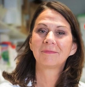 Alexandra Durr合作论文数Faculté De Médecine, l’Institut du Cerveau et de la Moelle Épinière6
 José Berciano合作论文数Department of Medicine and Psychiatry, Universidad de Cantabria6Tanja Schmitz-Hübsch合作论文数6Elzbieta Zdzienicka合作论文数6A. Seroka合作论文数5Jolanta Kubalska合作论文数5Robyn Labrum合作论文数5




