Diabetic retinopathy (DR) causes vision loss due to sustained inflammation and vascular damage. The vascular damage is evident by fibrinogen leakage, angiogenesis, and hypoxia. Neuronal regulation of microglia via the CX3CL1 (Fractalkine or FKN)-CX3CR1 pathway plays a significant role in retinal pathology. Defects in FKN or CX3CR1 exacerbate inflammation, vascular damage, and vision impairment. However, the contribution of hypoxic astrocytes to the pathological process of DR is unclear. A hypoxic model (7 days of systemic 7.5% O2) was utilized to induce retinal damage in adult mice in the absence of systemic inflammatory signals. This model induced vascular and microglial responses similar to 10 weeks of STZ-induced hyperglycemia. The goal of this study is to characterize retinal damage in WT and mice with defects in the FKN-CX3CR1 signaling axis and hence assess the impact of the microglial inflammatory responses to hypoxic retinopathy. Tissues were analyzed by immunostaining, RNA sequencing, and cytokine quantification. We found that CX3CR1 deficiency in hypoxic animals induced reactive astrogliosis and that Müller glial responses to hypoxia and systemic inflammation were dependent on FKN signaling. Exacerbated microglial reactivity to hypoxic conditions significantly altered the expression of HIF transcripts. Microglial dysregulation was found to reduce the anti-inflammatory response to hypoxic conditions, downregulate hypoxia-responsive gene expression, and restrained LPS-induced inflammatory responses. We found that microglia dysregulation alters the hypoxic response by inhibiting the upregulation of HIF2α/3α, increasing CD31 immunoreactivity, and altering the expression of ECM-associated transcripts such as type I, III, and XVIII collagens to hypoxic conditions.
Diabetic retinopathy, a microvascular disease characterized by irreparable vascular damage, neurodegeneration and neuroinflammation, is a leading complication of diabetes mellitus. There is no cure for DR, and medical interventions marginally slow the progression of disease. Microglia-mediated inflammation in the diabetic retina is regulated via CX3CR1-FKN signaling, where FKN serves as a calming signal for microglial activation in several neuroinflammatory models. Polymorphic variants of CX3CR1, hCX3CR1I249/M280, found in 25% of the human population, result in a receptor with lower binding affinity for FKN. Furthermore, disrupted CX3CR1-FKN signaling in CX3CR1-KO and FKN-KO mice leads to exacerbated microglial activation, robust neuronal cell loss and substantial vascular damage in the diabetic retina. Thus, studies to characterize the effects of hCX3CR1I249/M280-expression in microglia-mediated inflammation in the diseased retina are relevant to identify mechanisms by which microglia contribute to disease progression. Our results show that hCX3CR1I249/M280 mice are significantly more susceptible to microgliosis and production of Cxcl10 and TNFα under acute inflammatory conditions. Inflammation is exacerbated under diabetic conditions and coincides with robust neuronal loss in comparison to CX3CR1-WT mice. Therefore, to further investigate the role of hCX3CR1I249/M280-expression in microglial responses, we pharmacologically depleted microglia using PLX-5622, a CSF-1R antagonist. PLX-5622 treatment led to a robust (~70%) reduction in Iba1+ microglia in all non-diabetic and diabetic mice. CSF-1R antagonism in diabetic CX3CR1-WT prevented TUJ1+ axonal loss, angiogenesis and fibrinogen deposition. In contrast, PLX-5622 microglia depletion in CX3CR1-KO and hCX3CR1I249/M280 mice did not alleviate TUJ1+ axonal loss or angiogenesis. Interestingly, PLX-5622 treatment reduced fibrinogen deposition in CX3CR1-KO mice but not in hCX3CR1I249/M280 mice, suggesting that hCX3CR1I249/M280 expressing microglia influences vascular pathology differently compared to CX3CR1-KO microglia. Currently CX3CR1-KO mice are the most commonly used strain to investigate CX3CR1-FKN signaling effects on microglia-mediated inflammation and the results in this study indicate that hCX3CR1I249/M280 receptor variants may serve as a complementary model to study dysregulated CX3CR1-FKN signaling. In summary, the protective effects of microglia depletion is CX3CR1-dependent as microglia depletion in CX3CR1-KO and hCX3CR1I249/M280 mice did not alleviate retinal degeneration nor microglial morphological activation as observed in CX3CR1-WT mice.
CX3CR1, one of the highest expressed genes in microglia in mice and humans, is implicated in numerous microglial functions. However, the molecular mechanisms underlying Cx3cr1 signaling are not well understood. Here, we analyzed transcriptomes of Cx3cr1-deficient microglia under varying conditions by RNA-sequencing (RNA-seq). In 2-mo-old mice, Cx3cr1 deletion resulted in the down-regulation of a subset of immune-related genes, without substantial epigenetic changes in markers of active chromatin. Surprisingly, Cx3cr1-deficient microglia from young mice exhibited a transcriptome consistent with that of aged Cx3cr1-sufficient animals, suggesting a premature aging transcriptomic signature. Immunohistochemical analysis of microglia in young and aged mice revealed that loss of Cx3cr1 modulates microglial morphology in a comparable fashion. Our results suggest that CX3CR1 may regulate microglial function in part by modulating the expression levels of a subset of inflammatory genes during chronological aging, making Cx3cr1-deficient mice useful for studying aged microglia.
Remyelination is the regenerative process whereby myelin sheaths are restored around axons following nervous system injury, allowing reinstatement of electrical impulse conduction, trophic/metabolic support, and axon health. Failure of remyelination in progressive multiple sclerosis is considered to contribute to axon loss, a correlate of clinical decline. Lack of approved pro-regenerative therapies for MS highlights the need to understand the cellular and molecular mechanisms underpinning successful remyelination. One approach is to conduct nonbiased gene expression analyses of cell types which regulate remyelination, such as microglia and monocyte-derived macrophages. Recent technological advances address the challenges of RNA sequencing of small tissue samples, thus allowing relatively small numbers of cells to be isolated from discrete lesions for analysis. Here, we present methods for FACS-based isolation of cells from focal remyelinating lesions of the adult mouse brain and subsequent RNA extraction for sequencing, using isolation of microglia/macrophages as an example.
Objective: To address the hypothesis that physiologic interactions between astrocytes and endothelial cells (EC) at the blood–brain barrier (BBB) are afflicted by pathogenic inflammatory signaling when astrocytes are exposed to aquaporin-4 (AQP4) antibodies present in the immunoglobulin G (IgG) fraction of serum from patients with neuromyelitis optica (NMO), referred to as NMO-IgG. Methods: We established static and flow-based in vitro BBB models incorporating co-cultures of conditionally immortalized human brain microvascular endothelial cells and human astrocyte cell lines with or without AQP4 expression. Results: In astrocyte–EC co-cultures, exposure of astrocytes to NMO-IgG decreased barrier function, induced CCL2 and CXCL8 expression by EC, and promoted leukocyte migration under flow, contingent on astrocyte expression of AQP4. NMO-IgG selectively induced interleukin (IL)-6 production by AQP4-positive astrocytes. When EC were exposed to IL-6, we observed decreased barrier function, increased CCL2 and CXCL8 expression, and enhanced leukocyte transmigration under flow. These effects were reversed after application of IL–6 neutralizing antibody. Conclusions: Our results indicate that NMO-IgG induces IL-6 production by AQP4-positive astrocytes and that IL-6 signaling to EC decreases barrier function, increases chemokine production, and enhances leukocyte transmigration under flow.
[This corrects the article DOI: 10.1371/journal.pone.0013693.].
Neuromyelitis optica (NMO) is an inflammatory disorder mediated by antibodies to aquaporin-4 (AQP4) with prominent blood-brain barrier (BBB) breakdown in the acute phase of the disease. Anti-AQP4 antibodies are produced mainly in the periphery, yet they target the astrocyte perivascular end feet behind the BBB. We reasoned that an endothelial cell-targeted autoantibody might promote BBB transit of AQP4 antibodies and facilitate NMO attacks. Using monoclonal recombinant antibodies (rAbs) from patients with NMO, we identified two that strongly bound to the brain microvascular endothelial cells (BMECs). Exposure of BMECs to these rAbs resulted in nuclear translocation of nuclear factor κB p65, decreased claudin-5 protein expression, and enhanced transit of macromolecules. Unbiased membrane proteomics identified glucose-regulated protein 78 (GRP78) as the rAb target. Using immobilized GRP78 to deplete GRP78 antibodies from pooled total immunoglobulin G (IgG) of 50 NMO patients (NMO-IgG) reduced the biological effect of NMO-IgG on BMECs. GRP78 was expressed on the surface of murine BMECs in vivo, and repeated administration of a GRP78-specific rAb caused extravasation of serum albumin, IgG, and fibrinogen into mouse brains. Our results identify GRP78 antibodies as a potential component of NMO pathogenesis and GRP78 as a candidate target for promoting central nervous system transit of therapeutic antibodies.
The ability of the Blood Brain Barrier (BBB) to maintain proper barrier functions, keeping an optimal environment for central nervous system (CNS) activity and regulating leukocytes' access, can be affected in CNS diseases. Endothelial cells and astrocytes are the principal BBB cellular constituents and their interaction is essential to maintain its function. Both endothelial cells and astrocytes express the receptors for the bioactive sphingolipid S1P. Fingolimod, an immune modulatory drug whose structure is similar to S1P, has been approved for treatment in multiple sclerosis (MS): fingolimod reduces the rate of MS relapses by preventing leukocyte egress from the lymph nodes. Here, we examined the ability of S1P and fingolimod to act on the BBB, using an in vitro co-culture model that allowed us to investigate the effects of S1P on endothelial cells, astrocytes, and interactions between the two. Acting selectively on endothelial cells, S1P receptor signaling reduced cell death induced by inflammatory cytokines. When acting on astrocytes, fingolimod treatment induced the release of a factor, granulocyte macrophage colony-stimulating factor (GM-CSF) that reduced the effects of cytokines on endothelium. In an in vitro BBB model incorporating shear stress, S1P receptor modulation reduced leukocyte migration across the endothelial barrier, indicating a novel mechanism that might contribute to fingolimod efficacy in MS treatment.
Background: Residual CXCR2 expression on CNS cells in Cxcr2(-/+) -> Cxcr2(-/-) chimeric animals slowed remyelination after both experimental autoimmune encephalomyelitis and cuprizone-induced demyelination.Methods: We generated Cxcr2(fl/-): PLPCre-ER(T) mice enabling an inducible, conditional deletion of Cxcr2 on oligodendrocyte lineage cells of the CNS. Cxcr2(fl/-): PLPCre-ER(T) mice were evaluated in 2 demyelination/remyelination models: cuprizone-feeding and in vitro lysophosphatidylcholine (LPC) treatment of cerebellar slice cultures.Results: Cxcr2(fl/-): PLPCre-ER(T)(+) (termed Cxcr2-cKO) mice showed better myelin repair 4 days after LPC-induced demyelination of cerebellar slice cultures. Cxcr2-cKOs also displayed enhanced hippocampal remyelination after a 2-week recovery from 6-week cuprizone feeding.Conclusion: Using 2 independent demyelination/remyelination models, our data document enhanced myelin repair in Cxcr2-cKO mice, consistent with the data obtained from radiation chimerism studies of germline CXCR2. Further experiments are appropriate to explore how CXCR2 function in the oligodendrocyte lineage accelerates myelin repair.
OBJECTIVE: We evaluated how Neuromyelitis optica (NMO)-IgG induced the leukocyte transmigration in in vitro Blood-Brain Barrier (BBB) model. BACKGROUND: Neuromyelitis optica (NMO), an autoimmune inflammatory astrocytopathy, is caused by antibodies to the astrocyte water channel aquaporin 4 (AQP4). AQP4 is expressed exclusively on astrocytes and absent from endothelial cells. The IgG plasma fraction of NMO patients (NMO-IgG) contains AQP4 antibodies. It remains uncertain the mechanisms by which inflammatory cells migrate across the BBB. DESIGN/METHODS: NMO-IgG was pooled from therapeutic plasma exchange from total 50 NMO patients. We established in vitro static and flow based BBB models incorporating a conditionally immortalized human brain microvascular endothelial cell line (EC) and human astrocyte cell lines with (A4) or without (A) AQP4 expression. EC and A4 (EC/A4) or A (EC/A) were co-cultured on each side of polycarbonate membranes. After exposing NMO-IgG to A4 or A (Brain parenchymal) side, peripheral blood mononuclear cells flowed over EC with physiological shear force. Migrated cells were enumerated. We screened EC, A4 and A pre-exposed to NMO-IgG for cytokines and chemokines. RESULTS: Parenchymal application of NMO-IgG induced CCL2 and CXCL8 in EC and increased leukocyte migration (especially monocyte) in EC/A4 but not EC/A. Of 20 cytokines, NMO-IgG selectively induced IL-6 in A4 but not A. IL-6 application increased CCL2 and CXCL8 in EC and induced leukocyte migration. IL-6 receptor blockade inhibited NMO-IgG induced leukocyte migration. CONCLUSIONS: NMO-IgG induces IL-6 in astrocytes via AQP4 and IL-6 signaling to ECs increases chemokines and drives leukocyte migration. Study Supported by: the Guthy Jackson Charitable Foundation and the National Institutes of Health.
Introduction: We previously reported on the relationship between unbound CSF iron and brain injury following SAH. The ferroxidase ceruloplasmin (Cp) regulates iron levels in the CNS and prevents free radical injury. Paradoxically, oxidative stress is known to cause structural modifications that result in decreased enzymatic activity of Cp. We sought to determine if CSF of patients with SAH exhibits impaired ferroxidase activity (FA). Methods: Samples of CSF were obtained on day 5 following aneurysm rupture and levels of malondialdehyde (MDA; a marker of lipid peroxidation) and ferroxidase activity (defined as the amount of enzyme that converts 1 μM of substrate into product per minute) were determined by means of absorbance and colorimetric assays. Ceruloplasmin concentration in CSF was measured with ELISA. Demographic and clinical data were recorded. Values are expressed as mean ± SEM. Comparisons were made using Wilcoxon rank sum test. Simple linear regression analysis was performed for comparison of continuous variables. Results: A total of 5 patients and 4 control subjects were included in this pilot study. Median Hunt-Hess and modified Fisher scores were 4 (IQR 1.5) and 4 (IQR 1), respectively. Levels of MDA in CSF (9.2 ± 0.9 vs. 0.09 ± 0.01 μM, p =0.02) were significantly higher in SAH patients, while FA was significantly reduced (64.8 ± 17.1 vs. 284.1 ± 64.9 U/L, p =0.01). Ceruloplasmin concentration was higher in SAH samples (49.4 ± 12 vs. 1.72 ± 0.7 mg/L, p =0.04), and patients who developed delayed cerebral ischemia had lower CSF FA (43.2 ± 6.3 vs. 97.2 ± 32.4 U/L, p =0.07), compared to those who did not. Conclusions: The ferroxidase activity of CSF following SAH is significantly impaired, in spite of higher ceruloplasmin concentrations. Evidence of increased CNS oxidative damage was also demonstrated in our cohort. We postulate that structural modifications in Cp mediated by an oxidative milieu occur in SAH and are associated with decreased enzymatic function. There is a possible inverse relationship between ferroxidase activity and risk of development of delayed cerebral ischemia. Further studies are needed to explore the significance of these findings.
Variants in triggering receptor expressed on myeloid cells 2 (TREM2) confer high risk for Alzheimer's disease (AD) and other neurodegenerative diseases. However, the cell types and mechanisms underlying TREM2's involvement in neurodegeneration remain to be established. Here, we report that TREM2 is up-regulated on myeloid cells surrounding amyloid deposits in AD mouse models and human AD tissue. TREM2 was detected on CD45(hi)Ly6C(+) myeloid cells, but not on P2RY12(+) parenchymal microglia. In AD mice deficient for TREM2, the CD45(hi)Ly6C(+) macrophages are virtually eliminated, resulting in reduced inflammation and ameliorated amyloid and tau pathologies. These data suggest a functionally important role for TREM2(+) macrophages in AD pathogenesis and an unexpected, detrimental role of TREM2 in AD pathology. These findings have direct implications for future development of TREM2-targeted therapeutics.
Background: In vitro blood-brain barrier (BBB) models can be useful for understanding leukocyte-endothelial interactions at this unique vascular-tissue interface. Desirable features of such a model include shear stress, non-transformed cells and co-cultures of brain microvascular endothelial cells with astrocytes. Recovery of transmigrated leukocytes for further analysis is also appealing.New methods: We report an in vitro BBB model for leukocyte transmigration incorporating shear stress with co-culture of conditionally immortalized human endothelial cell line (hBMVEC) and human astrocyte cell line (hAST). Transmigrated leukocytes can be recovered for comparison with input and non-transmigrated cells.Result: hBMVEC and hAST exhibited physiological and morphological BBB properties when cocultured back-to-back on membranes. In particular, astrocyte processes protruded through 3 mu m membrane pores, terminating in close proximity to the hBMVEC with a morphology reminiscent of end-feet. Co-culture with hAST also decreased the permeability of hBMVEC. In our model, astrocytes promoted transendothelial leukocyte transmigration.Comparison with existing methods: This model offers the opportunity to evaluate whether BBB properties and leukocyte transmigration across cytokine-activated hBMVEC are influenced by human astrocytes.Conclusions: We present a BBB model for leukocyte transmigration incorporating shear stress with co-culture of hBMVEC and hAST. We demonstrate that hAST promoted leukocyte transmigration and also increased certain barrier functions of hBMVEC. This model provides reproducible assays for leukocyte transmigration with robust results, which will enable further defining the relationships among leukocytes and the cellular elements of the BBB. (C) 2014 Published by Elsevier B.V.
Iron-mediated oxidative damage has been implicated in the genesis of cerebral vasospasm in animal models of SAH. We sought to explore the relationship between levels of non-protein bound iron in cerebrospinal fluid and the development of brain injury in patients with aneurysmal SAH.
In the human disorder multiple sclerosis (MS) and in the model experimental autoimmune encephalomyelitis (EAE), macrophages predominate in demyelinated areas and their numbers correlate to tissue damage. Macrophages may be derived from infiltrating monocytes or resident microglia, yet are indistinguishable by light microscopy and surface phenotype. It is axiomatic that T cell-mediated macrophage activation is critical for inflammatory demyelination in EAE, yet the precise details by which tissue injury takes place remain poorly understood. In the present study, we addressed the cellular basis of autoimmune demyelination by discriminating microglial versus monocyte origins of effector macrophages. Using serial block-face scanning electron microscopy (SBF-SEM), we show that monocyte-derived macrophages associate with nodes of Ranvier and initiate demyelination, whereas microglia appear to clear debris. Gene expression profiles confirm that monocyte-derived macrophages are highly phagocytic and inflammatory, whereas those arising from microglia demonstrate an unexpected signature of globally suppressed cellular metabolism at disease onset. Distinguishing tissue-resident macrophages from infiltrating monocytes will point toward new strategies to treat disease and promote repair in diverse inflammatory pathologies in varied organs.
 Richard Ransohoff合作论文数Third Rock Ventures;Abata Therapeutics24
 Birgit Obermeier合作论文数Department of Neuroimmunology, Max-Planck-Institute of Neurobiology10
 Yasuteru Sano合作论文数10Yukio Takeshita合作论文数10Takashi Kanda合作论文数9
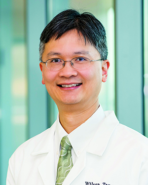 Wai Hong Wilson Tang合作论文数Department of Cardiovascular & Metabolic Sciences, Lerner Research Institute, Cleveland Clinic;Genomic Medicine Institute, Lerner Research Institute, Cleveland Clinic;Cleveland Clinic Lerner College of Medicine of School of Medicine, Case Western Reserve University8
 Clara Pelfrey合作论文数Department of Neurosciences, Cleveland Clinic Foundation8
 Anna Love合作论文数Dept Chem, Univ Calif Irvine7
 Simona Federica Spampinato合作论文数Department of Clinical and Molecular Biomedicine, University of Catania7
 Astrid E. Cardona合作论文数Department of Neuroscience;Cleveland Clinic;Department of Neuroscience, Cleveland Clinic5Israel F. Charo合作论文数4Liping Liu合作论文数4Jason M. Lappe合作论文数4Fumitaka Shimizu合作论文数4
 Stefka Gyoneva合作论文数4Kelsey Croft合作论文数3
 Chia-Lin Tsou合作论文数3Saederup Noah合作论文数3Kryzer T J合作论文数3
 Vanda A. Lennon合作论文数Department of Immunology, Mayo Clinic;Department of Neurology, Mayo Clinic;Division of Clinical Biochemistry & Immunology, Department of Laboratory Medicine and Pathology, Mayo Clinic3Natacha Zamor合作论文数3Makiko Mizutani合作论文数3Joao A. Gomes合作论文数2Difernando Vanegas合作论文数2Carmel Halley合作论文数2Lisa C Spangler合作论文数2Derek Rodriguez合作论文数2Meizhang Li合作论文数2Kaira A. Church合作论文数2Schuster Andres合作论文数2Moldovan Ioana合作论文数2Robert S. Butler合作论文数2Sandra M. Cardona合作论文数2
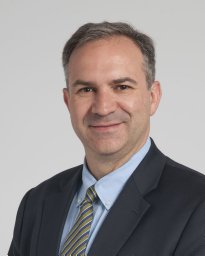 J. Javier Provencio合作论文数Center for Brain Immunology and Glia, Department of Neurology, University of Virginia2Lindsey Darnall合作论文数2
 Oleg Butovsky合作论文数Ann Romney Center for Neurologic Diseases, Department of Neurology, Brigham and Women's Hospital, Harvard Medical School;Evergrande Center for Immunologic Diseases, Brigham and Women's Hospital, Harvard Medical School2Jar-Chi Lee合作论文数2Gregory P. Owens合作论文数1Nozomu Sato合作论文数1Grahame Kidd合作论文数1Colin Rorex合作论文数1Ravi Challa合作论文数1Janet Schaefer-Klein合作论文数1Pauline M Wu合作论文数1Kristin L. Schaller合作论文数1Tracy Kovach合作论文数1Kelly Miao合作论文数1Anna Rietsch合作论文数1Anjli Maroo合作论文数1




