OBJECTIVES:To assess the influence of crown design, cement type, and margin depth on the removal efficiency of residual cement around single implant-supported restorations. METHODS:Twelve mandibular models with a missing first molar were fabricated and provided with single implant analogs (centrally/distally placed) and two different crown designs (non-cantilever/cantilever). Three of each design had either a superficial (SM = -1 mm) or a deep (DM = -2 mm) submucosal margin. Crown restorations were made from CAD/CAM titanium-base abutments and monolithic zirconia (n = 120). Three different cements (glass ionomer [GI], polycarboxylate [PC], dual-curing resin cement [RC]) were used in a standardized way for crown cementation. Ten dentists removed excess cement using metal scalers. The volumetric values of residual cement were calculated (mm3). Statistical analyses assessed the effects of the investigated variables on residual cement volume (p < 0.05). RESULTS:The overall analysis showed no significant differences in the volume of cement remnants between both crown designs (non-cantilever: 0.42 ± 0.96 mm3; cantilever: 0.52 ± 0.98 mm3; p > 0.05). For crown margin depth, a significantly higher cement volume for deeper margins (SM: 0.28 ± 0.62 mm3; DM: 0.68 ± 1.19 mm3; p < 0.05) was found. The highest volume of cement remnants was found in the non-cantilever_DM_RC (1.19 ± 1.83 mm3), whereas the lowest value was found in non-cantilever_SM_GI and cantilever_SM_GI (0.00 ± 0.00 mm3). While crown design alone showed no statistically significant effect, its interaction with cement type was associated with improved cement removal in the cantilever group. CONCLUSION:Crown margin depth emerged as the dominant factor influencing cement removal. Although cement type also affected residual cement, with glass ionomer showing lower values overall, crown design did not provide a consistent advantage.
The aim of the study was to evaluate antagonist enamel wear caused by posterior monolithic zirconia crowns, and the wear of contralateral enamel/enamel tooth pairs over a 36-month observation period. Out of thirty-two patients, each provided with one 5 mol
OBJECTIVES:To evaluate the survival rates and marginal bone loss of narrow-diameter titanium-zirconium implants supporting complete maxillary and mandibular overdentures up to 3 years after loading. MATERIALS AND METHODS:Ten completely edentulous patients who were dissatisfied with their complete dentures were enrolled. Two narrow-diameter implants were placed in the canine region of the maxilla and mandible. After second-stage surgery, implant-supported overdentures (palatal-free) attached by parallel alignable stud-attachments were placed. Patients were followed periodically for up to 36 months. Standardized radiographs were taken at baseline, 12 and 36 months to analyze mean marginal bone level changes around the implants. RESULTS:The Kaplan-Meier survival rates were 100% for mandibular and 68.0% (SE ± 10.9%) for maxillary implants at 36 months (p = .008). Six maxillary implants failed after loading; no mandibular implants were lost. Five implants failed due to loss of osseointegration. One implant fractured. The mean marginal bone level changes around the analyzed implants (n = 28, 9 patients) were -0.71 ± 0.82 mm in the mandible and -2.08 ± 1.52 mm in the maxilla at the 36-month follow-up. The difference in marginal bone level changes between the maxilla and mandible was significant (p = .019) at the 12- and 36-month follow-ups. CONCLUSION:Two narrow-diameter titanium-zirconium implants with stud-attachments showed a highly satisfactory outcome in the mandible. The maxillary implants showed a high failure rate and significantly more bone loss over time than the mandibular implants. The minimal concept of two implants and an overdenture should be limited to the edentulous mandible.
Objectives A serious game application was developed to train factual knowledge and for self-assessment. The aim of the present study was to compare the effects of a game application (intervention group) or paper scripts (control group) on knowledge acquisition and to evaluate the acceptance of the new application among dental students. Methods The 4th semester students of the second preclinical prosthodontics course were randomly assigned to one of the two groups ( n = 58/51) for two consecutive years. The study was conducted in two phases: First, all participants took a pretest, with the intervention group using the game application and the control group receiving the same set of questions in a paper script. In the second phase, all participants took a post-test. After the post-test, both groups had access to the application for another three weeks. After that, all participants completed standardized questionnaires and a scale to evaluate the usability of the system. Usage statistics were also tracked. Differences between groups were evaluated together and for both years separately in terms of pretest and posttest scores and learning success. Results There was no significant difference between the groups with regard to the posttest and learning success. A significant improvement in knowledge between pretest and posttest ( p < 0.05) was demonstrated in both groups. Each student played approximately 350 questions. Participants rated the application with the German school grade "good". Participants appreciated the application and rated it positively. They stated that the game motivated them to learn and that they spent more time with the learning content. Conclusion Due to the positive perception achieved through the game, this application is able to motivate students to learn. The learning effect achieved is similar to learning on paper.
Purpose To evaluate the radiographic and peri-implant outcomes of intramobile cylinder implants (IMZs) and the feasibility of long-term follow-up studies after nearly 30 years. Methods Of the 94 patients treated with IMZ implants between 1981 and 1995, 39 patients were successfully contacted (contact group, CG), of which 15 patients with a total of 32 implants agreed to participate in the present follow-up study (clinical evaluation group, CEG). The overall implant survival rate was calculated. Information on implant status and oral and general health data was collected. Marginal bone level was evaluated and then compared to the patients’ baseline data. Possible risk factors for peri-implantitis were also identified. Results In total, 16 implants in seven patients were lost, amounting to an overall survival rate of 79.5% after 30 years with a mean follow-up time of 24 ± 10 years (CG). Eight patients were treated with bar-retained mandibular overdentures and seven patients had fixed partial dentures. After a mean observation time of 29 ± 3 years, the surviving implants showed a peri-implantitis rate of 9.4% with a mean marginal bone loss of 2.5 ± 1.8 mm (CEG). No significant correlation between peri-implantitis and possible risk factors could be found. Conclusions Long-term follow-up studies with acceptable response rates after nearly 30 years are not feasible. Contact was only possible with 41% of the patients. This contact group showed a high implant survival rate. Due to the retrospective study design, additional risk factors could not be considered in a conclusive analysis. Graphical Abstract
OBJECTIVES:To summarize the existing evidence on patient-reported outcome measures (PROMs) of implant-supported restorations fabricated using a digital workflow in comparison to conventional manufacturing procedures. METHODS:A PICO strategy was executed using an electronic and manual search focusing on clinical studies evaluating PROMs of implant-supported restorations. Only clinical trials assessing conventional versus digital workflows for implant-supported restorations were included. PROMS on implant impression procedures and fabrication of final restorations were evaluated using random and fixed effects meta-analyses, while implant planning/placement was reported descriptively. RESULTS:Among 1062 titles identified, 14 studies were finally included, and only seven studies were eligible for meta-analysis. For implant planning and placement, only a qualitative analysis was possible due to heterogeneity between the studies. For impression procedures, the random effects model revealed statistically significant differences in taste, anxiety, nausea, pain, shortness of breath, and discomfort in favor of optical impressions. No significant difference in the subjective perception of the duration of an impression could be reported. For the final fabrication of restorations, no significant difference between veneered and monolithic posterior restorations was found in terms of esthetic, function, and general satisfaction. CONCLUSION:Most of the studies reporting about PROMs were published during the last ten years and limited to implant-supported single crowns in the posterior region. Based on PROMs, no scientifically proven recommendation for guided implant placement could be given at this time. Patients showed high preference for optical impressions, whereas no differences between veneered and monolithic restorations could be reported.
BackgroundLocal anaesthesia plays a key role in many aspects of a dentist's work. The required skills to perform anaesthesia successfully are acquired at university. To take advantage of the possibilities for new teaching formats, a blended learning concept for the local anaesthesia course was developed. The aim of the study was to compare the effectiveness of face-to-face, blended and e-learning in teaching in local anaesthesia by assessing students' knowledge gain, performance of practical skills and satisfaction with the course.MethodsAll participants (n=37) were randomly allocated into three groups. After acquiring the theoretical background in the blended learning, e-learning or lecture groups, a test to assess knowledge gain was performed. The performance of the practical skills was assessed in a small-group seminar. Student attitudes were evaluated with a questionnaire.ResultsThe blended group showed significantly better results (mean=17, SD =1.5) in theoretical knowledge gain than the other two groups (e-learning group: mean=14.7, SD=2.2; lecture group: mean=14.8, SD =2.3). When comparing the results of the clinical skills assessment, there was no significant difference among all three groups (p>0.017). The participants confirmed a high overall satisfaction with the course, in particular with the blended learning approach.ConclusionThis study indicates that blended learning improves the learning outcome for theoretical knowledge in teaching local anaesthesia more than either face-to-face learning or e-learning alone. Furthermore, the blended learning approach is highly appreciated by the students. For acquiring practical skills, this study shows that blended learning is as effective as other teaching methods.
OBJECTIVES:To compare the incidence of biological and technical complications of cemented and screw-retained monolithic lithium-disilicate implant-supported posterior single crowns.MATERIAL AND METHODS:Forty-one subjects with a total of 56 implants received randomly allocated 28 cemented and 28 screw-retained crowns. In the screw-retained group, monolithic lithium-disilicate restorations were luted to titanium bases extraorally. In the cemented group, monolithic lithium-disilicate crowns were cemented on individualized titanium abutments intraorally. All restorations were examined according to modified FDI criteria within 2 weeks of inserting the crowns (baseline) and after 12 (n = 46) and 24 (n = 43) months. Bone loss was evaluated by standardized radiographs at baseline and 12 months.RESULTS:After 12 months, the incidence of mucositis (positive bleeding on probing) was 14.2% (screw-retained) and 17.9% (cement-retained). The gingival and plaque index and a mean marginal bone loss between 0.03-0.15 mm showed no significant difference between the groups. In the cemented group, cement residues were detected at baseline at two restorations (6.9%) by radiographic examination. A complete digital workflow was realized in most cases (85.7%). At 24 months, no restoration had failed, and no chipping of the ceramic had occurred. In the screw-retained group, screw loosening occurred in one implant. In both groups, there was obvious deterioration in the quality of 32% of the occlusal and of 18% of the proximal contact points.CONCLUSIONS:The type of retention mode of monolithic implant-retained lithium-disilicate posterior crowns had no influence on the biological and technical complication rate.
OBJECTIVE To compare the radiographic marginal bone loss and clinical parameters of splinted and non-splinted fixed dental prostheses on short implants in the posterior region of the lower jaw 3 years after loading. MATERIAL AND METHODS Twenty patients, 15 female and five males, with uni- or bilateral free-end situations in the mandible participated in the study. Two short implants (7 mm) in the posterior mandible were placed and patients were randomized to receive splinted (n = 11) or non-splinted (n = 13) cemented crowns. Marginal bone loss (MBL) was assessed on radiographs taken with customized positioning jigs at baseline, 1 and 3 years after loading. Plaque index (PI), gingival index (GI), probing depth (PD), and bleeding on probing (BOP) were measured. (ClinicalTrials.gov; identifier: NCT03558347). RESULTS After 3-year survival rate of altogether 48 implants was 100% for both groups. Success rate (according to Papaspyridakos, Chen, Singh, Weber, & Gallucci, 2012) was 84.6% for non-splinted and 86.4% for splinted implants. At restoration level survival rate was 100% for both groups. Marginal bone level changes showed mean gain of 0.3 ± 0.8 mm for non-splinted and 0.1 ± 0.5 mm for splinted implants 3 years after loading. Statistical analysis showed no significant difference in PI, GI, PD, BOP, and marginal bone loss between both groups (p > .05). CONCLUSION Within the limitations of this study it can be concluded that splinting crowns on short implants neither seems to affect the amount of marginal bone loss nor peri-implant health 3 years after loading.
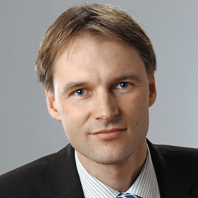 Stefan Wolfart合作论文数Department of Prosthodontics and Biomaterials, Faculty of Medicine, RWTH University of Aachen;Clinic of Prosthodontics and Biomaterials, Center for Implantology, University Hospital Aachen7
 Taskin Tuna合作论文数School of Dentistry, Albert-Ludwigs University of Freiburg3
 Martin Lemos合作论文数Audiovisual Media Ctr, Rhein Westfal TH Aachen2
 Frank Hölzle合作论文数2Oliver Hartkamp合作论文数1Christoph Mautsch合作论文数1Dirk Elvers合作论文数1Annabelle von der Stueck合作论文数1Thomas Kern合作论文数1Shaza Bishti合作论文数1Anna Bock合作论文数1
 Kristian Kniha合作论文数1Goloborodko Evgeny合作论文数1Rafai Nicole合作论文数1Modabber Ali合作论文数1Möhlhenrich Stephan Christian合作论文数Department of Oral and Maxillofacial Surgery, RWTH Aachen University1
 Jaana-Sophia Kern合作论文数1
 Stefan Raith合作论文数1Karin Gross合作论文数1Sven Reich合作论文数1




