Ocular surface neuropeptides are vital molecules primarily involved in maintaining ocular surface integrity and homeostasis. They also serve as communication channels between the nervous system and the immune system, maintaining the homeostasis of the ocular surface. Tear film and ocular surface neuropeptides have a role in disease often due to abnormalities in their synthesis (either high or low production), signaling through defective receptors, or both. This creates imbalances in otherwise normal physiological processes. They have been observed to be altered in many ocular surface and systemic diseases including dry eye disease, ocular allergy, keratoconus, LASIK-induced dry eye, pterygium, neurotrophic keratitis, corneal graft rejection, microbial keratitis, headaches and diabetes. This review examines the characteristics of neuropeptides, their synthesis and their signaling through G-protein coupled receptors. The review also explores the types of neuropeptides within the tears and ocular surface, and how they change in ocular and systemic diseases.
IMR Press is a leading publisher of open access peer-reviewed biomedical and life sciences journals. We aim to facilitate the dissemination of high-quality research in the area of biomedical science. With a long tradition and wide readership, IMR Press is dedicated to making positive contributions to academics, corporate libraries as well as to readers and authors. All the editors in IMR Press will provide the best service for researchers, allowing them to have a easy and smooth publication experience and helping maximize the impact and visibility of their research.
Systemic dehydration due to inadequate water intake or excessive water loss, is common in the elderly and results in a high morbidity and significant mortality. Diagnosis is often overlooked and there is a need for a simple, bedside diagnostic test in at-risk populations. Body hydration is highly regulated with plasma osmolality (pOsm) being tightly controlled over a wide range of physiological conditions. By contrast, normal tear osmolarity (tOsm) is more variable since the tear film is exposed to evaporation from the open eye. While plasma hyperosmolality is a diagnostic feature of systemic dehydration, tear hyperosmolality, with other clinical features, is diagnostic of dry eye. Studies in young adults subjected to exercise and water-deprivation, have shown that tOsm may provide an index of pOsm, with the inference that it may provide a simple measure to diagnose systemic dehydration. However, since the prevalence of both dry eye and systemic dehydration increases with age, the finding of a raised tOsm in the elderly could imply the presence of either condition. This diagnostic difficulty can be overcome by measuring tear osmolality after a period of evaporative suppression (e.g., a 45 min period of lid closure) which drives tOsm osmolality down to a basal level, close to that of the pOsm. The arguments supporting the use of this basal tear osmolarity (BTO) in the diagnosis of systemic dehydration are reviewed here. Further studies are needed to confirm that the BTO can act as a surrogate for pOsm in both normally hydrated subjects and in patients with systemic dehydration and to determine the minimum period of lid closure required for a simple, "point-of-care" test.
La sécheresse oculaire est une maladie multifactorielle de la surface oculaire caractérisée par une perte de l’homéostasie du film lacrymal, et accompagnée de symptômes oculaires dans lesquels l’instabilité et l’hyperosmolarité du film lacrymal, l’inflammation et les lésions de la surface oculaire, et des anomalies neurosensorielles jouent des rôles étiologiques (voir le rapport du sous-comité Définition et classification).
PURPOSE:Tear osmolarity (tOsm) is used as a measure of severity in dry eye disease (DED) and has been proposed as an index of body hydration. In DED the level of tear hyperosmolarity is compared with that of a control population. It is proposed here that a better index of body hydration and a more valid reference point in DED can be acquired by measuring the tOsm after a period of evaporative suppression.METHOD:8 normal and DED subjects were recruited, their tOsm measured in uncontrolled environmental 'clinic conditions'. Then in experiment 1 they entered a controlled environment chamber and had tOsm measured after 45 minutes of eye closure and then, with the eyes open, at 15 minute intervals for a further 45 minutes, at a relative humidity (RH) of 45%. Alternatively, in experiment 2, they had tOsm measured every 15 minutes for 45 minutes during exposure to 70% RH, as a separate measure to suppress evaporation.RESULTS:A significant decrease in tOsm occurred in both normal and DED subjects after lid closure in experiment 1 (normal RE p=0.015; normal LE p=0.006; DED RE p=0.0002; DED LE p=0.01). The tOsm also fell slightly after exposure to 70% RH in experiment 2 significant in the LE of normal group only (normal LE p=0.045).CONCLUSIONS:Suppression of tear evaporation resulted in a fall in tOsm, close to that of plasma osmolarity (285-295mOsm/L). It is proposed that this new measure, termed Basal Tear Osmolarity (BTO), could provide a valuable index of plasma osmolarity and hence of body hydration and in DED, a personal baseline against which to gauge the severity of tear hyperosmolarity.
The TFOS DEWS II Pathophysiology Subcommittee reviewed the mechanisms involved in the initiation and perpetuation of dry eye disease. Its central mechanism is evaporative water loss leading to hyperosmolar tissue damage. Research in human disease and in animal models has shown that this, either directly or by inducing inflammation, causes a loss of both epithelial and goblet cells. The consequent decrease in surface wettability leads to early tear film breakup and amplifies hyperosmolarity via a Vicious Circle. Pain in dry eye is caused by tear hyperosmolarity, loss of lubrication, inflammatory mediators and neurosensory factors, while visual symptoms arise from tear and ocular surface irregularity. Increased friction targets damage to the lids and ocular surface, resulting in characteristic punctate epithelial keratitis, superior limbic keratoconjunctivitis, filamentary keratitis, lid parallel conjunctival folds, and lid wiper epitheliopathy. Hybrid dry eye disease, with features of both aqueous deficiency and increased evaporation, is common and efforts should be made to determine the relative contribution of each form to the total picture. To this end, practical methods are needed to measure tear evaporation in the clinic, and similarly, methods are needed to measure osmolarity at the tissue level across the ocular surface, to better determine the severity of dry eye. Areas for future research include the role of genetic mechanisms in non-Sjögren syndrome dry eye, the targeting of the terminal duct in meibomian gland disease and the influence of gaze dynamics and the closed eye state on tear stability and ocular surface inflammation.
This article presents an Executive Summary of the conclusions and recommendations of the 10-chapter TFOS DEWS II report. The entire TFOS DEWS II report was published in the July 2017 issue of The Ocular Surface. A downloadable version of the document and additional material, including videos of diagnostic and management techniques, are available on the TFOS website: www.TearFilm.org.
PURPOSE:To assess the psychometric properties of Chinese versions of the Ocular Comfort Index (OCI), Ocular Surface Disease Index (OSDI) and McMonnies questionnaires. Further, to assess the correlation between questionnaire scores and objective dry eye disease (DED) clinical tests. METHODS:Translated versions of the OCI, OSDI and McMonnies questionnaires were completed in a random order by 238 participants with DED. Objective clinical tests included visual acuity (VA), fluorescein tear film break-up time (TBUT), corneal fluorescein staining, Schirmer I testing and meibomian gland grading. Rasch analysis was used to assess questionnaire psychometrics and spearman rank for correlations. RESULTS:For the OCI, the person separation was 2.31, item infit and outfit statistics ranged from 0.74-1.14 and 0.75-1.32, respectively, and targeting 1.54 logits. For the OSDI, person separation was 0.94. None of the three subscales provided valid measurements based on Rasch analysis. For the McMonnies questionnaire, person separation was 1.17, item infit and outfit statistics ranged from 0.7 to 1.21 and 0.51-3.49, respectively. There were weak correlations between questionnaire scores and clinical tests. There were weak correlations between OSDI scores and VA, fluorescein TBUT, Schirmer I testing and corneal fluorescein staining. There were weak correlations between McMonnies scores and VA, fluorescein TBUT, Schirmer I testing, and corneal fluorescein staining and meibomian gland grading. CONCLUSIONS:The OCI questionnaire was the only questionnaire that provided valid measurement on the basis of Rasch analysis, although slight multidimensionality was found. There were weak correlations between OCI scores and fluorescein TBUT, Schirmer I testing, and corneal fluorescein staining. Due to this paradoxical disconnect between symptoms and signs and the repeatability of tests, the use of both subjective and objective markers in the clinical management of patients or as endpoints in clinical trials would appear prudent.
PURPOSE:To study the contribution of each eye to the reflex tear response, after unilateral and bilateral topical anesthesia.METHOD:A closed-eye, modified Schirmer test was performed bilaterally in 8 normal subjects, in a controlled environment chamber set to 23°C, 45% relative humidity, and 0.08 m/s airflow. Eye drops were instilled into each eye 10 minutes before the Schirmer test. Experiments were as follows: 1) bilateral saline (control), 2) unilateral anesthesia (ipsilateral anesthetic; contralateral saline), and 3) bilateral anesthesia.RESULTS:There was no difference in between-eye wetting lengths in the saline control eyes (P = 0.394) or the bilaterally anesthetized eyes (P = 0.171). The wetting length was reduced in both eyes after bilateral anesthesia compared with saline controls (P = 0.001; P ≤ 0.0005). After unilateral anesthesia, the wetting length was reduced in the anesthetized eye compared with its saline control by 51.4% (P ≤ 0.0005) and compared with its fellow, unanesthetized eye (P = 0.005). The fellow eye value was also reduced compared with its saline control (P = 0.06).CONCLUSIONS:The wetting length was reduced by topical anesthesia, when instilled bilaterally and ipsilaterally. The latter response implies an ipsilateral, reflex sensory drive to lacrimal secretion. In the unanesthetized fellow eye, the reduction compared with its saline control was not quite significant. This implies a relative lack of central, sensory, reflex cross-innervation, although the possibility cannot entirely be ruled out. These results are relevant to the possibility of reflex lacrimal compensation from a normal fellow eye, in cases of unilateral corneal anesthesia.
La malattia dell’occhio secco (DED) colpisce centinaia di milioni di persone in tutto il mondo, ed è una delle cause più frequenti delle visite dei pazienti a professionisti di oftalmologia. Si tratta di una malattia sintomatica, caratterizzata da un circolo vizioso di instabilità ed iperosmolarità del film lacrimale, che provoca un aumento di infiammazione della superficie oculare, lesioni ed anomalie neurosensoriali. La DED moderata o grave è associata a dolori significativi, limitazioni nelle attività quotidiane, riduzione della vitalità, cattiva salute generale e spesso depressione.Al fine di aumentare la nostra comprensione della DED, la Tear Film & Ocular Surface Society (TFOS), un’organizzazione senza scopo di lucro, ha lanciato il secondo Seminario sull’occhio secco della TFOS (TFOS DEWS II) nel marzo 2015 [1]. Tale iniziativa rispecchiava la missione della TFOS, che è quella di promuovere la ricerca, la competenza e gli aspetti educativi del campo scientifico che si occupa di film lacrimale e superficie oculare. L’obiettivo del TFOS DEWS II era quello di raggiungere un consenso globale per quanto riguarda i
On March 21, 2015, a meeting was held in London, United Kingdom, to address the progress in targeting the unmet need for dry eye disease (DED) treatment. The meeting, which launched the i(2) = initiating innovation series, was sponsored by the Tear Film & Ocular Surface Society (TFOS; www.TearFilm.org) and supported by Dompé. The TFOS i(2) meeting was designed to review advances in the understanding of DED since publication of the 2007 TFOS International Dry Eye WorkShop (DEWS) report, and to help launch the highly anticipated sequel, DEWS II. The meeting was structured to discuss the scope of the DED problem, to review the clinical challenges of DED, and to consider the treatment challenges of DED. This article provides a synopsis of the presentations of this TFOS i(2) meeting.
PURPOSE:The aims of this study were (i) to develop a new portable slit-lamp mounted digital meniscometer (PDM) and (ii) to test its accuracy and repeatability compared to the existing Yokoi et al. videomeniscometer (VM).METHODS:We developed a novel application for an iPod or iPhone, which created an illuminated target of parallel black and white bands. This was used as a portable device with which to perform reflective meniscometry. The medians of three consecutive measurements on five glass capillaries (internal radii, 0.100 to 0.505 mm) were compared between VM and PDM at two different sessions. Also, the central lower tear meniscus radius (TMR) in 20 normal subjects (10 males and 10 females; mean [SD] age, 32.3 [9.3] years) was measured using both techniques. Correlations between the instruments were analyzed using the Pearson coefficient. Differences between sessions and instruments were analyzed using Bland-Altman plots, coefficient of repeatability, and paired t-tests.RESULTS:The PDM and VM were accurate in vitro (95% confidence interval [CI] of difference: PDM -0.0134 to +0.0074 mm, p = 0.468; VM -0.0282 to + 0.0226 mm; p = 0.775) and reproducible between sessions (95% coefficient of repeatability, 0.019 and 0.018, respectively). The mean difference between the PDM and VM in vitro was 0.0002 mm (95% CI, -0.0252 to + 0.0256; p = 0.984). In human subjects, mean (SD) TMR measured with the PDM (0.34 [0.10] mm) and VM (0.36 [0.11] mm) was significantly correlated (r = 0.940; p < 0.001), and there was no statistically significant difference between the measured TMR of the instruments (p = 0.124).CONCLUSIONS:This new slit-lamp mounted digital meniscometer produces accurate and reliable measurements and provides similar values for tear meniscus radius, in human studies, to the existing VM. The instrument is suitable for use in both research and clinical practice.
Publication of the DEWS report in 2007 established the state of the science of dry eye disease (DED). Since that time, new evidence suggests that a rethinking of traditional concepts of dry eye disease is in order. Specifically, new evidence on the epidemiology of the disease, as well as strategies for diagnosis, have changed the understanding of DED, which is a heterogeneous disease associated with considerable variability in presentation. These advances, along with implications for clinical care, are summarized herein. The most widely used signs of DED are poorly correlated with each other and with symptoms. While symptoms are thought to be characteristic of DED, recent studies have shown that less than 60% of subjects with other objective evidence of DED are symptomatic. Thus the use of symptoms alone in diagnosis will likely result in missing a significant percentage of DED patients, particularly with early/mild disease. This could have considerable impact in patients undergoing cataract or refractive surgery as patients with DED have less than optimal visual results. The most widely used objective signs for diagnosing DED all show greater variability between eyes and in the same eye over time compared with normal subjects. This variability is thought to be a manifestation of tear film instability which results in rapid breakup of the tearfilm between blinks and is an identifier of patients with DED. This feature emphasizes the bilateral nature of the disease in most subjects not suffering from unilateral lid or other unilateral destabilizing surface disorders. Instability of the composition of the tears also occurs in dry eye disease and shows the same variance between eyes. Finally, elevated tear osmolarity has been reported to be a global marker (present in both subtypes of the disease- aqueous-deficient dry eye and evaporative dry eye). Clinically, osmolarity has been shown to be the best single metric for diagnosis of DED and is directly related to increasing severity of disease. Clinical examination and other assessments differentiate which subtype of disease is present. With effective treatment, the tear osmolarity returns to normal, and its variability between eyes and with time disappears. Other promising markers include objective measures of visual deficits, proinflammatory molecular markers and other molecular markers, specific to each disease subtype, and panels of tear proteins. As yet, however, no single protein or panel of markers has been shown to discriminate between the major forms of DED. With the advent of new tests and technology, improved endpoints for clinical trials may be established, which in turn may allow new therapeutic agents to emerge in the foreseeable future. Accurate recognition of disease is now possible and successful management of DED appears to be within our grasp, for a majority of our patients.
 Norihiko Yokoi合作论文数16
 David A Sullivan合作论文数Department of Ophthalmology, Harvard Medical School;Schepens Eye Research Institute14John M. Tiffany合作论文数12
 KELLY K. NICHOLS合作论文数School of Optometry, University of Alabama at Birmingham11Gary N Foulks合作论文数Department of Ophthalmology and Visual Sciences, University of Louisville9Nicholas P. Brown合作论文数9
 Maria Markoulli合作论文数School of Optometry and Vision Science, Faculty of Science, The University of New South Wales8Erich Knop合作论文数7De Paiva Cintia S合作论文数7
 Mark Willcox合作论文数School of Optometry and Vision Science, The University of New South Wales7
 Sunil K. Chauhan合作论文数Department of Ophthalmology, Medical School, Harvard University;Harvard Stem Cell Institute;Schepens Eye Research Institute, Massachusetts Eye and Ear7
 Lyndon Jones合作论文数Centre for Ocular Research & Education, School of Optometry & Vision Science, University of Waterloo;Centre for Ocular Research & Education6Stefano Bonini合作论文数5J Daniel Nelson合作论文数5
 Jennifer Craig合作论文数Department of Ophthalmology, School of Medicine, Faculty of Medical and Health Sciences, The University of Auckland5
 Murat Dogru合作论文数5
 Sandeep Jain合作论文数Ophthalmol & Visual Sci, Univ Illinois5
 Driss Zoukhri合作论文数Department of General Dentistry, Tufts University School of Dental Medicine5
 Victor Perez合作论文数Foster Center for Ocular Immunology, Department of Ophthalmology, Duke University School of Medicine4
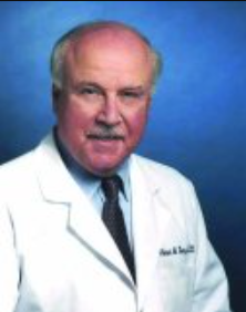 Michael A. Lemp合作论文数Schools of Medicine, George Washington University;Department of Ophthalmology, Georgetown University4
 Shigeru Kinoshita (木下茂)合作论文数Kyoto Prefectural University of Medicine4
 James Wolffsohn合作论文数School of Optometry, Aston University;University of Houston4Gary D. Novack合作论文数4
 Alan Tomlinson合作论文数Glasgow Caledonian University4
 Dimitri Azar合作论文数Department of Ophthalmology and Visual Sciences, College of Medicine, University of Illinois4
 Carlos Belmonte合作论文数Instituto de Neurociencias, Universidad Miguel Hernández de Elche4
 Fiona Jane Stapleton合作论文数School of Optometry and Vision Science, Faculty of Science, The University of New South Wales4Yoko Ogawa合作论文数4Yuichi Uchino合作论文数Ryogoku Eye Clinic4
 Saeed Akhtar合作论文数Faculty of Medicine, Kuwait University4
 Eric Gabison合作论文数Université Paris Cité;Banque Française des Yeux4
 JASON J. NICHOLS合作论文数School of Optometry, University of Alabama at Birmingham4John M Sparrow合作论文数3Catherine Willshire合作论文数3
 Gaffney A. Eamonn合作论文数Mathematical Institute, University of Oxford3Georgi As Georgiev合作论文数University of Sofia3
 Katherine Morland Hammitt合作论文数3
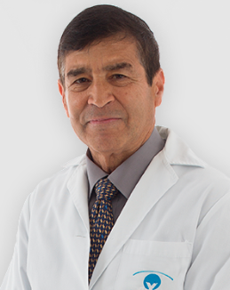 Neville N. Osborne合作论文数Department of Ophthalmology, University of Oxford;Fundación de Investigación Oftalmológica3
 José Álvaro Pereira Gomes合作论文数Departamento de Oftalmologia, Universidade Federal de São Paulo3
 Stefano Barabino合作论文数3Janine A. Smith合作论文数3Debra A. Schaumberg合作论文数3M B Abelson合作论文数2William Ayliffe合作论文数2Rieko Sakai合作论文数2Jose M. Benitez del Castillo合作论文数2chris chew合作论文数2Oliver Schein合作论文数Epidemiology, Bloomberg School of Public Health, Johns Hopkins University2Begley Carolyn G合作论文数2




