Objective: Neuropsychiatric symptoms (NPS) constitute a major challenge in Alzheimer disease (AD). We applied a component-based symptom paradigm by deriving Neuropsychiatric Inventory Questionnaire (NPI-Q) clusters and evaluating their longitudinal associations with regional brain volumes and functional outcomes (instrumental activities of daily living, activities of daily living [ADLs]). Methods: Participants with AD (N=111) were from the Ontario Neurodegenerative Disease Research Initiative. NPS were assessed using the NPI-Q. Symptom clusters were identified via principal components analysis at baseline. Magnetic resonance imaging-derived volumes for 34 cortical and 9 subcortical regions were obtained annually over 3 years. Longitudinal associations between NPS clusters and functional outcomes were examined using linear mixed-effects models adjusting for age, sex, Montreal Cognitive Assessment (MoCA), education, visit number, and cholinesterase inhibitor use. Results: Four clusters explained 62% of variance: hyperactivity (disinhibition, irritability, motor disturbance, agitation), psychosis (hallucinations, delusions, euphoria), neurovegetative (apathy, appetite), and affective (depression, anxiety, nighttime behavior). The hyperactivity cluster was associated with the left middle temporal (β=-0.24, P=.025) and right nucleus accumbens (β=-0.28, P=.007). The neurovegetative cluster was associated with the left middle temporal (β=-0.50, P<.001) and right nucleus accumbens (β=-0.55, P<.001). The affective cluster showed the strongest associations with the left rostral anterior cingulate (β=-0.42, P=.002) and right medial orbitofrontal cortex (β=-0.47, P=.001). All clusters predicted iADL outcomes; clusters 1, 3, and 4 also predicted ADL outcomes. Greater NPS burden, male sex, age, lower MoCA, and later visits predicted worse function. Conclusion: NPS in AD separate into hyperactivity, psychosis, neurovegetative, and affective clusters, supporting a cluster-based paradigm linking co-occurring behavioral symptoms with brain structure and functional decline.
We investigated the role of copy number variations (CNVs) in Parkinson's disease (PD) using genotyping data from 10,815 patients (2731 early-onset PD, EOPD) and 8901 controls from the COURAGE-PD consortium. CNVs were analyzed using a sliding window genome-wide association and burden approach. No genome-wide significant CNVs were detected in the overall cohort, but a robust deletion spanning exons 2-6 of PRKN was identified in EOPD cases, validated by MLPA, and replicated in the GP2 dataset (23,089 cases, 18,824 controls). CNV burden was significantly enriched in PD-related genes, primarily driven by PRKN, with the strongest effect observed in EOPD. PRKN CNV carriers showed earlier age at onset, confirmed by survival analysis. No association was observed for genome-wide or large CNV burden. Our findings reinforce the pivotal role of PRKN deletions in early-onset PD and highlight the need for high-resolution CNV analysis in large cohorts to uncover additional rare contributors to PD risk.
Parkinson's disease (PD) is increasingly recognized as a heterogeneous neurodegenerative entity with diverse clinical presentations, genetic contributors, and neuropathological features. Central to its pathogenesis is misfolded and aggregated α-synuclein, which collectively form Lewy pathology. Recent advances in biomarker and genetic research have enabled biologically grounded models of PD classification, diagnosis and staging. This review summarizes key principles, differences, and ongoing challenges of two emerging research frameworks: the SynNeurGe criteria and the Neuronal α-Synuclein Disease Integrated Staging System (NSD-ISS)-the former proposed a biologically based classification, while the latter proposed a more restrictive biological definition and staging schema. SynNeurGe incorporates synucleinopathy (S), neurodegeneration (N), genetic risk (G) and clinical status (C) to classify etiologic subtypes across the disease spectrum, emphasizing clinical heterogeneity and multifaceted underlying biological processes. In contrast, the NSD-ISS defines "neuronal α-synuclein disease" (NSD) based on specific molecular (S) and dopaminergic dysfunction (D) markers and a single genetic anchor (SNCA) (G), and maps disease progression across seven clinical stages. While both aim to improve early detection and to advance PD research, they differ in scope, operational definitions, implementation principles, and intended applications. Prevailing challenges include current limitations in mechanistic insights, biomarker standardization and accessibility, underrepresentation of genetic diversity, and ethical considerations around disease labeling and risk disclosure, particularly in asymptomatic cases. These frameworks represent a pivotal shift toward biologically based concepts of PD and related disorders, with future success contingent on continued refinement, validation, and equitable implementation.
BACKGROUND:Chorea-acanthocytosis (ChA) clinically presents with motor and non-motor symptoms. Some features of the disease and their intercorrelations have remained insufficiently characterized. Thus, we aimed to perform a multidimensional assessment of ChA patients, focusing on motor, behavioral, cognitive and olfactory domains. METHODS:A cross-sectional clinical assessment of 23 genetically confirmed ChA patients was conducted from 2022 to 2024. Patients were evaluated using the Unified Huntington's Disease Rating Scale (UHDRS) and Mini-Mental State Examination. A case-control comparison of olfactory function was also performed between eligible patients (n = 20) and age-and gender-matched controls (n = 20) using the Sniffin' Sticks test. RESULTS:The mean age and disease duration were 38.57 ± 7.26 years and 7.86 ± 6.04 years, respectively. Chorea (91.3%), dystonia (82.6%) and oculomotor abnormalities (78.3%), particularly impaired vertical saccades and smooth pursuit, were frequent. All patients exhibited at least one behavioral disorder, most commonly anxiety, mood disorders (including sad mood and low self-esteem) and obsessive-compulsive features. Suicidal thoughts were found in 21.7% of the patients. Among patients with preserved cognition, 50% demonstrated hyposmia (sum of odor identification, discrimination and threshold scores [TDI] < 30.75), with significantly reduced odor discrimination, identification and TDI scores versus controls. Disease duration was negatively associated with olfactory function. Higher UHDRS motor scores were associated with poorer olfactory, cognitive and functional outcomes. CONCLUSION:These findings demonstrated the varied manifestations of ChA, identifying olfactory impairment as a prevalent, underrecognized non-motor symptom, with potential implications for clinical evaluation and management. Further multicenter studies with larger cohorts are required to confirm these observations and clarify their prognostic value.
This Viewpoint reports on the potential of multimodal approaches that include validation, standardization, and ethical considerations in diagnosing neurodegenerative diseases, many of which have overlapping clinical phenotypes.
BACKGROUND AND OBJECTIVES:Plasma biomarkers of Alzheimer disease (AD), neuroinflammation, and neurodegeneration are increasingly being used in clinical trials for diagnosis and monitoring of dementia. However, their association with longitudinal structural brain MRI changes, an important outcome measure across neurodegenerative and cerebrovascular diseases, is less known. We investigated how baseline plasma biomarkers reflect MRI markers of progression over time in patients with neurodegenerative and cerebrovascular diseases. METHODS:This longitudinal cohort study included patients from the Ontario Neurodegenerative Disease Research Initiative diagnosed with AD or mild cognitive impairment (AD/MCI), Parkinson disease (PD), frontotemporal dementia spectrum disorders (FTD), or cerebrovascular disease (CVD), followed annually for 2 years. Recruitment took place at specialized university-based dementia, movement disorders, and/or stroke clinics in the province of ON, Canada. MRI outcomes included markers of cerebral atrophy (ventricular CSF and regional gray matter volumes) and of small vessel disease pathology (white matter hyperintensity [WMH], perivascular spaces, and lacunar volumes). Hemorrhagic markers at baseline were also included. Plasma levels of glial fibrillary acidic protein (GFAP), neurofilament light chain (NfL), phosphorylated tau181 and tau217 (p-tau181, p-tau217), and β-amyloid (Aβ42/40) were quantified from blood samples collected at baseline using Simoa and used as predictors in linear mixed models adjusted for time (months), age, sex, apolipoprotein E (APOE)-ε4 carrier status, kidney function, vascular risk factors, microtubule-associated protein tau (MAPT) diplotypes, waist-hip circumference ratio, and disease duration. RESULTS:We analyzed 1,240 MRIs from 473 patients (age: 69.2 ± 7.4 [range: 49-87]; 32.8% women). Elevated baseline levels of GFAP, NfL, p-tau181, and p-tau217, and to a lesser extent decreased levels of Aβ42/40, were significantly associated with more cerebral atrophy and WMH burden at baseline (|B| = 0.02 to 1.69, p = 0.044 to <0.001) and with progression over time (|B| = 0.001 to 0.028, p = 0.049 to <0.001) in the pooled disease-agnostic group. Within disease-specific cohorts, GFAP and NfL were associated with cerebral atrophy and/or small vessel disease copathology in AD/MCI, PD, FTD, or CVD. P-tau181 and p-tau217 were associated with cerebral atrophy and/or small vessel disease copathology in AD/MCI, CVD, PD-MCI, or PD-dementia. DISCUSSION:Selected plasma biomarkers seem useful as prognosis and monitoring tools of longitudinal imaging changes within real-world populations of neurodegenerative and/or cerebrovascular diseases, and provide insight into overlap across diseases in shared pathologic burden.
BACKGROUND:A satisfying sexual life is essential for maintaining good quality of life and healthy relationships for all individuals, including those with movement disorders and their partners. However, its significance is often overlooked in neurological clinics. METHODS:We review the literature and draw from our own experience to offer a practical approach for assessing and managing sexual health issues in patients with movement disorders. RESULTS:We begin by introducing fundamental concepts of sexual medicine, emphasizing models of sexual response that offer useful frameworks for diagnostic classification and management practices. We then address the most common sexual problems and disorders, including sexual dysfunction, sexual pain disorders, sexual compulsivity, and paraphilic behaviors, and their connections to different movement disorders. Next, we discuss assessment approaches that encompass biological, psychological, and social factors related to sexual health, including investigations for structural, vascular, or hormonal causes. Brief assessment questionnaires are also discussed. In addition to specific sexual therapeutic practices and supportive psychotherapy, we cover adjustments to pharmacological treatments related to movement disorders, as well as the use of mechanical devices. We also address the need for individualized management in specific cases, such as for patients undergoing deep brain stimulation surgery. CONCLUSION:Neurologists can play a key role in addressing sexual health concerns, considering the impact of both the disorder and its treatments. A comprehensive approach includes assessing biological, psychological, and sociocultural factors, with interventions like sexual therapy, supportive psychotherapy, pharmacological treatments and medical devices. Further research is needed to establish evidence-based treatments in this population.
Tics are repetitive and intrusive movements, including vocalizations, typically characterized by premonitory urge with associated relief.1 People with tic disorders have reported heightened sensitivity to sensations, including auditory stimuli, aggravating tics.2 Misophonia is a form of auditory hypersensitivity characterized by severe dislike of specific sounds with intense emotional responses.3 Although misophonia is a well-established phenomenon in autism spectrum disorders (ASD) and other neuropsychiatric disorders such as obsessive-compulsive disorder (OCD),3, 4 its interaction with tic disorders remains unacknowledged. We present 6 cases illustrating the clinical spectrum of misophonia-associated tics and their neuropsychiatric associations. The first case was an 18-year-old patient with chronic tic disorder (CTD), ASD, and OCD. He had motor and vocal tics, including coprolalia (the "F-word") triggered by specific sounds. Misophonia to swallowing and mouth sounds, especially from his mother, triggered intense distress and tics. Avoiding proximity to his mother while eating significantly decreased misophonia and tics. Similarly, the second case was a 32-year-old woman with CTD, ASD, and depression, with misophonia to chewing that worsened all tics, whereas the third case was a 26-year-old woman with OCD, notably without ASD, and chewing misophonia that triggered violent thoughts and facial and shoulder shrugging tics. The fourth case was a 14-year-old boy with CTD, ASD, and OCD. Despite tolerance to most noxious sounds, hearing the specific words "right" and "sorry" was so disturbing that rapid neck extension, aggressive head nodding, and trunk flexion tics occurred in intense bouts. Volume, tone, and word context were not modifying factors apart from greater impact with maternal origin of speech. The fifth case was a 27-year-old man with CTD, OCD, ASD, and chromosome 2p16.3 deletion. Motor tics began at age 11, later developing prominent misophonia and hyperacusis with most sounds perceived as painful, including speech and environmental sounds like air-conditioning humming (Video 1, Segment 1). He required almost-constant noise-canceling headphones, causing extreme social isolation and impairment. Audiology testing was unrevealing. Cognitive behavioral therapy mildly improved misophonia. The sixth case was a 19-year-old woman with a functional tic disorder. She had stereotyped repetitive shouting of phrases, including "who makes the noise?" and "stop it!," triggered by subtle background noises like breathing (including from examiners), squeaking chair noises, and door opening (Video 1, Segment 2). These cases illustrate the range and severity of misophonia in CTDs and their neuropsychiatric associations, as well as in functional tics. Misophonia can occur with any sound but often with oral or environmental sounds3 and can be a debilitating symptom, commonly associated with neurodevelopmental and neuropsychiatric conditions.3, 4 Indeed, patients with primary tic disorders here also had comorbid ASD or OCD. Distinguishing their contribution from CTDs itself is challenging, but awareness that misophonia may be a tic-modifying factor is prudent for both education and behavioral therapies. Importantly, most patients experienced misophonia that itself could trigger tics, whereas avoiding misophonia triggers resulted in the absence of or decreased tics. Abrupt emotional dysregulation may cause misophonia5; however, the pathophysiology remains unclear.4 Misophonia may also occur in functional neurological disorders (FND) (case 5), potentially with different underpinnings. Although misophonia lacks evidence-based treatments, behavioral management is preferred over neuropharmacology.3 The response of misophonia-associated tics to therapy beyond trigger avoidance is unclear; however, we guided patients for Comprehensive Behavioral Intervention for Tics (cases 1–4), behavior therapy (case 5), and FND programs (case 6). Recognition will inform clinical and pathophysiological research to develop targeted interventions for this underexplored symptom. (1) Research project: A. Conception, B. Organization, C. Execution; (2) Analysis: A. Design, B. Execution, C. Review and critique; (3) Manuscript: A. Writing of the first draft, B. Review and critique. A.B.: 1C, 2A, 2B, 3A T.C.G.: 1A, 2B, 2C, 3A, 3B W.K.L.: 1A, 1B, 2C, 3A, 3B I.A.M.: 1A, 1B, 2C, 3B A.E.L.: 1A, 1B, 2C, 3B C.G.: 1A, 1B, 2C, 3B Ethical Compliance Statement: We confirm that we have read the journal's position on issues involved in ethical publication and affirm that this work is consistent with those guidelines. The authors confirm that the approval of the University Health Network Research Ethics Board was obtained for this work. Informed consent was obtained for all cases in this case report. Funding Sources and Conflicts of Interest: No specific funding was received for this work. The authors declare that there are no conflicts of interest relevant to this work. Financial Disclosures for the Previous 12 Months: Arjun Balachandar, Talyta Cortez Grippe, Wei Kang Lim, Irene A. Malaty, Anthony E. Lang, and Christos Ganos report no relevant funding sources in the past 12 months. The data that support the findings of this study are available from the corresponding author upon reasonable request.
Progressive supranuclear palsy (PSP) is a neurodegenerative disorder characterized by four-repeat (4R) tau protein deposition. The substantia nigra (SN) and midbrain tegmentum nuclei (MBT) are consistently affected. Lymphocyte infiltrates are scarce in the brains of patients with neurodegenerative diseases, although a few reports have described their presence in the α-synucleinopathy Parkinson's disease (PD). To evaluate the cytotoxic T-cell response, serial sections spanning 120 μm of the SN were immunostained consecutively for phosphorylated tau (p-tau, AT8) or α-synuclein, cytotoxic T-cell marker and microglia marker HLA-DR. Sections were analysed with stereology software in 9 patients with PSP, 10 with PD and 6 healthy controls. We semiquantitatively scored CD8-positive cells in further brain regions. CD8 lymphocyte cell counts and microglial activation in the SN were higher in PSP than PD and controls. Furthermore, T-cell/neuron contact was observed in PSP. In multivariate models, CD8 counts were not predicted by disease duration, younger age at death or the amount of p-tau pathology. The SN and midbrain tegmentum showed more CD8 cells than the cortex. A more prominent nigral cytotoxic T-cell response in PSP than PD supports the suggestion that p-tau neuropathology in PSP might have potential relationships with autoimmune mechanisms.
The ordered assembly of α-synuclein protein encoded by SNCA into filaments characterizes neurodegenerative synucleinopathies. Lewy body disease (LBD) shows predominantly neuronal and multiple system atrophy (MSA), predominantly oligodendrocytic α-synuclein pathology affecting subcortical brain structures. Based on cryo-electron microscopy, it was reported that the structures of α-synuclein filaments from LBD differ from MSA and juvenile-onset synucleinopathy (JOS). The rare atypical MSA subtype shows abundant neuronal argyrophilic α-synuclein inclusions in the limbic system. Current concepts indicate that disease entities are characterized by unique protofilament folds. Here we demonstrate that α-synuclein can form a Lewy-MSA hybrid fold, leading to the atypical histopathological form of MSA. Distinct biochemical characteristics of α-synuclein, as demonstrated by protease-sensitivity digestion assay, seed amplification assays (SAAs), and conformational stability assays (CSA), are also linked to cytopathological differences. We expand the current structure-based classification of α-synucleinopathies and propose that cell-specific protein pathologies can be associated with distinct filament folds.
Vascular risk factors are common in older adults and contribute to brain damage, can manifest as increased white matter hyperintensities (WMH), and associated with future risk of stroke and dementia. However, their prevalence, effect across different neurodegenerative diseases, and association with WMH remains underexplored. To investigate the association between vascular risk burden, and brain white matter integrity, across five neurodegenerative conditions. Cross-sectional study including 520 participants from the Ontario Neurodegenerative Disease Research Initiative (ONDRI) cohorts: 126 with amnestic Mild Cognitive Impairment/Alzheimer’s Disease (MCI/AD), 53 with Frontotemporal Dementia (FTD), 161 with Cerebrovascular Disease (CVD), 140 with Parkinson’s Disease (PD), and 40 with Amyotrophic Lateral Sclerosis (ALS), along with 41 cognitively healthy controls. A vascular risk index (VRI, range 0–5) assessed hypertension, diabetes, dyslipidemia, obesity (BMI ≥ 30), and smoking history. Macro (WMH volume) and micro (Diffusion tensor imaging) white matter integrity were evaluated using 3-Tesla MRI. Associations were analyzed using multinomial logistic regression and ANCOVA, adjusting for age, sex, education, and APOE ε4 allele status. Vascular risk factors, particularly hypertension and hypercholesterolemia, were more prevalent in the disease cohorts than controls. A higher VRI was significantly associated with MCI/AD (1.5-fold, p = 0.05), FTD (1.7-fold, p =0 .02), and CVD (2.6-fold, p < 0.005) cohorts. High VRI was associated with reduced macro and microstructural white matter integrity in the pooled sample (macro: p = 0.005; micro: p = 0.003), and separately in CVD (macro: p = 0.04; micro: p = 0.002). APOE ε4 status only mildly attenuated these associations. Vascular risk burden is prevalent in neurocognitive syndromes including MCI/AD, FTD and CVD, and impacts white matter integrity. Future studies are needed to explore if vascular risk management may mitigate the consequences of neurodegeneration in these clinical groups.
Protein misfolding and aggregation is a major pathological hallmark in a variety of human conditions, including cancer, diabetes, and neurodegeneration. However, we still do not fully understand the role of protein accumulation in disease. Interestingly, recent breakthroughs in artificial intelligence (AI) are having a tremendous impact on our ability to predict three-dimensional protein structures and understand the molecular rules governing protein folding/misfolding. This progress will enable us to understand how intrinsic and extrinsic factors trigger protein misfolding, thereby changing protein function. These changes, in some cases, are related to normal biological responses and, in other cases, associated with pathological alterations, such as those found in many neurodegenerative disorders. Here, we provide a brief historical perspective of how findings in the field of prion diseases and prion biology have enabled tremendous advances that are now forming the basis for our understanding of disease processes and discuss how this knowledge is now emerging as central for our ability to classify, diagnose, and treat devastating neurodegenerative disorders such as Parkinson’s and Alzheimer’s diseases.
Language impairment is common in progressive supranuclear palsy (PSP) and is often overlooked due to the severity of the motor symptoms. We investigated whether language can be used to predict PSP prognosis. One hundred-forty-six patients with a diagnosis of possible or probable PSP from the Tilavonemab (ABBV-8E12) clinical trial were evaluated at baseline and week 32 using the Repeatable Battery for the Assessment of Neuropsychological Status (RBANS), the PSP rating scale (PSPRS) and the Schwab and England Activities of Daily Living Scale (SEADL). Percentage of change was calculated for each measure. Using correlations, we evaluated relationships between all RBANS-subscores (language, attention, memory and visuoconstructional), the PSPRS and SEADL scores at baseline; p-values were FDR corrected. Linear regression analyses were performed between the RBANS-language, RBANS-delayed-memory and RBANS-executive at baseline and the percentage of change over time. Grey matter volumes were extracted from three regions of interest based on language (bilateral temporal poles and inferior frontal gyri), executive (bilateral dorsolateral and superior frontal gyri and frontal pole) and memory (bilateral hippocampus, inferior, middle and superior temporal gyri) areas. Mean age of PSP patients: 68.8 years (49-86 years), 57 (39%) females. The RBANS language, executive and delayed memory scores at baseline were positively correlated with each other and with visuoconstructional and immediate memory score (all p<0.05). Only the RBANS-language score at baseline predicted percentage of change in PSPRS (B = -0.63, p = 0.003). The percentage of change in the RBANS-language score was predicted by the RBANS-language at baseline (B = -0.59, p<0.001). Lower age at baseline was associated with a worsening in language score over time (B = 0.51, p = 0.009). Language grey matter volume was associated with the change in RBANS-language score (B = 0.01, p = 0.02). language impairment at baseline, in contrast to memory and executive functions, was predictive of functional decline as measured by PSPRS. Atrophy in language areas at baseline predicted language decline. Language impairment may be an independent prognostic factor in PSP. *Based on research using data from AbbVie that has been made available through Vivli, Inc. Vivli has not contributed to or approved, and is not in any way responsible for, the contents of this publication.
BACKGROUND:Tremor is a hyperkinetic movement disorder defined as a rhythmic, involuntary, oscillatory movement of a body part. It can present as early as in the neonatal period and may be an isolated finding or part of an associated neurological or systemic disorder. Although it is commonly seen in childhood, it is not frequently described in the literature. OBJECTIVES:This review aims to provide a clinically useful overview of pediatric tremors based on the current literature. METHODS:We identified relevant studies published in English by searching PubMed (until February 2024), using the following subject headings: pediatric/tremor, deep brain stimulation, and focused ultrasound. RESULTS:Data from 65 articles were critically reviewed with focus on tremor classification, etiologies, clinical features, and management. CONCLUSION:Some tremor disorders in children can persist into adulthood, however, others are exclusively seen in the pediatric population. The lack of epidemiological and clinical studies in the pediatric population creates substantial challenges for pediatricians, and medical decisions are mostly guided by adult data.
Mild Behavioral Impairment (MBI) is a condition characterized by neuropsychiatric symptoms (NPS) in older adults without dementia, serving as a precursor to various forms of dementia. This study explores the association between NPS and functional connectivity (FC) within the default mode network (DMN), executive control network (ECN), and salience network (SN) across three high-risk cohorts: mild cognitive impairment (due to Alzheimer’s) (MCI, n = 79), cerebrovascular disease (CVD, n = 144), and Parkinson’s disease (PD, n = 132). A total of 367 participants were recruited from the Ontario Neurodegenerative Disease Research Initiative (ONDRI). The assessment of NPS utilized the Neuropsychiatric Inventory Questionnaire (NPI-Q), with symptom severity rated on a scale from 1 to 3 (mild, moderate, severe). Resting-state FC was analyzed for the DMN, ECN, and SN, using dual regression analysis to generate subject-specific whole-brain FC maps for each network. The association between FC maps and NPS scores was examined using FSL’s randomise with 5,000 permutations, while controlling for age, sex, and education. Results are presented following cluster False Discovery Rate (FDR) correction. The study revealed significant associations between NPS and FC specific to each cohort. In the MCI group, disturbed appetite and nighttime behaviors were correlated with increased FC of the dorsal DMN (p<0.05, R = 0.47, and p = 0.01, R = 0.47). The CVD group exhibited correlations between higher levels of anxiety and decreased FC of the dorsal DMN (p<0.05, R = -0.4), ventral DMN (p<0.05, R = -0.33), and bilateral ECN (p<0.05, R = -0.35 and R = -0.33). The PD group showed disturbed nighttime behavior associated with increased FC in ventral DMN (p<0.05, R = 0.35) and bilateral ECN (p<0.05, R = 0.43 and R = 0.37). This research underscores disorder-specific correlations between specific NPS domains and FC in MCI, CVD, and PD, emphasizing the unique neural underpinnings of symptomatology in each group. Furthermore, it is essential to note the inherent heterogeneity in all groups. Overall, the pathological substrates of neurodegenerative disorders likely play a pivotal role in shaping the neural correlates of MBI within each disorder. These findings provide valuable insights into targeted interventions and avenues for future research in neurodegenerative disorders.
Progressive supranuclear palsy (PSP) is a main form of idiopathic tauopathy characterized neuropathologically by subcortical neurofibrillary tangles in neurons, oligodendroglial coiled bodies, and tufted astrocytes, which follow sequential distribution in the human brain. Mitochondrial dysfunction is thought to be a contributor to many neurodegenerative diseases, but its role in PSP at the cellular level remains incompletely understood. To address this, we performed cell-specific morphometric analysis of mitochondrial markers in post-mortem tissues from motor cortex of PSP patients and non-diseased controls (n = 5 each) followed by single-nuclear transcriptomics (n = 3 each) to identify changes in genes that regulate mitochondrial function. We treated iCell astrocytes with PSP brain homogenates and isolated viable astrocytes from multiple regions of PSP-affected brains. We found that PSP is characterized by significant mitochondrial changes in neurons and astrocytes at the immunohistochemical level, particularly in complex I, with distinct transcriptomic responses across cell types. Glial cells exhibited upregulation of pathways associated with mitochondrial function. In contrast, excitatory and inhibitory neurons showed downregulation in these pathways, indicating impaired mitochondrial function. Astrocytes derived from different human brain regions express varied levels of GFAP and EAAT1 immunoreactivity. Astrocytic tau pathology in cell culture derived from postmortem PSP brains mirrors that seen in corresponding brain tissue histology. Tau pathology in human astrocyte cell culture is associated with clumps of mitochondria potentially associated with impairment in their neuron supportive function. Our results underscore selective complex I damage and cell-type specific patterns that differentiate PSP from other neurodegenerative diseases.
The sudden death on October 25, 2024 of C. Warren Olanow produced an outpouring of grief by his many friends and colleagues in the International Parkinson and Movement Disorder Society (MDS), and the movement disorders world in general. Accompanying the sadness, however, was a sense of gratitude from those whose personal and professional lives he touched and selflessly supported. Many of Warren's academic achievements are described in detail in the accompanying obituary,1 in this issue of the Journal, from his longstanding close friends and collaborators José Obeso, Anthony Schapira, and Fabrizio Stocchi. After attending medical school at the University of Toronto, he completed his neurology residency at the New York Neurological Institute at Columbia University. Warren's interest in Parkinson's disease (PD) was influenced by Melvin Yahr and Roger Duvoisin, as well as Stanley Fahn, who had just returned to Columbia around that time. After further postdoctoral work on the basal ganglia at Columbia with the leading neuroanatomist, Malcolm Carpenter, Warren secured his first faculty position at McGill University and the Montreal Neurological Institute, where he taught the neuroanatomy course and set up a Parkinson's clinic.2 He was then recruited to Duke University as Head of Clinical Neurology. At first, his major interests were in both myasthenia gravis and PD, but he settled on a career focusing on movement disorders. Collaborative work with Burton Drayer, who discovered that excessive brain iron led to characteristic magnetic resonance imaging changes, stirred an interest in laboratory science and exploring the pathophysiology of disease. He went on to pursue this as Professor of Neurology at the University of South Florida, where he helped pioneer a program in cell transplantation into the basal ganglia as therapy for PD. In 1994, he became Chairman of the Department of Neurology at the Mount Sinai School of Medicine in New York, where he remained until his retirement from academic medicine, and was then appointed Emeritus Professor in both the Departments of Neurology and Neuroscience. Even in academic retirement, he appeared on the 2024 Clarivate list of Highly Cited Researchers. The Movement Disorder Society (MDS) was created in 1992 from the formal merger of the International Medical Society for Motor Disturbances (ISMD) and MODIS (the 'original' Movement Disorder Society), of which Stanley Fahn was the founding President (1988–1991). Warren was the last President of the ISMD, from 1993 to 1994, and concurrently the first Treasurer of the newly merged MDS, working with his friend and colleague, C. David Marsden (deceased), who was its inaugural President from 1991 to 1994. As Treasurer, Warren played a crucial role in navigating the financial aspects of the merger of the two societies, and thus played an integral role in the birth of the MDS.3 Warren became the 7th President of the MDS, a position he held from 2003 to 2004 (Fig. 1). He played a prominent role in several key milestones in the evolution of the Society, including the change from biannual to annual International Congresses from 2004, the deliberate pivot towards publishing basic, translational, and clinical science as Co-Editor with Jose Obeso of the Movement Disorder Journal from 2010 to 2014, and in the establishment of its companion journal, Movement Disorders Clinical Practice, in 2014. For his contributions to the MDS, he received the President's Distinguished Service Award in 2007 and Honorary Membership Award in 2015. Warren was also honored with board positions or awards from many other societies and organizations, including being a Past Treasurer of the American Neurological Association, an Honorary Member of the French Neurological Society, an Honorary Fellow of the Royal College of Physicians (UK), an Honorary Professor at the University of London (Royal Free Hospital), and a recipient of the Movement Disorder Research Award from the American Academy of Neurology. He served on numerous medical, scientific advisory, and editorial boards including being a founding member of the Parkinson Study Group. Warren was close friends and colleagues with, and mentored and supported, a large number of the organizational and scientific leadership (Data S1) and members of the MDS, too many to mention comprehensively. His involvement with the birth of the MDS required him to work closely with early Presidents such as Joe Jankovic and Mark Hallett. He actively encouraged and supported future Presidents such as Anthony Lang, Philip Thompson, Matthew Stern, Christopher Goetz, and David Burn early in their careers, without any anticipated benefit to himself. He nurtured the careers of MDS Journal Editors such as Jon Stoessl, Kailash Bhatia, and Marcelo Merello, and leading clinician scientists like Karl Kieburtz and Jeff Kordower. A couple of anecdotes from his interactions with Jeff help to exemplify Warren's qualities as a mentor and person. As an unknown postdoc at the University of Rochester, Jeff was encouraged by Ira Shoulson to speak to Warren who was visiting to deliver Grand Rounds. Jeff introduced himself and told Warren that he did PD research; Warren spontaneously prioritized time for a meeting, leading to a lifetime of collaboration. Many years later, when they were already longstanding friends and colleagues, at a brief encounter with Jeff's mother, Warren took care to use the moment to say: "Mrs. Kordower it is wonderful to meet you; you must be so proud of your son", sensing how much that would mean to her. Warren was known for his keen intellect and incisive thinking, that was enabled by his extensive clinical and scientific knowledge. He was also known for his astute financial and negotiating skills. He was a hard taskmaster, expecting those around him to pursue excellence and achievement rigorously, with the same drive as himself. He was always willing to listen, and if he disagreed but thought your argument was reasonable (even if wrong), enjoyed a debate. He did not suffer fools gladly, but once you gained his respect, even if you had a different viewpoint from him, you became a friend, both as a sparring, but also likely a dining, partner. Warren was known for his passion not just for science, but for life, especially as a connoisseur of travel, food, and wine. He had a keen sense of humor and the absurd, and a unique grin and method of laughter. At times, his grin would appear before his punchline. Warren presented himself as a warm, big-hearted, and inclusive leader. He was careful, however, in choosing his close friendships, and these were strong and few because of his intense fidelity to his inner circle. Many of us cherish fond memories of the warm and generous hospitality extended by Warren and Mariana at their home in Rye, NY during visits to New York conducting MDS business. Nowhere was his nurturing nature, loyalty, and dedication greater displayed than in his lifelong devotion to his wife Mariana, and their children and families. He spoke of them to his friends frequently and with love, and the void left by the death of Mariana in 2019 was never filled and his sadness never erased. Warren is survived by his children Edward, James, Alessandra, and Andrew, and his six grandchildren. Warren continued his passion for life and movement disorders right up until his unexpected death. He leaves us with the lives of patients, clinicians, researchers, and the MDS immeasurably richer for his contributions. Our guess is that Warren would appreciate ending his obituary with a positive message from him, his advice to those contemplating a career in movement disorders: "Go into the field, this is a great field, and I would really encourage you to go in with your heart, and to go in with passion…".2 Data sharing is not applicable to this article as no new data were created or analyzed in this study. Data S1. Warren Olanow Individual Reminiscences.docx. Please note: The publisher is not responsible for the content or functionality of any supporting information supplied by the authors. Any queries (other than missing content) should be directed to the corresponding author for the article.
 Andres Lozano合作论文数Department of Surgery, Temerty Faculty of Medicine, University of Toronto189
 Connie Marras合作论文数Institute of Health Policy, Management and Evaluation, University of Toronto;Department of Neurology, University of Toronto138
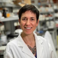 Carmela Tartaglia合作论文数Tanz Centre for Research in Neurodegenerative Diseases, Temerty Faculty of Medicine, University of Toronto89
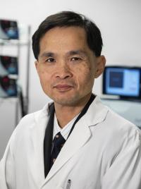 Robert Chen合作论文数Institute of Medical Sciences, Temerty Faculty of Medicine, University of Toronto86
 Ekaterina Rogaeva合作论文数Department of Neurology, Faculty of Medicine, University of Toronto;Tanz Centre for Research in Neurodegenerative Disease, University of Toronto83
 Alberto J. Espay合作论文数College of Medicine, University of Cincinnati81
 Mario Masellis合作论文数68
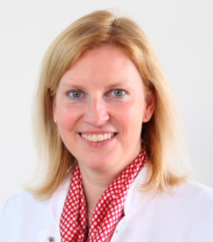 Christine Klein合作论文数Section of Clinical and Molecular Neurogenetics, Department of Neurology, University of Lubeck;Institute of Neurogenetics, University of Lubeck62
 Alfonso Fasano合作论文数Institute of Medical Science, Temerty Faculty of Medicine, University of Toronto;Krembil Research Institute;Department of Medicine, Temerty Faculty of Medicine, University of Toronto59
 Antonio P. Strafella合作论文数Krembil Research Institute, University Health Network;Division of Neurology, Department of Medicine, University of Toronto;Institute of Medical Science, University of Toronto;Research Imaging Centre, Centre for Addiction & Mental Health59
 Sandra Black合作论文数Institute of Medical Science, Temerty Faculty of Medicine, University of Toronto;Sunnybrook Health Science Centre58
 David France Tang-Wai合作论文数Division of the Vice-President and Provost, University of Toronto;Krembil Research Institute, University Health Network52
 William D. Hutchison合作论文数Division of Neurosurgery, Department of Surgery, Temerty Faculty of Medicine, University of Toronto;Toronto Western Research Institute49
 Gabor Geza Kovacs合作论文数Department of Laboratory Medicine & Pathobiology, Temerty Faculty of Medicine, University of Toronto48
 Jonathan Dostrovsky合作论文数Department of Physiology, Temerty Faculty of Medicine, University of Toronto;Department of Oral Physiology, Faculty of Dentistry, University of Toronto48
 Susan Fox合作论文数Division of Neurology, Department of Medicine, Temerty Faculty of Medicine, University of Toronto;Toronto Western Hospital46
 Irene Litvan合作论文数Department of Neurosciences, University of California, San Diego;Parkinson and Other Movement Disorder Center, University of California, San Diego45Fox Susan H合作论文数42
 Janis M Miyasaki合作论文数From the Department of Neurology (M.N.),, Icahn School of Medicine at Mount Sinai;Department of Neurology (J. McFarlin),, University of Kentucky;Medical Center, University of Rochester;Medical Center, University of Rochester;Department of Medicine (J. Miyasaki),, University of Alberta;Department of Neurology, Rush University;and Department of Internal Medicine (N.M.K.),, Rush University;Rush University, Rush University41
 Richard Swartz合作论文数Institute of Medical Science, Temerty Faculty of Medicine, University of Toronto;Division of Neurology, Temerty Faculty of Medicine, University of Toronto;Sunnybrook Health Sciences Centre41
 Werner Poewe合作论文数Department of Neurology, Medical University Innsbruck40
 Naomi P. Visanji合作论文数Toronto Western Research Institute40E. Moro合作论文数37
 Mestre Tiago A合作论文数Neurology Department, Hospital of Santa Maria37
 Morris Freedman合作论文数Department of Medicine, Temerty Faculty of Medicine, University of Toronto36Grimes D A合作论文数36
 Joseph Jankovic合作论文数Parkinson's Disease Center and Movement Disorders Clinic, Baylor College of Medicine;Department of Neurology, Baylor College of Medicine34
 Caroline Tanner合作论文数School of Medicine, University of California, San Francisco;University of California San Francisco Health33Elizabeth Finger合作论文数Univ Western Ontario, Dept Clin Neurol Sci, London, ON, Canada32
 Renato Munhoz合作论文数Division of Neurology, Department of Medicine, University of Toronto;Krembil Research Institute;Neuromodulation Unit and Ataxia Clinic, Toronto Western Hospital, University Health Network;Division of Neurology, Toronto Western Hospital, University Health Network32Brian Tan合作论文数31
 Valerie Voon合作论文数Department of Psychiatry, School of Clinical Medicine, University of Cambridge;Fudan University31Sean P. Symons合作论文数Sunnybrook Health Sciences Centre, University of Toronto31
 Paula McLaughlin合作论文数Nova Scotia Hlth30
 Shen-Yang Lim合作论文数Department of Medicine, Faculty of Medicine, University of Malaya30
 Joel Ramirez合作论文数Sunnybrook Health Sciences Centre30
 Ronald B Postuma合作论文数Department of Neurology and Neurosurgery, Faculty of Medicine and Health Sciences, McGill University;Montreal Neurological Institute, McGill University;McGill University Health Centre29
 Thomas Gasser合作论文数Center of Neurology, Hertie Institute for Clinical Brain Research, University of Tübingen29Malcolm A. Binns合作论文数29
 Donna Kwan合作论文数Ctr Neurosci Studies, Queens Univ28
 Eduard Tolosa合作论文数Department of Neurology, Faculty of Medicine, University of Barcelona;Parkinson Disease and Movement Disorder Research Unit, University of Barcelona Hospital28
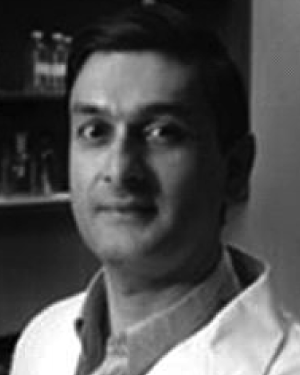 Mandar Jog合作论文数Clinical Neurological Sciences, University of Western Ontario Health Centre–University Hospital28
 David Breen合作论文数Centre for Clinical Brain Sciences, Edinburgh Medical School,The University of Edinburgh27Sanjeev Kumar合作论文数Institute of Medical Science, Temerty Faculty of Medicine, University of Toronto27
 Robert Bartha合作论文数Robarts Research Institute27
 Andrew Lees合作论文数Reta Lila Weston Institute of Neurological Studies, University College London;National Hospital for Neurology and Neurosurgery27Elena Moro合作论文数27Jean-Christophe Corvol合作论文数26
 Alexis Brice合作论文数Institut du Cerveau, Hôpital Pitié-Salpêtrière à Paris;Institut Hospitalo-Universitaire-A-ICM à l’Hôpital Pitié-Salpêtrière à Paris26




