Deposits of hydroxyapatite called Randall's plaques are found in the renal papilla of calcium oxalate kidney stone formers and likely serve as the nidus for stone formation, but their pathogenesis is unknown. Claudin-2 is a paracellular ion channel that mediates calcium reabsorption in the renal proximal tubule. To investigate the role of renal claudin-2, we generated kidney tubule-specific claudin-2 conditional KO mice (KS-Cldn2 KO). KS-Cldn2 KO mice exhibited transient hypercalciuria in early life. Normalization of urine calcium was accompanied by a compensatory increase in expression and function of renal tubule calcium transporters, including in the thick ascending limb. Despite normocalciuria, KS-Cldn2 KO mice developed papillary hydroxyapatite deposits, beginning at 6 months of age, that resembled Randall's plaques and tubule plugs. Bulk chemical tissue analysis and laser ablation-inductively coupled plasma mass spectrometry revealed a gradient of intrarenal calcium concentration along the corticomedullary axis in normal mice that was accentuated in KS-Cldn2 KO mice. Our findings provide evidence for the "vas washdown" hypothesis for Randall's plaque formation and identify the corticomedullary calcium gradient as a potential target for therapies to prevent kidney stone disease.
The goal of this retrospective study was to determine whether having added tiletamine–zolazepam to an anesthetic cocktail of ketamine and xylazine (KX) during an ongoing series of studies of renal function in domestic pigs changed baseline renal hemodynamics. Group A (10 pigs) had been anesthetized with KX, group B (25 pigs) was anesthetized with tiletamine–zolazepam combined with KX (TKX), and group C (10 pigs) was anesthetized with KX. Measurements of baseline glomerular filtration rate (GFR; inulin clearance), effective renal plasma flow (eRPF; para-aminohippuric acid clearance), and mean blood pressure (BP) were made during three 15-min urine collection periods. GFR and eRPF were lower in group B (TKX) than in groups A and C (KX only) by 34% to 40% and 39% to 49%, respectively. BP did not differ between the 3 groups. GFR and eRPF in groups A and C were not different from each other. These findings suggest that adding tiletamine–zolazepam to an anesthetic cocktail can cause an acute decline in GFR and eRPF independent of arterial BP in laboratory swine.
Introduction: About 1 in 11 Americans will experience a kidney stone, but underlying causes remain obscure. The objective of the present study was to separate idiopathic calcium oxalate stone formers by whether or not they showed positive evidence of forming a stone on Randall's plaque (RP).Materials and Methods: In patients undergoing either percutaneous or ureteroscopic procedures for kidney stone removal, all stone material was extracted and analyzed using micro-CT imaging to identify those attached to RP. Twenty-four-hour urine samples were collected weeks after the stone removal procedure and patients were off of medications that would affect urine composition. The endoscopic video was analyzed for papillary pathology (RP, pitting, plugging, dilated ducts, and loss of papillary shape) by an observer blinded to the data on stone type. The percent papillary area occupied by RP and ductal plugging was quantified using image analysis software.Results: Patients having even one stone on RP (N = 36) did not differ from non-RP patients (N = 37) in age, sex, BMI, or other clinical characteristics. Compared with the non-RP group, RP stone formers had more numerous, but smaller, stones, more abundant papillary RP formation, and fewer ductal plugs, both by quantitative measurement of surface area (on average, three times more plaque area, but only 41% as much plug area as in non-RP patients) and by semiquantitative visual grading. Serum and blood values did not differ between RP and non-RP stone formers by any measure.Conclusions: Growth of many small stones on plaque seems the pathogenetic scheme for the RP stone-forming phenotype, whereas the non-RP phenotype stone pathogenesis pathway is less obvious. Higher papillary plugging in non-RP patients suggests that plugs play a role in stone formation and that these patients have a greater degree of papillary damage. Underlying mechanisms that create these distinctive phenotypes are presently unknown.
Chronic kidney disease (CKD) leads to musculoskeletal impairments that are impacted by muscle metabolism. We tested the hypothesis that 10-weeks of voluntary wheel running can improve skeletal muscle mitochondria activity and function in a rat model of CKD. Groups included (n = 12–14/group): (1) normal littermates (NL); (2) CKD, and; (3) CKD-10 weeks of voluntary wheel running (CKD-W). At 35-weeks old the following assays were performed in the soleus and extensor digitorum longus (EDL): targeted metabolomics, mitochondrial respiration, and protein expression. Amino acid-related compounds were reduced in CKD muscle and not restored by physical activity. Mitochondrial respiration in the CKD soleus was increased compared to NL, but not impacted by physical activity. The EDL respiration was not different between NL and CKD, but increased in CKD-wheel rats compared to CKD and NL groups. Our results demonstrate that the soleus may be more susceptible to CKD-induced changes of mitochondrial complex content and respiration, while in the EDL, these alterations were in response the physiological load induced by mild physical activity. Future studies should focus on therapies to improve mitochondrial function in both types of muscle to determine if such treatments can improve the ability to adapt to physical activity in CKD.
You have accessJournal of UrologyStone Disease: Basic Research & Pathophysiology (MP07)1 Sep 2021MP07-15 CALCIUM OXALATE STONE FORMERS WHO MAKE EVEN ONE STONE ON RANDALL'S PLAQUE HAVE SMALLER STONE BURDENS AND DISTINCTIVE PAPILLARY MINERAL PATHOLOGY James Williams, Haider Al-Awadi, Manognya Muthenini, Sharon Bledsoe, Tarek El-Achkar, Andrew Evan, Fredric Coe, Elaine Worcester, and James Lingeman James WilliamsJames Williams More articles by this author , Haider Al-AwadiHaider Al-Awadi More articles by this author , Manognya MutheniniManognya Muthenini More articles by this author , Sharon BledsoeSharon Bledsoe More articles by this author , Tarek El-AchkarTarek El-Achkar More articles by this author , Andrew EvanAndrew Evan More articles by this author , Fredric CoeFredric Coe More articles by this author , Elaine WorcesterElaine Worcester More articles by this author , and James LingemanJames Lingeman More articles by this author View All Author Informationhttps://doi.org/10.1097/JU.0000000000001980.15AboutPDF ToolsAdd to favoritesDownload CitationsTrack CitationsPermissionsReprints ShareFacebookLinked InTwitterEmail Abstract INTRODUCTION AND OBJECTIVE: The purpose of this study was to compare idiopathic calcium oxalate stone formers with (RP) and without (Non-RP) positive evidence for formation of stones on Randall’s (interstitial) plaque. METHODS: Patients undergoing stone removal by percutaneous (PNL) or ureteroscopic (URS) endoscopy were consented for study. Included patients had stone analysis of >50% calcium oxalate, with no brushite, medullary sponge, bariatric surgery, primary hyperoxaluria or hyperparathyroidism. all stone material was imaged by micro CT and stones growing on Randall’s plaque were identified by adherent regions of apatite that showed characteristics of calcified tissue (including lumens of vessels/tubules in 76% of the cases). Endoscopic papillary appearance was graded and measured on endoscopic video by an investigator blinded to the patient classification. RESULTS: 32 RP patients had on average 5±6 stones on Randall’s plaque, with none in the 42 Non-RP patients. The RP group was younger (48±17 v 56±14, p=0.04), had more stones (8±8 v 5±4 stones/kidney, p=0.02), less total stone removed (80±120 v 480±1200 cubic mm/kidney), and smaller stones (160±230 v 560±1200 cubic mm per stone, p=0.01) than did Non-RP patients, and RP were more likely to have undergone URS. RP had on average 240% more Randall's plaque area (5.2±4.3 v 1.5±1.7 as percent of papilla area, p <0.0001) and 77% less ductal plugging area (0.17±0.38 v 0.73±0.77, p <0.0001). In semi-quantitative scores, RP had more plaque and pitting, less plugging/dilated ducts, and less loss of papillary contour (figure 1). 24-hour urine values did not differ between RP and Non-RP on any measure. CONCLUSIONS: Calcium oxalate stone formers who showed positive evidence of having even a single stone on plaque had small stone burdens and were more likely to have undergone URS. Their renal papillae showed much more plaque and much less ductal plugging than the Non-RP group. These data support RP as a distinct phenotype among calcium oxalate stone formers. Source of Funding: NIH P01 DK056788; NIH R01 DK124776 © 2021 by American Urological Association Education and Research, Inc.FiguresReferencesRelatedDetails Volume 206Issue Supplement 3September 2021Page: e145-e145 Advertisement Copyright & Permissions© 2021 by American Urological Association Education and Research, Inc.MetricsAuthor Information James Williams More articles by this author Haider Al-Awadi More articles by this author Manognya Muthenini More articles by this author Sharon Bledsoe More articles by this author Tarek El-Achkar More articles by this author Andrew Evan More articles by this author Fredric Coe More articles by this author Elaine Worcester More articles by this author James Lingeman More articles by this author Expand All Advertisement Loading ...
Kidney stones frequently develop as an overgrowth on Randall's plaque (RP) which is formed in the papillary interstitium. The organic composition of RP is distinct from stone matrix in that RP contains fibrillar collagen; RP in tissue has also been shown to have two proteins that are also found in stones, but otherwise the molecular constituents of RP are unstudied. We hypothesized that RP contains unique organic molecules that can be differentiated from the stone overgrowth by fluorescence. To test this, we used micro-CT-guided polishing to expose the interior of kidney stones for multimodal imaging with multiphoton, confocal and infrared microscopy. We detected a blue autofluorescence signature unique to RP, the specificity of which was also confirmed in papillary tissue from patients with stone disease. High-resolution mineral mapping of the stone also showed a transition from the apatite within RP to the calcium oxalate in the overgrowth, demonstrating the molecular and spatial transition from the tissue to the urine. This work provides a systematic and practical approach to uncover specific fluorescence signatures which correlate with mineral type, verifies previous observations regarding mineral overgrowth onto RP and identifies a novel autofluorescence signature of RP demonstrating RP's unique molecular composition.
Introduction: We have previously found that papillary histopathology differs greatly between calcium oxalate and brushite stone formers (SF); the latter have much more papillary mineral deposition, tubular cell injury, and tissue fibrosis. Methods: In this study, we applied unbiased orthogonal omics approaches on biopsied renal papillae and extracted stones from patients with brushite or calcium oxalate (CaOx) stones. Our goal was to discover stone type-specific molecular signatures to advance our understanding of the underlying pathogenesis. Results: Brushite SF did not differ from CaOx SF with respect to metabolic risk factors for stones but did exhibit increased tubule plugging in their papillae. Brushite SF had upregulation of inflammatory pathways in papillary tissue and increased neutrophil markers in stone matrix compared with those with CaOx stones. Large-scale 3-dimensional tissue cytometry on renal papillary biopsies showed an increase in the number and density of neutrophils in the papillae of patients with brushite versus CaOx, thereby linking the observed inflammatory signatures to the neutrophils in the tissue. To explain how neutrophil proteins appear in the stone matrix, we measured neutrophil extracellular trap (NET) formation-NETosis-and found it significantly increased in the papillae of patients with brushite stones compared with CaOx stones. Conclusion: We show that increased neutrophil infiltration and NETosis is an unrecognized factor that differentiates brushite and CaOx SF and may explain the markedly increased scarring and inflammation seen in the papillae of patients with brushite stones. Given the increasing prevalence of brushite stones, the role of neutrophil activation in brushite stone formation requires further study.
Renal stones are common, with a prevalence of 5 to 10% worldwide. Acute stone passage almost always produces the severe pain of renal colic, but stones are often asymptomatic and discovered incidentally on imaging. Prevalence of both symptomatic and asymptomatic disease appears to be rising, although the relative contributions of increasing use of more sensitive imaging modalities and real changes relating to diet and lifestyle are debated. The initial evaluation of patients with renal colic optimally includes noncontrast CT to accurately visualize the size and location of stones in the urinary tract. Initial management of stones less than 5 mm in diameter in patients without anatomical abnormalities of the urinary tract is to provide adequate analgesia coupled with α-blockade, followed by watchful waiting to allow time for stone passage. The presence of urinary tract infection, inability to take oral fluids, or obstruction of a single functioning kidney requires hospitalization and active management. Once the acute episode of stone passage or removal is over, thought should be given to diagnosis of the underlying causes and steps taken towards prevention. Since stone analysis is the cornerstone of diagnosis, the patient should be encouraged to collect any stones passed and retain them for analysis. Most stones (66–76%) are formed of calcium oxalate: other types include calcium phosphate (12–17%), uric acid (7–11%), struvite (magnesium ammonium phosphate, 2–3%), and cystine (1–2%). They form because urine becomes supersaturated with respect to the solute, and treatment to lower its concentration can prevent recurrence. This chapter describes the aetiology, pathogenesis, diagnosis and treatment of calcium oxalate stones, calcium phosphate stones, uric acid stones, struvite stones, cystine stones, and nephrocalcinosis.
The major risk factor for kidney stone disease is idiopathic hypercalciuria. Recent evidence implicates a role for defective calcium reabsorption in the renal proximal tubule. We hypothesized that claudin-2, a paracellular cation channel protein, mediates proximal tubule calcium reabsorption. We found that claudin-2-null mice have hypercalciuria due to a primary defect in renal tubule calcium transport and papillary nephrocalcinosis that resembles the intratubular plugs in kidney stone formers. Our findings suggest that a proximal tubule defect in calcium reabsorption predisposes to papillary calcification, providing support for the vas washdown hypothesis. Claudin-2-null mice were also found to have increased net intestinal calcium absorption, but reduced paracellular calcium permeability in the colon, suggesting that this was due to reduced intestinal calcium secretion. Common genetic variants in the claudin-2 gene were associated with decreased tissue expression of claudin-2 and increased risk of kidney stones in 2 large population-based studies. Finally, we describe a family in which males with a rare missense variant in claudin-2 have marked hypercalciuria and kidney stone disease. Our findings indicate that claudin-2 is a key regulator of calcium excretion and a potential target for therapies to prevent kidney stones.
Objectives: To describe the papillary pathology found in uric acid (UA) stone formers, and to investigate the mineral form of tissue deposits. Materials and Methods: We studied eight UA stone formers treated with percutaneous nephrolithotomy. Papillae were imaged intraoperatively using digital endoscopy, and cortical and papillary biopsies were taken. Biopsies were analyzed by light microscopy, micro-CT, and microinfrared spectroscopy. Results: As expected, urine pH was generally low. UA supersaturation exceeded one in all but one case, compatible with the stone material. By intraoperative imaging, the renal papillae displayed a heterogeneous mixture of plaque and plugging, ranging from normal to severe. All patients had mineral in ducts of Bellini and inner medullary collecting ducts, mainly apatite with lesser amounts of urate and/or calcium oxalate in some specimens. Papillary and cortical interstitial tissue injury was modest despite the tubule plugging. No instance was found of a stone growing attached to either plaque or plugs. Conclusions: UA stone formers resemble those with ileostomy in having rather low urine pH while forming tubule plugs that contain crystals that can only form at pH values above those of their bulk urine. This discrepancy between tissue mineral deposits and stone type suggests that local tubular pH exceeds that of the bulk urine, perhaps because of localized tubule injury. The manner in which UA stones form and the discordance between tubule crystals and stone type remain open research questions.
Abstract Stone formation is a complex process, mainly because stone disease is a polygenic, multifactorial disorder that involves an interrelationship between the kidney, bone, and intestine. Although great progress has been made in recent years to delineate the exact processes that lead to the formation of renal calculi, there are many incompletely answered questions regarding pathogenesis of stone formation. There are distinct stone phenotypes and the cascade of events leading to kidney stone formation varies depending on this phenotype. Different mechanisms of stone formation have been described for numerous stone types and clinical situations. Herein, we reviewed the current knowledge about the basic pathophysiologic theories involved in the formation of different renal calculi.
Introduction: There is evidence that patients with a history of ileostomies, who produce acidic urine and form uric acid or calcium oxalate stones, may plug some collecting ducts with calcium phosphate (CaP) and urate crystals. This is a paradoxical finding as such minerals should not form at an acid pH. One possible explanation is the presence of acidification defects due to focal damage to inner medullary collecting duct and Bellini duct (BD) cells. We sought to further investigate this hypothesis through direct measurement of ductal pH in dilated BDs in patients with ileostomies undergoing percutaneous nephrolithotomy (PCNL) for stone removal. Methods: After obtaining institutional review board approval, we used a fiber-optic pH microsensor with a 140-mu m-diameter tip to measure intraluminal pH from the bladder, saline irrigant, and dilated BDs of patients undergoing PCNL. Results: Measurements were taken from three patients meeting inclusion criteria. Measured pH of bladder urine ranged from 4.97 to 5.58 and pH of saline irrigant used during surgery ranged from 5.17 to 5.75. BD measurements were achieved in 11 different BDs. Mean intraductal BD pH was more than 1 unit higher than bulk urine (6.43 +/- 0.22 vs 5.31 +/- 0.22, p < 0.01). Conclusions: This is the first evidence for focal acidification defects within injured/dilated BDs of human kidneys producing highly acidic bulk phase urine. These results may help explain the paradoxical finding of CaP and urate plugs in dilated ducts of patients with stone-forming diseases characterized by highly acidic urine.
OBJECTIVE To determine if an innovative extracorporeal electrohydraulic shock wave (SW) device (sparker array [SPA]) can effectively fracture artificial stones in vitro and in vivo, and if SPA treatment produces a renal lesion in our pig model of lithotripsy injury. Results of these experiments will be used to help evaluate the suitability of this device as a clinical lithotripter. MATERIALS AND METHODS Ultracal-30 artificial stones were placed in a holder at the focus of the SPA and treated with 600 SWs (21.6 kV, 60 shocks/min). Stone fragments were collected, dried, and weighed to determine stone breakage. In vivo stone breakage entailed implanting stones into pigs. These stones were treated with 600 or 1200 SWs and the fragments were collected for analysis. Lesion analysis consisted of treating the left kidney of pigs with 1200 or 2400 SWs and quantitating the hemorrhagic lesion. RESULTS In vitro, 71% +/- 2% of each artificial stone was fractured to < 2 mm in size. In vivo stone breakage averaged 63%. Renal injury analysis revealed that only 1 of 7 kidneys showed evidence of hemorrhagic injury in the treated area. CONCLUSION The SPA consistently comminuted artificial stones demonstrating its ability to fracture stones like other lithotripters. Also, the SPA caused little to no renal injury at the settings used in this study. These findings suggest further research is warranted to determine the potential of this device as a clinical lithotripter. (C) 2018 Elsevier Inc.
PURPOSE:Mechanisms of early stone retention in the kidney are under studied and poorly understood. To date attachment via Randall's plaque is the only widely accepted theory in this regard, which is best described in idiopathic calcium oxalate stone formers. Brushite stone formers are known to have distinct papillary morphology relative to calcium oxalate stone formers. As such we sought to determine whether stone attachment mechanisms in such patients may be similarly unique.MATERIALS AND METHODS:Patients undergoing percutaneous and or ureteroscopic procedures for stone removal consented to endoscopic renal papillary examination and individual stone collection. Each removed stone was processed using micro computerized tomography to assess the 3-dimensional microstructure and the minerals contained, and search for common structural features indicative of novel mechanisms of early growth and attachment to renal tissue.RESULTS:A total of 25 intact brushite stones were removed from 8 patients and analyzed. Video confirmed attachment of 13 of the 25 stones with the remainder believed to have been accidently dislodged during the procedure. Microscopic examination by light and computerized tomography failed to show evidence of Randall's plaque associated with any stone containing brushite. Conversely each brushite stone demonstrated microstructural evidence of having grown attached to a ductal plug formed of apatite.CONCLUSIONS:Three-dimensional analysis of small brushite stones suggests overgrowth on ductal apatite plugs as a mechanism of early stone growth and retention. Such findings represent what is to our knowledge the initial supporting evidence for a novel mechanism of stone formation which has previously been hypothesized but never verified.
Randall's plaque, an attachment site over which calcium oxalate stones form, begins in the basement membranes of thin limbs of the loop of Henle. The mechanism of its formation is unknown. Possibly, enhanced delivery of calcium out of the proximal tubule, found in many stone formers, increases reabsorption of calcium from the thick ascending limb into the interstitium around descending vasa recta, which convey that calcium into the deep medulla, and raises supersaturations near thin limbs ("vas washdown"). According to this hypothesis, plaque should form preferentially on ascending thin limbs, which do not reabsorb water. We stained serial sections of papillary biopsies from stone-forming patients for aquaporin 1 (which is found in the descending thin limb) and the kidney-specific chloride channel ClC-Ka (which is found in the ascending thin limb). Plaque (which is detected using Yasue stain) colocalized with ClC-Ka, but not with aquaporin 1 (χ2 = 464, P < 0.001). We conclude that plaque forms preferentially in the basement membranes of ascending thin limbs, fulfilling a critical prediction of the vas washdown theory of plaque pathogenesis. The clinical implication is that treatments such as a low-sodium diet or thiazide diuretics that raise proximal tubule calcium reabsorption may reduce formation of plaque as well as calcium kidney stones.
You have accessJournal of UrologyStone Disease: Shock Wave Lithotripsy1 Apr 2017MP62-10 PRELIMINARY REPORT ON STONE BREAKAGE AND LESION SIZE EVALUATION OF A NEW ELECTROHYDRAULIC (SPARKER ARRAY) DISCHARGE DEVICE Bret Connors, Ray Schaefer, John Gallagher, Cynthia Johnson, and Andrew Evan Bret ConnorsBret Connors More articles by this author , Ray SchaeferRay Schaefer More articles by this author , John GallagherJohn Gallagher More articles by this author , Cynthia JohnsonCynthia Johnson More articles by this author , and Andrew EvanAndrew Evan More articles by this author View All Author Informationhttps://doi.org/10.1016/j.juro.2017.02.1941AboutPDF ToolsAdd to favoritesDownload CitationsTrack CitationsPermissionsReprints ShareFacebookTwitterLinked InEmail INTRODUCTION AND OBJECTIVES An innovative electrohydraulic discharge device has been developed to fracture kidney stones. This device is composed of an array of 18 sparker units, each generating an acoustic pulse, which coalesce to produce a single shock wave front. We report on preliminary tests to determine if the sparker array can fracture stones (in vitro) and if sparker array treatment produces a renal hemorrhagic lesion in our pig model of lithotripsy injury (in vivo). METHODS U-30 model stones were placed at the focus of the sparker array in a holder with 2-mm mesh openings. 600 shock waves (SWs) were then administered to each stone using a charging voltage of 21.6 kV at 60 SWs/min. All large stone fragments (≥ 2-mm) retained in the holder were collected, dried and weighed to determine percent of stone breakage. The renal lesion analysis portion of the study consisted of treating the left kidney of anesthetized female pigs (35-40 kg) with 2400 SWs using 21.6 kV at 60 SWs/min. Immediately after sparker array treatment the kidneys were perfusion fixed with glutaraldehyde, excised and processed to quantitate the parenchymal hemorrhagic lesion of that kidney. RESULTS On average, 71.3±1.9% (mean±S.E., n=6) of each model stone was fractured to < 2-mm in size. Renal injury analysis revealed that none of the kidneys treated with 2400 SWs (n=3) showed evidence of hemorrhagic injury. CONCLUSIONS The sparker array consistently comminuted model stones demonstrating its ability to fracture stones similar to other lithotripters. However, unlike most other lithotripters, the sparker array did not cause renal injury at the settings used in this study. These findings suggest further research is warranted to determine the potential of this device. © 2017FiguresReferencesRelatedDetails Volume 197Issue 4SApril 2017Page: e831 Advertisement Copyright & Permissions© 2017MetricsAuthor Information Bret Connors More articles by this author Ray Schaefer More articles by this author John Gallagher More articles by this author Cynthia Johnson More articles by this author Andrew Evan More articles by this author Expand All Advertisement Advertisement PDF downloadLoading ...
In the 1980s shockwave lithotripsy emerged as a revolutionary advancement for the treatment of kidney stones. Initial studies with patients showed SWL to be highly effective. The technology was elegant, outcomes exceptionally positive and early tests suggested treatment was safe. As experience with SWL grew, limitations surfaced. A key finding was that SWs have the potential to induce significant trauma to the kidney. Our group convinced the NIH it was time to conduct a rigorous assessment to characterize the adverse effects of SWL and determine the mechanisms of SW action in stone breakage and tissue injury. The NIH Program Project Grant mechanism mandated we establish a panel of external advisors to help guide our work. We needed expertise in physical acoustics, cavitation and animal models of ultrasound exposure. We wanted a leading expert. We were extremely fortunate to land Ed Carstensen. Ed worked with us for nearly 15 years, well into our third renewal cycle. He was a brilliant scientist, a man dedicated to the highest standards of conduct in research. Ed taught us a great deal, he inspired by example and had an exceptional influence on our work and on the greater field of lithotripsy research. [Work supported by NIH-DK43881.]
In this paper, an extracorporeal shock wave source composed of small ellipsoidal sparker units is described. The sparker units were arranged in an array designed to produce a coherent shock wave of sufficient strength to fracture kidney stones. The objective of this paper was to measure the acoustical output of this array of 18 individual sparker units and compare this array to commercial lithotripters. Representative waveforms acquired with a fiber-optic probe hydrophone at the geometric focus of the sparker array indicated that the sparker array produces a shock wave (P+ ∼40-47 MPa, P- ∼2.5-5.0 MPa) similar to shock waves produced by a Dornier HM-3 or Dornier Compact S. The sparker array's pressure field map also appeared similar to the measurements from a HM-3 and Compact S. Compared to the HM-3, the electrohydraulic technology of the sparker array produced a more consistent SW pulse (shot-to-shot positive pressure value standard deviation of ±4.7 MPa vs ±3.3 MPa).
You have accessJournal of UrologyStone Disease: Basic Research & Pathophysiology I1 Apr 2017MP12-17 PROTEOMIC ANALYSIS OF KIDNEY STONES FROM MALE AND FEMALE BRUSHITE (M-BR AND F-BR) AND MALE CALCIUM OXALATE (M-CAOX) PATIENTS Frank Witzmann, James Lingeman, Andrew Evan, Fredric Coe, Elaine Worcester, and James Williams Frank WitzmannFrank Witzmann More articles by this author , James LingemanJames Lingeman More articles by this author , Andrew EvanAndrew Evan More articles by this author , Fredric CoeFredric Coe More articles by this author , Elaine WorcesterElaine Worcester More articles by this author , and James WilliamsJames Williams More articles by this author View All Author Informationhttps://doi.org/10.1016/j.juro.2017.02.439AboutPDF ToolsAdd to favoritesDownload CitationsTrack CitationsPermissionsReprints ShareFacebookTwitterLinked InEmail INTRODUCTION AND OBJECTIVES Brushite stone disease is rare, but apparently increasing in Western countries. Why some patients form their stones of brushite is unknown. We sought to identify proteins that might be involved uniquely in brushite stone formation. METHODS Micro-CT characterized stone specimens from 15 patients (5 M-BR, 5 F-BR, 5 M-CaOx) were used, 2 from each patient except for 1 M-CaOx, for which enough material was available for only 1 specimen. These 29 stone specimens were ground to fine powder and protein was extracted using sonication in 8M urea and 10 mM dithiothreitol. Protein extracts were digested with trypsin and analyzed using label-free quantitative mass spectrometry via LC-MS/MS. The acquired data were searched against the UniProt protein sequence database of HUMAN using X!Tandem algorithms in the Trans-Proteomic Pipeline. RESULTS 1,941 unique protein database entries representing 1,812 unique gene products were identified, quantified, and statistically compared. Of these, 1,004 proteins were detected and quantified in all three groups, mean abundances compared by ANOVA, and fold-differences calculated. M-CaOx stone matrix had the largest number (305) of unique protein types while M-BR and F-BR proteins were least different. Significant differences in the abundance of both commonly detected and previously unreported stone matrix proteins were observed between groups that suggest differential stone-formation mechanisms. Overall, proteins in brushite stones were more likely to be of cellular origin, while proteins in CaOx stones were more likely to be those typically found in urine. CONCLUSIONS We found a number of proteins that differed significantly between CaOx and BR stones. Most of these were membrane or cytoplasmic proteins that were higher in brushite stones than in CaOx. These proteins could simply reflect the increased cell damage that has been shown to be a part of brushite stone disease, but they are also candidates to explain the formation of this unusual mineral in this aggressive form of stone disease. © 2017FiguresReferencesRelatedDetails Volume 197Issue 4SApril 2017Page: e151 Advertisement Copyright & Permissions© 2017MetricsAuthor Information Frank Witzmann More articles by this author James Lingeman More articles by this author Andrew Evan More articles by this author Fredric Coe More articles by this author Elaine Worcester More articles by this author James Williams More articles by this author Expand All Advertisement Advertisement PDF downloadLoading ...
 James E. Lingeman合作论文数Indiana Clinic Urology Methodist Professional Center195
 James A. Mcateer合作论文数135
 James C. Williams合作论文数Department of Anatomy and Cell Biology, Indiana University School of Medicine105Bret A. Connors合作论文数84
 Fredric Coe合作论文数Department of Medicine, University of Chicago69Rajash K. Handa合作论文数57Sharon B. Bledsoe合作论文数School of Medicine, Indiana University54BA CONNORS合作论文数52
 Elaine M. Worcester合作论文数Biological Sciences Division, The University of Chicago49
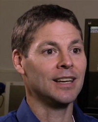 Michael Bailey合作论文数Department of Mechanical Engineering, College of Engineering, University of Washington;Applied Physics Laboratory, University of Washington48Lynn R. Willis合作论文数48Philip M. Blomgren合作论文数36
 Andre Sommer合作论文数36Ryan F. Paterson合作论文数30LR Willis合作论文数28
 Ramsay L. Kuo合作论文数Comprehensive Kidney Stone Center, Duke University Medical Center26
 Friedrich C. Luft合作论文数Max-Delbrück-Center for Molecular Medicine (MDC)24
 Yuri Pishchalnikov合作论文数Applaud Medical24Samuel C. Kim合作论文数24
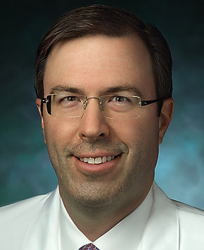 Brian Richard Matlaga合作论文数Stephens Center for Stone Disease, School of Medicine, Johns Hopkins University;James Buchanan Brady Urological Institute, School of Medicine, Johns Hopkins University;Bayview Medical Center, School of Medicine, Johns Hopkins University23
 Elaine Worcester合作论文数Department of Medicine, The University of Chicago22Vincent H. Gattone合作论文数22
 Robin Cleveland合作论文数Department of Engineering Science, Magdalen College, University of Oxford20
 Lawrence Crum合作论文数Department of Bioengineering, College of Engineering and UW Medicine, University of Washington;Applied Physics Laboratory, University of Washington19
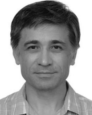 Oleg A. Sapozhnikov合作论文数18Cynthia D. Johnson合作论文数16
 Nicole Miller合作论文数16
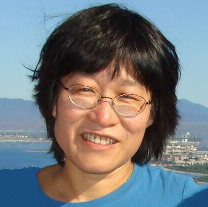 Sujuan Gao合作论文数Department of Biostatistics and Health Data Science, Richard M. Fairbanks School of Public Health, Indiana University;School of Medicine, Indiana University15Marla Paun合作论文数14J H Parks合作论文数13Kd Gardner合作论文数13
 Cynthia J Johnson合作论文数13YZ Shao合作论文数13La Crum合作论文数12Ro Cleveland合作论文数11
 Ehud Gnessin合作论文数Indianapolis11Frank Starr合作论文数10
 Ryan Hsi合作论文数10
 Liu Ziyue合作论文数10
 Michael S Borofsky合作论文数10
 Jay Tischfield合作论文数Department of Genetics, School of Arts and Sciences, Rutgers, The State University of New Jersey;Human Genetics Institute of New Jersey, Rutgers, The State University of New Jersey10H G Bohlen合作论文数9
 Amy E Krambeck合作论文数Department of Urology, Feinberg School of Medicine, Northwestern University9
 Julianna C Simon合作论文数9Jonathan D Harper合作论文数9Naomi S. Fineberg合作论文数9
 Tim Colonius合作论文数Andrew and Peggy Cherng Department of Medical Engineering, Division of Engineering and Applied Science, California Institute of Technology;Department of Mechanical and Civil Engineering, Division of Engineering and Applied Science, California Institute of Technology9
 Bryan W. Cunitz合作论文数9




