Importance Active surveillance is noninferior to standard surgery for 2-year survival and improves short-term health-related quality of life among patients with a complete clinical response (CCR) after neoadjuvant chemoradiotherapy (nCRT) for esophageal cancer. Although active surveillance reduces the upfront costs of surgery and hospital stay, it requires repeated diagnostic tests and, for some patients, delayed surgery and hospitalization during follow-up. Objective To assess the cost-effectiveness of active surveillance compared with standard surgery after nCRT. Design, Setting, and Participants This prespecified cost-effectiveness analysis from a health care perspective conducted at 12 hospitals in the Netherlands as a secondary analysis of the Surgery as Needed for Oesophageal Cancer (SANO) trial, a noninferiority, cluster randomized study, enrolled patients with esophageal cancer who achieved a CCR after nCRT between November 8, 2017, and January 17, 2021, with follow-up for up to 5 years. Data were analyzed on June 1, 2025. Interventions Active surveillance, consisting of repeated response evaluations at 6, 9, 12, 16, 20, 24, 30, 36, 48, and 60 months after nCRT, compared with standard surgery. Main Outcome and Measures Incremental cost-effectiveness of active surveillance vs standard surgery and quality-adjusted life-years (QALYs) with 95% CIs up to 5 years were derived with bootstrapping, with 80% of patients (247 of 309) having complete follow-up. Incremental net monetary benefit (iNMB) was calculated at varying willingness-to-pay thresholds. All analyses followed the modified intention-to-treat principle. Costs are given in Euros (currency exchange rate of €1 = US $1.16 as of June 11, 2026). Results Among 309 patients (198 in the active surveillance group; median age, 69 years [IQR, 63-74 years]; 156 men [79%]; and 111 in the standard surgery group; median age, 68 years [IQR, 61-73 years]; 86 men [77%]), those in the active surveillance group had a mean of 2.99 QALYs (95% CI, 2.73-3.26) at 5 years vs 2.88 QALYs (95% CI 2.69-3.06) in the standard surgery group. Mean health care costs per patient at 5 years were €36 733 (95% CI, €33 530-€40 009) in the active surveillance group vs €45 106 (95% CI, €39 449-€51 545) in the standard surgery group. The incremental QALY for active surveillance was 0.11 (95% CI, −0.10 to 0.33) and mean costs were €8374 lower (95% CI, €1792-€15 355) compared with standard surgery. At a willingness-to-pay threshold of €80 000 per QALY, the mean iNMB was €17 568 (95% CI, −€725 to €37 497), indicating that active surveillance is cost-effective. Bootstrap analysis showed that 97% of replications fell in the cost-effective region. Conclusions and Relevance In this secondary analysis of a randomized clinical trial of patients with esophageal cancer achieving a CCR after nCRT, active surveillance was cost-effective over a 5-year horizon compared with standard surgery. Broader implementation of this strategy among appropriately selected patients would most likely reduce health care costs without compromising health outcomes. Trial Registration The Dutch Trial Register: NTR 6803
BACKGROUND:Endoscopic closure techniques are essential for management of endoscopic defects and help prevent delayed bleeding and perforation. The SutuArt endoscopic hand-suturing system (EHS) is an innovative method for defect closure, but clinical data remain limited. METHODS:In a multicenter European registry study, all procedures using EHS from 14 centers were evaluated. The primary endpoint was technical success, defined as complete defect closure without additional closure techniques. Secondary endpoints were in-hospital clinical success (absence of post-interventional complications); overall defect closure rate (closure achieved with additional devices) and procedure time. RESULTS:Between February 2023 and January 2026, 338 procedures were included. Indications comprised 240 ESD (Endoscopic Submucosal Dissection), 32 EID (Endoscopic Intermuscular Dissection), 41 G-POEM (Gastric Peroral Endoscopic Myotomy), 10 fistulas, 6 EMR (Endoscopic Mucosa Resection), 3 STERs (Submucosal Tunneling Endoscopic Resection), 2 NEWS (Non-exposed Endoscopic Wall inversion Surgery), 1 anastomotic leak, 1 perforation, 1 EFTR (Endoscopic Full Thickness Resection) and 1 endoscopic hemostasis. The median defect size was 35 mm (range 4-155 mm), the median suturing time was 25 minutes (range: 4-120). Technical success was 96.2% (325/338). Overall defect closure rate was 96.7% (327/338). In-hospital clinical success was 97.9% (331/338). Defect size did not predict suturing time in the global size-time model, indicating that procedural complexity was not explained by size alone. Procedure-related adverse events occurred in 5/338 (1.5%), overall AEs were 10/338 (3%). CONCLUSION:EHS demonstrated high technical and in-hospital clinical success with a low observed short-term adverse event rate. Further studies are needed to assess the performance for complex defects.
BACKGROUND:Low grade dysplasia (LGD) diagnosed on multiple occasions is considered the strongest risk factor for neoplastic progression in patients with Barrett's esophagus (BE); however, recent studies have shown that aberrant p53 expression may be a more powerful risk factor for progression. This study aimed to compare the association between aberrant p53 expression versus LGD diagnosed on two or more occasions and neoplastic progression risk. METHODS:Patients with a BE segment length ≥ 2 cm were included in a multicenter prospective cohort. p53 expression was determined by immunohistochemistry staining. Neoplastic progression was defined as development of high grade dysplasia (HGD) or esophageal adenocarcinoma (EAC). Multivariable Cox regression analyses were used to determine the association between selected variables and neoplastic progression risk. RESULTS:960 patients (median age 62; 73 % men) were included. During a median (interquartile range) follow-up of 6.3 (3.5-11.9) years, 81 patients (8 %) developed HGD/EAC. p53 expression was aberrant in 24 % of patients, and was strongly associated with an increased risk of progression (hazard ratio [HR] 20.0, 95 %CI 10.0-40.0). In contrast, LGD on multiple occasions was not similarly associated (HR 0.84, 95 %CI 0.47-1.53), after adjustment for age, sex, and segment length. Progression-free survival was better in patients with normal versus aberrant p53 expression, regardless of the histopathological diagnosis (P < 0.001). CONCLUSIONS:Aberrant p53 expression is strongly associated with an increased neoplastic progression risk in BE patients, regardless of the histopathological findings. It may therefore be a better parameter than confirmed LGD on multiple occasions to identify patients who might benefit from ablation therapy.
Verrucous carcinoma of the esophagus (VCE) is a rare variant of squamous cell carcinoma that poses diagnostic challenges due to nonspecific endoscopic and histologic findings. Its typical Candida overgrowth and hyperkeratotic, non-malignant superficial layer, combined with biopsy results showing fungal hyphae and minimal cytological atypia, are often misleading. Consequently, VCE is frequently diagnosed at advanced stages, limiting curative treatment options and contributing to poor outcomes. Clinicians should consider VCE in patients with persistent verrucous or hyperkeratotic esophageal lesions, especially in the setting of chronic Candida esophagitis. In this commentary, we reflect on seven cases diagnosed at a Dutch tertiary referral center to illustrate diagnostic pitfalls, discuss various treatment options, and offer guidance for improved recognition of VCE. By facilitating earlier diagnosis, curative treatment through endoscopic resection becomes more feasible, ultimately improving patient outcomes.
Females with Barrett’s esophagus (BE) are thought to be less likely to develop neoplasia. This study assessed neoplastic progression risk in females to inform surveillance management. Patients were included from three prospective BE cohorts. Multivariable Cox regression models were used to assess risk factors (including sex) for neoplastic progression (i.e., the development of high-grade dysplasia or esophageal adenocarcinoma). 3099 patients were included, of whom 866 (28%) were females. Fifty-two females (6.0%) and 206 males (9.2%) developed neoplasia during a median follow-up of 6.2 years (IQR 3.3–10.9). Although progression occurred less frequently in females than in males (0.82% vs 1.20% per patient-year), sex was just weakly associated with progression risk in multivariable analysis (HR 1.38, 95% CI 1.00–1.89). Moreover, progression rates in females and males with a BE segment ≥3 cm were similar. Furthermore, mean time to progression, tumor stage, and treatment did not differ between sexes. Therefore, sex-specific surveillance cannot be recommended.
BACKGROUND & AIMS:Endoscopic eradication therapy is standard of care for T1a esophageal adenocarcinoma. There are limited data regarding long-term outcomes of endoscopic eradication therapy for T1b esophageal adenocarcinoma. The aim of this study was to assess clinical outcomes after endoscopic management of low-risk and high-risk T1b esophageal adenocarcinoma via a pooled analysis of individual patient-level data. METHODS:A pooled analysis of studies reporting endoscopic management of T1b esophageal adenocarcinoma was conducted. Inclusion required endoscopic resection revealing T1b esophageal adenocarcinoma with negative deep margins. High-risk T1b esophageal adenocarcinoma was defined by presence of: submucosal 2/3 or deep (>500 μm) submucosal invasion, lymphovascular invasion, or poor grade of differentiation. Clinical outcomes included all-cause and esophageal adenocarcinoma-related mortality, intraluminal recurrence, and extraluminal metastases. RESULTS:Of 418 studies identified, 7 with 216 patients were included (84% men; 94% White). The median follow-up was 48.1 months (interquartile range, 25.5-68.1 months). High-risk T1b esophageal adenocarcinoma was observed in 114 patients (53%). Baseline clinical characteristics were mostly similar between groups. Although the rates of intraluminal recurrence in the low-risk and high-risk groups were comparable (15% vs 22%; P = .30), the high-risk group had a 5-fold significantly higher risk of extraluminal metastases (11% vs 2%; P = .01). Both all-cause and esophageal adenocarcinoma-related mortality were significantly higher in the high-risk group (log rank P = .04). CONCLUSIONS:In this international pooled analysis, rates of extraluminal metastases and esophageal adenocarcinoma-related mortality were very low in patients with low-risk T1b esophageal adenocarcinoma, supporting the role of endoscopic eradication therapy in this group. However, outcomes were significantly worse for the high-risk T1b group, suggesting careful patient selection and shared decision-making in this population.
Background and Aims Endoscopic eradication therapy (EET) for Barrett’s esophagus (BE) with high-grade dysplasia (HGD) aims to prevent progression to life-limiting cancer. However, HGD patients with a short life expectancy realize limited gains from this approach owing to competing mortality. Clinicians are poorly equipped to counsel such patients, because there are little data regarding survival in untreated HGD. We aimed to evaluate existing literature and describe a case series of patients with untreated HGD. Methods We systematically reviewed Medline, Embase, and Cochrane Library, selecting studies describing the natural history of untreated HGD in BE. The primary outcomes were symptomatic esophageal adenocarcinoma (EAC) and EAC-related death. In addition, cases in which HGD was left untreated were retrospectively identified in the Netherlands. We assessed the time until progression to clinically evident EAC. Results A total of 3229 studies were identified, of which 3 were included. In 1 study, progression from HGD to clinically evident EAC occurred in 4 subjects after a median of 34 months. The remaining 2 cases progressed to clinically evident EAC after 70 and 115 months. In our previously unreported case series, 11 Dutch patients with flat HGD (n = 3) or HGD in a visible abnormality (n = 8) were included. Four of these 11 patients progressed to clinically evident EAC after a median 52 months (range, 17-78 months). Conclusions The lag-time between the diagnosis of HGD and progression to clinically evident EAC varied from 1.5 to 10 years. EET for BE with HGD in patients with less than 3 years of life expectancy seems unlikely to be beneficial. These results may guide management decisions for patients with BE. (Netherlands Trial Registry, NL7039; NL-OMON29089.)
BACKGROUND AND OBJECTIVES:Surveillance after gastric endoscopic submucosal dissection (ESD) is crucial due to the high risk of metachronous gastric lesions (MGL), although this risk may differ between patients. We sought to validate the FAMISH score - a prediction score to estimate MGL risk after gastric ESD - within a multicentric framework. MATERIALS AND METHODS:Performance measures of the FAMISH score were assessed in a retrospective analysis of a multicenter cohort, which included consecutive adult patients undergoing ESD for a primary gastric superficial lesion at 15 international centers, with a minimum endoscopic follow-up of at least 3 years. RESULTS:A total of 855 individuals were included, with 20% of them considered low-risk according to the FAMISH score. After a mean follow-up time of 5 years (SD ± 2 years), 168 patients (19.6%) developed MGL. At 3 years of follow-up, the score achieved 90.4% sensitivity and 93.9% NPV. At 5 years follow-up, the score achieved 89.1% sensitivity and 85.3% NPV. The FAMISH risk score achieved a fair diagnostic accuracy with an AUC of 0.618 at 3 years (p < 0.001) and 0.597 (p = 0.006) at 5 years of follow-up. The progression to MGL at 5 years of follow-up was significantly lower for the low-risk group (9.6% vs. 18.2%, p = 0.029). CONCLUSIONS:The FAMISH score achieved an acceptable diagnostic accuracy in a multicentric validation cohort from Western countries. This score is a useful tool to identify patients with low risk for MGL allowing to safely extend surveillance intervals and reduce the burden of care.
The incidence of oesophageal squamous cell carcinoma accounts for almost 85% of all oesophageal cancers worldwide. Patients with oesophageal cancer can present with clinical symptoms including dysphagia, weight loss, retrosternal discomfort or regurgitation. Nevertheless, most cancers remain asymptomatic and thereby undetected until the cancer has reached advanced or even incurable stages. This results in poor prognosis and relatively low long-term survival rates. The identification of patients at high risk for oesophageal cancer development is therefore an important cornerstone in the detection of oesophageal cancer in early and curable stages. This group contains patients with a history of cancer in the aerodigestive tract, squamous dysplasia, certain lifestyle aspects, three types of genetic syndromes, caustic or radiation induced injury to the oesophagus and achalasia. In this review, we evaluate different risk factors for the development of oesophageal squamous cell carcinoma and discuss whether screening of these patients might be justified.
 Roos Pouw合作论文数44
 E. J. (Erik) Schoon合作论文数Catharina Hospital Eindhoven42
 Bas Weusten合作论文数Department of Gastroenterology and Hepatology, University Medical Center Utrecht41
 M.J. (Marco) Bruno合作论文数Department of Gastroenterology and Hepatology, Erasmus MC37
 Jacques J.G.H.M. Bergman合作论文数32
 Wouter B Nagengast合作论文数32A. Alkhalaf合作论文数28
 Wouter Curvers合作论文数Sentio Ergo Sum;Catharina Ziekenhuis Specialist manager MDL;Barrett Expert Center CZE26Martin H M G Houben合作论文数24
 Sanne van Munster合作论文数24
 Steffi van de Ven合作论文数22Jessie Westerhof合作论文数22
 Manon Spaander合作论文数19
 Esther Nieuwenhuis合作论文数18Thjon J. Tang合作论文数17Laurelle van Tilburg合作论文数16
 Ernst J Kuipers合作论文数Lee Kong Chian School of Medicine, Nanyang Technology University16Paul Didden合作论文数14Robert A. De Man合作论文数14B E Schenk合作论文数12
 Eva Verheij合作论文数12L Alvarez Herrero合作论文数12P.J.F. de Jonge合作论文数12Michail Doukas合作论文数12Ekkelenkamp Vivian E合作论文数12
 Raf Bisschops合作论文数Department of Chronic Diseases and Metabolism, Faculty of Medicine, KU Leuven;UZ Leuven12
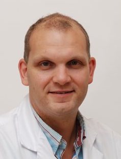 Leon Moons合作论文数Divison of Internal Medicine and Dermatology, University Medical Center Utrecht11
 Anniek W. Gotink合作论文数10Lorenza Alvarez Herrero合作论文数10
 Lodewijk Brosens合作论文数Department of Pathology, University Medical Center Utrecht;Radboud University Medical Centre Nijmegen9
 Sybren Meijer合作论文数Cancer Center Amsterdam, Amsterdam University Medical Center9
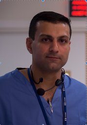 Rehan J. Haidry合作论文数9Sophia van der Wiel合作论文数8Jelle Haringsma合作论文数8
 J.W. Poley合作论文数Maastricht UMC8Pieter Jan F. de Jonge合作论文数7Auke Bogte合作论文数7
 Oliver Pech合作论文数7
 Jansen Marnix合作论文数Barts Cancer Institute, Queen Mary University of London7Torsten Beyna合作论文数7
 Horst Neuhaus合作论文数Department of Internal Medicine, Evangelisches Krankenhaus Düsseldorf, University of Düsseldorf7Michael J. Bourke合作论文数7
 James Hardwick合作论文数Department of Gastroenterology & Hepatology, Leiden University Medical Center7
 Kamar Belghazi合作论文数6Ricardo Küttner-Magalhães合作论文数6Wilmar de Graaf合作论文数6Martin H. Houben合作论文数6Stefan Seewald合作论文数6
 Judith Honing合作论文数University Medical Center Groningen, University of Groningen6




