RNA polymerase I inhibition affects rRNA synthesis from rDNA clusters residing in nucleolar organizer regions (NORs). Here we have demonstrated RNA Pol I inhibition disrupts nucleolar architecture, NPM1 localization, and alters the chromatin landscape using coordinated eraser and writer enzymes. siRNA mediated depletion or dissociation of NPM1 allows HDAC1 loading on the chromatin. HDAC1 mediated deacetylation of H3K9ac creates H3K9 that undegoes SUV39H1-mediated methylation. The stripping of active histone marks leads to enrichment of repressive H3K9me3 in the genome. These altered chromatin landscape corroborates with loss of genome-wide chromatin accessibility and DNA hypermethylation mediated by DNMT1. Chromatin architectural analysis revealed disrupted nucleolar associated domains (NADs) transforming to lamin associated domains (LADs) with specific histone signatures and repressive states. The 3D nuclear architecture was remodeled by A/B compartments reorganization and loss of Hi-C loops at the H3K9ac depleted sites.
Hypomethylating agents are frontline therapies for myelodysplastic neoplasms (MDS), yet clinical responses remain unpredictable. We conducted a phase 2 trial comparing injectable and oral azacitidine (AZA) administered over one or three weeks per four-week cycle, with the primary objective of investigating whether response is linked to in vivo drug incorporation or DNA hypomethylation. Our findings show that injection results in higher drug incorporation, but lower DNA demethylation per cycle, while global DNA methylation levels in mononuclear cells are comparable between responders and non-responders. However, hematopoietic stem and progenitor cells (HSPCs) from responders exhibit distinct baseline and early treatment-induced CpG methylation changes at regulatory regions linked to tissue patterning, cell migration, and myeloid differentiation. By cycle six-when clinical responses typically emerge-further differential hypomethylation in responder HSPCs suggests marrow adaptation as a driver of improved hematopoiesis. These findings indicate that intrinsic baseline and early drug-induced epigenetic differences in HSPCs may underlie the variable clinical response to AZA in MDS.
Hypomethylating agents are used as frontline therapy for myelodysplastic neoplasms (MDS), but clinical response is unpredictable. To determine whether response was associated with in vivo dynamics of DNA hypomethylation, we conducted a phase 2 trial for MDS using both injection and oral azacitidine (AZA). We established that global DNA methylation levels in peripheral blood and bone marrow mononuclear cells were comparable in AZA responders and non-responders during their course of treatment. However, there were distinct baseline and early drug induced differences in CpG methylation in haematopoietic stem and progenitor cells (HSPCs) in responders compared to non-responders that overlapped with regulatory regions of genes associated with tissue patterning, cell migration and myeloid differentiation. Following six cycles of therapy when clinical response typically manifests, differential hypomethylation in responder HSPCs pointed to marrow adaptation as a driver of enhanced haematopoiesis. Taken together, CpG methylation differences in HSPCs may explain variable response to AZA. ### Competing Interest Statement F.V. is affiliated with OmniOmics.AI Pty Ltd. C.F. is an advisory board member at Amgen, AbbVie, Adaptive Biotech, BeiGene, Pfizer, Otsuka, and Jazz, a consultant at Novotech, and received speaker fees from Amgen, Pfizer, Servier, BMS, and Astella. D.H. has consultancy agreements with GlaxoSmithKline and Pharming Corp. M.H. is a consultant/advisory board member at Roche, Gilead, Otsuka, Janssen, Beigene, and Takeda. M.N.P. received research funding and/or provision of drug for clinical trials (to institution) from AstraZeneca, BRII Biosciences, Celgene/BMS, CSL Behring, Eli Lilly, Emergent Biosciences, Gilead Pharmaceuticals, GlaxoSmithKline, Grifols, Janssen/Johnson and Johnson, Takeda, ViiV Pharmaceuticals and has advisory roles with Celgene/BMS, Gilead Pharmaceuticals, and ViiV Pharmaceuticals. J.E.P. received research funding and/or provision of drug for clinical trials (to institution) from Celgene/BMS, Astex, Verastem Oncology and received honoraria from Abbvie as an advisory board member. The remaining authors declare no competing financial interests. ### Clinical Trial NCT03493646 ### Funding Statement The investigator initiated clinical trial was funded in part by Celgene/BMS (RG172029) with research support from the National Health and Medical Research Council (RG170246, RG211412), Anthony Rothe Memorial Trust (RG182042, RG202657, RG213236), Leukaemia Foundation (RG231257). ### Author Declarations I confirm all relevant ethical guidelines have been followed, and any necessary IRB and/or ethics committee approvals have been obtained. Yes The details of the IRB/oversight body that provided approval or exemption for the research described are given below: The trial protocol received ethical approval from the South Eastern Sydney Local Health District Human Research Ethics Committee, and participating sites received Institution approval to conduct the trial prior to commencing recruitment. I confirm that all necessary patient/participant consent has been obtained and the appropriate institutional forms have been archived, and that any patient/participant/sample identifiers included were not known to anyone (e.g., hospital staff, patients or participants themselves) outside the research group so cannot be used to identify individuals. Yes I understand that all clinical trials and any other prospective interventional studies must be registered with an ICMJE-approved registry, such as ClinicalTrials.gov. I confirm that any such study reported in the manuscript has been registered and the trial registration ID is provided (note: if posting a prospective study registered retrospectively, please provide a statement in the trial ID field explaining why the study was not registered in advance). Yes I have followed all appropriate research reporting guidelines, such as any relevant EQUATOR Network research reporting checklist(s) and other pertinent material, if applicable. Yes All data produced in the present study are available upon reasonable request to the authors
Determinants of clinical response to hypomethylating agents (HMAs) in MDS/CMML are unclear. To address this knowledge gap, we enrolled 40 patients to NCT03493646 where they received 6 cycles of injection AZA (vidaza) or oral AZA (CC486) delivered for 7 or 21 days respectively in a 28-day cycle to study the kinetics of drug incorporation and DNA hypomethylation during treatment, and noted the following(i) Average DAC incorporation in DNA was higher with vidaza than CC486, but global DNA hypomethylation was greater with CC486. Although DAC incorporation was higher in responders (R) than in non-responders (NR) with vidaza, global DNA demethylation did not differ between response groups with either drug.(ii) DAC incorporation after vidaza correlated with the fraction of HPCs progressing through S/G2/M pre-treatment, but there were no pre-treatment differences between R and NR in the fraction of HSCs and HPCs progressing through S/G2/M. However, vidaza induced HSCs and HPCs to exit quiescence in R but not NR.(iii) Baseline CpG methylation was higher in CD34+ bone marrow cells in NR vs. R, and CpG sites that were associated with regulation of pattern specification (HOX cluster), epithelial cell migration/EMT and mesenchymal stromal cell differentiation were differentially hypomethylated in R vs. NR. Following vidaza, early drug-induced hypomethylation was seen at CpG sites in CD34+ that regulated genes associated with myeloid cell differentiation. By contrast, late drug-induced CpG hypomethylation in R was at sites associated with leukocyte development in response to marrow environmental changes.(iv) Variant alleles persist in R and NR following vidaza or CC486, and there was no association between change in variant allele frequencies and the degree of DNA hypomethylation.Site-specific differences in CpG hypomethylation in HSPCs may explain clinical response to HMAs.
Myelodysplastic neoplasms (MDSs) and chronic myelomonocytic leukemia (CMML) are clonal disorders driven by progressively acquired somatic mutations in hematopoietic stem cells (HSCs). Hypomethylating agents (HMAs) can modify the clinical course of MDS and CMML. Clinical improvement does not require eradication of mutated cells and may be related to improved differentiation capacity of mutated HSCs. However, in patients with established disease it is unclear whether (1) HSCs with multiple mutations progress through differentiation with comparable frequency to their less mutated counterparts or (2) improvements in peripheral blood counts following HMA therapy are driven by residual wild-type HSCs or by clones with particular combinations of mutations. To address these questions, the somatic mutations of individual stem cells, progenitors (common myeloid progenitors, granulocyte monocyte progenitors, and megakaryocyte erythroid progenitors), and matched circulating hematopoietic cells (monocytes, neutrophils, and naive B cells) in MDS and CMML were characterized via high-throughput single-cell genotyping, followed by bulk analysis in immature and mature cells before and after AZA treatment. The mutational burden was similar throughout differentiation, with even the most mutated stem and progenitor clones maintaining their capacity to differentiate to mature cell types in vivo. Increased contributions from productive mutant progenitors appear to underlie improved hematopoiesis in MDS following HMA therapy.
Figure S1, related to Figure 1: Expression and correlation of CHK1 and CIP2A in multiple glioma cohorts. Figure S2, related to Figure 1: Overall survival and mRNA expression of PP2A subunits in glioma patients. Figure S3, related to Figure 1: Role of CHK1-CIP2A in Glioma Stem cell lines. Figure S4, related to Figure 3: Effect of CHK1 or CIP2A expression on GBM cells. Figure S5, related to Figure 5: Depletion of CIP2A induces senescence in GBM cells. Figure S6, related to Figure 6: Regulation of CIP2A expression by CHK1 and STAT3.
Supplementary Figure 3, related to main Figure 5.
Supplementary Figure 1, related to main Figure 1
Supplementary Figure 2, related to main Figure 3
The homeobox gene, Hoxa1 , has two different isoforms generated by alternative splicing: a full-length homeodomain-containing Hoxa1 ( Hoxa1-FL ), and a truncated Hoxa1 ( Hoxa1-T ), that lacks the homeodomain. Oncoretroviral overexpression of wildtype Hoxa1 cDNA ( WT-Hoxa1 ), which generates both Hoxa1 isoforms, in murine hematopoietic stem and progenitor cells (HSPCs) perturbed hematopoiesis, resulting in myelodysplastic syndromes (MDS) in mice. Overexpression of a mutated Hoxa1 cDNA ( MUT-Hoxa1 ) that generates Hoxa1-FL but not Hoxa1-T led to a more severe MDS capable of transforming to secondary acute myeloid leukemia (sAML). Similar to human MDS, DNA damage repair pathways were downregulated in Hoxa1 -overexpressing hematopoietic progenitor cells. Conditional knock-in mouse models revealed a Hoxa1-FL dosage-dependent effect on MDS disease severity. Our data reveal that increased expression of Hoxa1-FL in HSPCs is sufficient to initiate MDS in mice. CD34+ cells from up to 50% of patients with MDS had elevated HOXA1-FL expression, highlighting the clinical relevance of our mouse models. Statement of Significance Our study demonstrates that Hoxa1 is a key regulator of HSPCs and that increased expression of the transcriptionally active Hoxa1-FL can initiate MDS in mice. Furthermore, HOXA1-FL expression is upregulated in a significant proportion of human MDS patients and likely contributes to the disease in these patients.
•ROS elevation in HT-1080 by Ce-based nanoparticle treatment.•H2DCFDA-mediated detection of ROS by laser scanning confocal microscopy.•Writing of custom-built MATLAB software for image analysis of ROS fluorescence intensities.•Processing of images for quantification of fluorescence intensities.•Statistical analysis by ANOVA to confirm statistical significance.
 John Pimanda合作论文数School of Biomedical Sciences, The University of New South Wales;Lowy Cancer Research Centre52
 Thoms Julie A I合作论文数28
 Jason Wing Hon Wong (黃永瀚)合作论文数School of Biomedical Sciences, Li Ka Shing Faculty of Medicine, The University of Hong Kong18
 Govardhan Anande合作论文数Prince Wales Clin Sch, Univ New South Wales16
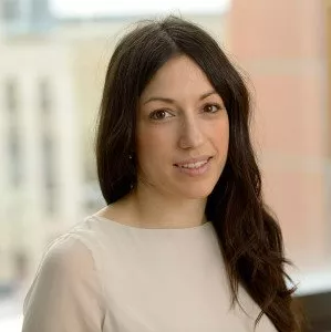 Elli Papaemmanuil合作论文数Computational Biology, Weill Cornell Graduate School of Medical Sciences, Cornell University15
 Dominik Beck合作论文数13
 Nandan P. Deshpande合作论文数School of Biotechnology & Biomolecular Sciences, Faculty of Science, The University of New South Wales12Henry R. Hampton合作论文数11
 Junyu Yang (杨君宇)合作论文数10Junsu Kwon合作论文数10Chong Gao合作论文数10
 Daniel G. Tenen合作论文数Division of Medical Sciences, Harvard Medical School;Beth Israel Deaconess Medical Center10Yao-Chung Liu合作论文数10Kathy Knezevic合作论文数10
 Li Chai合作论文数10Shenyi Yin合作论文数9
 Alicia Stein合作论文数9Miao Liu合作论文数9Silberstein Leslie E合作论文数9
 Jianzhong Jeff Xi (席建忠)合作论文数College of Future Technology, Faculty of Engineering, Peking University9
 Nikki Kong合作论文数9
 Jun Qi合作论文数Dana-Farber Cancer Institute9
 Marc Wilkins合作论文数The University of New South Wales9
 Peter Campbell合作论文数Quotient Therapeutics9Hannan Wong合作论文数8Kalpana Kumari合作论文数8Yue Wu合作论文数8Lachlin Vaughan合作论文数7Yanjing V. Liu合作论文数7Xi Tian合作论文数7Eva Hellström-Lindberg合作论文数7Lehmann Sören合作论文数7Tobiasson Magnus合作论文数Center for Hematology and Regenerative Medicine, Karolinska University Hospital Huddinge7Hertzberg Mark合作论文数7Stefan K. Bohlander合作论文数7
 Russell Pickford合作论文数Bioanalytical Mass Spectrometry Facility, The University of New South Wales7J. Pimanda合作论文数6
 Tobias Herold合作论文数6Hongbo Luo合作论文数6Fernando Roncolato合作论文数6Sarah Davidson合作论文数6
 Aarif Mohamed Nazeer Batcha合作论文数Data Integration for Future Medicine (DIFUTURE),, LMU Munich6Golam Sarower Bhuyan合作论文数6
 Polizzotto Mark N合作论文数Center for Cancer Research, National Cancer Institute;Center for Cancer Research, National Cancer Institute6
 Vashe Chandrakanthan合作论文数Adelaide Medical School, The University of Adelaide6Joshi Swapna合作论文数6
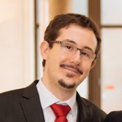 Mareschal Sylvain合作论文数Centre Henri Becquerel, Université de Rouen6
 Jake Olivier合作论文数School of Mathematics and Statistics, Faculty of Science, The University of New South Wales6Purvi Kakadiya合作论文数Department of Molecular Medicine and Pathology, School of Medical Sciences, Faculty of Medical and Health Sciences, The University of Auckland6




