Three-photon (3-P) fluorescence microscopy enables deep in vivo imaging with subcellular resolution, but its performance is fundamentally constrained by the maximum permissible laser power required to avoid tissue heating and photodamage. Under these power-limited conditions, fluorescence signal generation, image contrast, and achievable imaging depth are strongly affected by the illumination beam profile and aberration correction strategy. In this paper, we showed that using a fixed illumination beam size was suboptimal across different imaging depths. We further showed that conventional Zernike-based adaptive optics (AO) correction degrades under reduced Gaussian illumination beam sizes due to loss of modal orthogonality. This degradation results in slow convergence, unintended focal and field-of-view shifts, and excessive wavefront deformations. To overcome these limitations, we introduced a depth-adapted AO framework in which both the illumination beam profile and the aberration correction basis were dynamically matched to the imaging conditions. By combining depth-optimised beam underfilling with a bespoke set of illumination-matched aberration modes, we achieved faster and more stable AO convergence, enhanced fluorescence signal and image quality during deep in vivo multi-channel neuroimaging. Together, these results established a practical and robust AO-enabled three-photon microscopy strategy that maximised imaging performance under realistic power constraints.
Auditory learning is supported by long-term changes in the neural processing of sound. We examined these task-depend changes in the auditory cortex by mapping neural sensitivity to timbre, pitch, and location cues in cues in trained (n = 5) and untrained control female ferrets (n = 5). Trained animals either identified vowels in a two-alternative forced choice task (n = 3) or discriminated when a repeating vowel changed in identity or pitch (n = 2). Neural responses were recorded under anesthesia in two primary auditory cortical fields and two tonotopically organized nonprimary fields. In trained animals, the overall sensitivity to sound timbre was reduced across three cortical fields compared with control animals, but maintained in a nonprimary field (the posterior pseudosylvian field). While training did not increase sensitivity to timbre across the auditory cortex, it did change the way in which neurons integrated spectral information, with neural responses in trained animals increasing their sensitivity to first and second formant frequencies, whereas in control animals cortical sensitivity to spectral timbre depended mostly on the second formant. Animals trained on timbre identification were required to generalize across pitch when discriminating timbre, and their neurons became less modulated by fundamental frequency relative to control animals. Finally, both trained groups showed increased spatial sensitivity and an enhanced response to sound source locations close to the midline, where the loudspeaker was located in the training chamber. These results demonstrate that training elicited widespread alterations in the cortical representation of complex sounds.
Three-photon (3P) microscopy enables functional imaging at greater depths in the mammalian brain than any other technique with single-cell resolution, owing to greater penetration ability. One of the main challenges of such imaging is tissue-induced optical aberration, which inevitably reduces the excitation confinement at depth. Adaptive optics systems, by using deformable mirrors or other wavefront-shaping devices to compensate for optical distortions, enable real-time correction of these aberrations. In this study, we present a practical adaptive optics-assisted 3P imaging system optimized for in vivo functional recordings in the mouse cortex during behaviour. We introduce a hierarchical, three-level aberration correction strategy that sequentially targets aberration caused by the microscope system, the cranial window, and tissue depth. We demonstrate the application of this aberration correction strategy in two anatomically distinct regions—the prelimbic cortex, adjacent to the superior sagittal sinus, and the somatosensory cortex as a representative lateral cortical area—highlighting how aberration sources vary with imaging geometry. Adaptive optics significantly improved imaging performance in both contexts: restoring cellular visibility adjacent to large vascular structures in the prelimbic cortex, and enhancing signal-to-noise ratio during deep imaging in the lateral somatosensory cortex. Together, our work provides a practical framework for implementing adaptive optics-assisted 3P imaging and optimizing deep in vivo functional imaging performance across diverse cortical environments. ### Competing Interest Statement The authors have declared no competing interest. Wellcome Trust, 222807/Z/21/Z, 213465/Z/18/Z UK Research and Innovation, EP/X026655/1, EP/W024047/1 European Research Council Advanced Grant, AdOMiS 695140 Schmidt Sciences LLC
The claustrum is a densely interconnected structure involved in cognitive functions, but its influence on prefrontal circuits remains unclear. We conducted two-photon calcium imaging to assess mice dorsal prefrontal cortex (dPFC) cell responses during exposure to visual stimuli and widefield photostimulation of claustrum axons embedded in the dPFC. We identified three distinct subpopulations of neurons - sensory responsive, opto responsive, and opto-boosted cells-each exhibiting unique response dynamics to combined visual and optogenetic stimuli. Our findings reveal that claustrum stimulation increased single-cell variability while aligning average responses across neurons, thereby enhancing network homogeneity. During Pavlovian training, enhanced variability persisted, but homogeneity increased further, suggesting experience-dependent refinement. Finally, we performed claustrum axon silencing experiments which revealed that the claustrum may operate bidirectionally to maintain enhanced variability and homogeneity in the dPFC. These results highlight the crucial role of the claustrum in dynamically modulating dPFC activity, impacting both neuronal variability and network synchronization.
Economic decision-making requires evaluating information about available options, such as their expected value and economic risk. Previous studies have shown that frontal cortical neurons encode these variables, but how this encoding is structured across different frontal regions and projection pathways remains unclear. We developed a decision-making task for head-fixed mice in which we varied the expected value and risk associated with reward-predicting stimuli. Using large-scale electrophysiology, two-photon imaging, and projection-specific optotagging, we identified distinct spatial gradients for these variables, with stronger expected value coding in dorsal frontal regions and stronger risk coding in medial regions. We then demonstrated that this encoding further depends on the neuronal projections: frontal neurons projecting to the dorsomedial striatum and claustrum differentially encoded economic variables. Our findings illustrate that frontal cortical representation of economic variables is jointly determined by spatial organization and downstream connectivity of neurons, revealing a structured, multi-scale code for economic variables.
Adaptive optics (AO) was shown to improve microscope imaging quality. However, issues like prolonged sample exposures are often associated with sensorless AO implementation. We present an AO solution with embedded intelligence to achieve outstanding performance.
Transgenic mice engineered to express calcium indicators such as GCaMP have revolutionized exploration of neuronal circuit function. The latest development, GCaMP8 transgenic mice, exhibits enhanced temporal kinetics and sensitivity of neural signals, opening new avenues for studying neuronal dynamics within behaviorally relevant time frames. However, in initial attempts, it has been chalenging to train these mice in visual decision making tasks. Here we show that GCaMP8 transgenic mice, specifically TetO-jGCaMP8s x CaMK2a-tTA mice, learn to perform head-fixed visual decision tasks with a rate and accuracy comparable to wildtype mice. These proof-of-principle results enhance the utility of these transgenic animals in neuroscientific studies of learning and decision making.
The claustrum is recognized for its significant impact on various cognitive functions and its extensive connections with other brain regions, yet its functional role remains to be fully understood. Here, we utilized an optogenetic approach to investigate the claustrum's influence on neuronal activity within the dorsal prefrontal cortex (dPFC) of mice. We conducted two-photon calcium imaging to assess dPFC cell responses during exposure to visual stimuli and widefield photostimulation of claustrum axons embedded in the dPFC. We identified three distinct subpopulations of neurons - sensory responsive, opto responsive, and opto-boosted cells - each exhibiting unique response dynamics to combined visual and optogenetic stimuli. Our findings reveal that stimulation of claustrum axons can normalize neuronal responsiveness, while enhancing neural variability, and significantly increasing network homogeneity. Training in a Pavlovian task showed that while enhanced variability with claustrum axon stimulation in neural responses persists, training does not further increase this variability but instead leads to greater network homogeneity. Additionally, we also performed claustrum axon silencing experiments that revealed that the claustrum may operate bidirectionally to maintain enhanced variability and homogeneity in the prefrontal cortex. These results highlight the crucial role of the claustrum in dynamically modulating dPFC activity, impacting both neuronal variability and network synchronization. ### Competing Interest Statement The authors have declared no competing interest.
The resolution and contrast of microscope imaging is often affected by aberrations introduced by imperfect optical systems and inhomogeneous refractive structures in specimens. Adaptive optics (AO) compensates these aberrations and restores diffraction limited performance. A wide range of AO solutions have been introduced, often tailored to a specific microscope type or application. Until now, a universal AO solution - one that can be readily transferred between microscope modalities - has not been deployed. We propose versatile and fast aberration correction using a physics-based machine learning assisted wavefront-sensorless AO control (MLAO) method. Unlike previous ML methods, we used a specially constructed neural network (NN) architecture, designed using physical understanding of the general microscope image formation, that was embedded in the control loop of different microscope systems. The approach means that not only is the resulting NN orders of magnitude simpler than previous NN methods, but the concept is translatable across microscope modalities. We demonstrated the method on a two-photon, a three-photon and a widefield three-dimensional (3D) structured illumination microscope. Results showed that the method outperformed commonly-used modal-based sensorless AO methods. We also showed that our ML-based method was robust in a range of challenging imaging conditions, such as 3D sample structures, specimen motion, low signal to noise ratio and activity-induced fluorescence fluctuations. Moreover, as the bespoke architecture encapsulated physical understanding of the imaging process, the internal NN configuration was no-longer a "black box", but provided physical insights on internal workings, which could influence future designs.
Auditory learning is supported by long-term changes in the neural processing of sound. We mapped neural sensitivity to timbre, pitch and location in animals trained to discriminate the identity of artificial vowels based on their spectral timbre in a two-alternative forced choice (T2AFC, n=3, female ferrets) or to detect changes in fundamental frequency or timbre of repeating artificial vowels in a go/no-go task (n=2 female ferrets). Neural responses were recorded under anaesthesia in two primary cortical fields and two tonotopically organised non-primary fields. Responses were compared these data to that of naïve control animals. We observed that in both groups of trained animals the overall sensitivity to sound timbre was reduced across three cortical fields but enhanced in non-primary field PSF. Neural responses in trained animals were able to discriminate vowels that differed in either their first or second formant frequency unlike control animals whose sensitivity was mostly driven by changes in the second formant. Neural responses in the T2AFC animals, who were required to generalise across pitch when discriminating timbre, became less modulated by fundamental frequency, while those in the go/no-go animals were unchanged relative to controls. Finally, both trained groups showed increased spatial sensitivity and altered tuning. Trained animals showed an enhanced representation of the midline, where the speaker was located in the experimental chamber. Overall, these results demonstrate training elicited widespread changes in the way in which auditory cortical neurons represent complex sounds with changes in how both task relevant and task-irrelevant features were represented.### Competing Interest StatementThe authors have declared no competing interest.
Objectives/Aims The claustrum is a sheet-like bilateral brain region whose function remains unknown. We comprehensively examined cases of human claustrum lesions to assess their support for various hypotheses of function. To do so we searched the following terms on PubMed and Scopus: ‘claustrum AND (lesion OR contusion OR in- jury OR trauma)’. Results In total our search uncovered 103 cases, which were then screened for confirmed damage to the claustrum by neuroimaging. Thirty-eight individual cases and 14 cohort studies were included. Our results suggest human claustral lesions do not selectively impair a singular domain. Instead, they argue that human claustrum may have a more general function. The high incidence of seizures following claustral lesions suggests this may involve maintenance of excitation-inhibition balance. Conclusions The wide range of symptoms observed following claustral lesions do not provide compelling evidence to support prominent current theories of claustrum function such as spatial navigation, attentional allocation, cognition, multisensory integration, and salience computation. Conversely, the lesions studies support the hypothesis that the claustrum regulates cortical excitability.
There are often sudden changes in the state of environment. For a decision maker, accurate prediction and detection of change points are crucial for optimizing performance. Still unclear, however, is whether rodents are simply reactive to reinforcements, or if they can be proactive to estimate future change points during value-based decision making. In this study, we characterize head-fixed mice performing a two-armed bandit task with probabilistic reward reversals. Choice behavior deviates from classic reinforcement learning, but instead suggests a strategy involving belief updating, consistent with the anticipation of change points to exploit the task structure. Excitotoxic lesion and optogenetic inactivation implicate the anterior cingulate and premotor regions of medial frontal cortex. Specifically, over-estimation of hazard rate arises from imbalance across frontal hemispheres during the time window before the choice is made. Collectively, the results demonstrate that mice can capitalize on their knowledge of task regularities, and this estimation of future changes in the environment may be a main computational function of the rodent dorsal medial frontal cortex.
The claustrum is the most densely interconnected region in the human brain. Despite the accumulating data from clinical and experimental studies, the functional role of the claustrum remains unknown. Here, we systematically review claustrum lesion studies and discuss their functional implications. Claustral lesions are associated with an array of signs and symptoms, including changes in cognitive, perceptual and motor abilities; electrical activity; mental state; and sleep. The wide range of symptoms observed following claustral lesions do not provide compelling evidence to support prominent current theories of claustrum function such as multisensory integration or salience computation. Conversely, the lesions studies support the hypothesis that the claustrum regulates cortical excitability. We argue that the claustrum is connected to, or part of, multiple brain networks that perform both fundamental and higher cognitive functions. As a multifunctional node in numerous networks, this may explain the manifold effects of claustrum damage on brain and behaviour.
The claustrum is the most densely interconnected region in the human brain. Despite the accumulating data from clinical and experimental studies, the functional role(s) of the claustrum remain unknown. Here, we systematically review claustrum lesion studies and discuss their functional implications. Claustral lesions are associated with an array of signs and symptoms, including changes in cognitive, perceptual and motor abilities; electrical activity; mental state; and sleep. The wide range of symptoms observed following claustral lesions suggests that the claustrum may either have a number of distinct functions, or a global function that impacts many neural processes. We further discuss the implications of these lesions in the context of recent evidence linking the claustrum to sensory perception, sleep, and salience as well as highlighting an underexplored link between the claustrum and pain. We hypothesize that the claustrum is connected to multiple brain networks, both ancient and advanced, which underly fundamental functions as well as higher cognitive processes. Extensive evidence derived from human lesion studies and animal experiments provides unequivocal evidence for a key function of the claustrum as a multifunctional node in numerous networks.
The ability to use temporal relationships between cross-modal cues facilitates perception and behavior. Previously we observed that temporally correlated changes in the size of a visual stimulus and the intensity in an auditory stimulus influenced the ability of listeners to perform an auditory selective attention task (Maddox, Atilgan, Bizley, & Lee, 2015). Participants detected timbral changes in a target sound while ignoring those in a simultaneously presented masker. When the visual stimulus was temporally coherent with the target sound, performance was significantly better than when the visual stimulus was temporally coherent with the masker, despite the visual stimulus conveying no task-relevant information. Here, we trained observers to detect audiovisual temporal coherence and asked whether this changed the way in which they were able to exploit visual information in the auditory selective attention task. We observed that after training, participants were able to benefit from temporal coherence between the visual stimulus and both the target and masker streams, relative to the condition in which the visual stimulus was coherent with neither sound. However, we did not observe such changes in a second group that were trained to discriminate modulation rate differences between temporally coherent audiovisual streams, although they did show an improvement in their overall performance. A control group did not change their performance between pretest and post-test and did not change how they exploited visual information. These results provide insights into how crossmodal experience may optimize multisensory integration.
Learning from experience is essential to the optimization of behavior. In particular, we learn from past choices and outcomes to infer the predicted values of the actions to be taken. Then based on the values, we may select an informed choice. However, despite the many neural correlates identified, we still do not have a clear picture for how values are computed and translated into informed behavior. Here, we trained head-fixed mice to perform a two-armed bandit task. Animals based their decisions on past choices and reinforcements, consistent with having an internal representation of action values. To determine the causal contributions of the medial prefrontal cortex, we tested the animals before and after an excitotoxic lesion of the medial secondary motor cortex (M2). We found that unilateral M2 lesion led to side-specific effects on the animal’s ability to learn from past choices. To quantify the decision-making process, we fitted the animal’s choice behavior with Q-learning models to extract learning parameters such as learning rate, forgetting rate, and inverse temperature. Altogether, the results provide insights into the causal involvement of mouse mM2 in value-based decision making.
In an unfamiliar situation, animals display variable choice behavior. Based on computational modeling and empirical data, a new study suggests that the variability in decision-making across individuals is driven by differences in internal neural dynamics in the medial frontal cortex.
How and where in the brain audio-visual signals are bound to create multimodal objects remains unknown. One hypothesis is that temporal coherence between dynamic multisensory signals provides a mechanism for binding stimulus features across sensory modalities. Here, we report that when the luminance of a visual stimulus is temporally coherent with the amplitude fluctuations of one sound in a mixture, the representation of that sound is enhanced in auditory cortex. Critically, this enhancement extends to include both binding and non-binding features of the sound. We demonstrate that visual information conveyed from visual cortex via the phase of the local field potential is combined with auditory information within auditory cortex. These data provide evidence that early cross-sensory binding provides a bottom-up mechanism for the formation of cross-sensory objects and that one role for multisensory binding in auditory cortex is to support auditory scene analysis.
The objective of this study was to demonstrate the efficacy of acute inactivation of brain areas by cooling in the behaving ferret and to demonstrate that cooling auditory cortex produced a localisation deficit that was specific to auditory stimuli. The effect of cooling on neural activity was measured in anesthetized ferret cortex. The behavioural effect of cooling was determined in a benchmark sound localisation task in which inactivation of primary auditory cortex (A1) is known to impair performance. Cooling strongly suppressed the spontaneous and stimulus-evoked firing rates of cortical neurons when the cooling loop was held at temperatures below 10°C, and this suppression was reversed when the cortical temperature recovered. Cooling of ferret auditory cortex during behavioural testing impaired sound localisation performance, with unilateral cooling producing selective deficits in the hemifield contralateral to cooling, and bilateral cooling producing deficits on both sides of space. The deficit in sound localisation induced by inactivation of A1 was not caused by motivational or locomotor changes since inactivation of A1 did not affect localisation of visual stimuli in the same context.
To recognize and understand the auditory environment, the listener must first separate sounds that arise from different sources and capture each event. This process is known as auditory scene analysis. The aim of this thesis is to investigate whether and how visual information can influence auditory scene analysis. The thesis consists of four chapters. Firstly, I reviewed the literature to give a clear framework about the impact of visual information on the analysis of complex acoustic environments. In chapter II, I examined psychophysically whether temporal coherence between auditory and visual stimuli was sufficient to promote auditory stream segregation in a mixture. I have found that listeners were better able to report brief deviants in an amplitude modulated target stream when a visual stimulus changed in size in a temporally coherent manner than when the visual stream was coherent with the non-target auditory stream. This work demonstrates that temporal coherence between auditory and visual features can influence the way people analyse an auditory scene. In chapter III, the integration of auditory and visual features in auditory cortex was examined by recording neuronal responses in awake and anaesthetised ferret auditory cortex in response to the modified stimuli used in Chapter II. I demonstrated that temporal coherence between auditory and visual stimuli enhances the neural representation of a sound and influences which sound a neuron represents in a sound mixture. Visual stimuli elicited reliable changes in the phase of the local field potential which provides mechanistic insight into this finding. Together these findings provide evidence that early cross modal integration underlies the behavioural effects in chapter II. Finally, in chapter IV, I investigated whether training can influence the ability of listeners to utilize visual cues for auditory stream analysis and showed that this ability improved by training listeners to detect auditory-visual temporal coherence.
 Jennifer Bizley合作论文数Ear Institute, Faculty of Brain Sciences, University College London8
 Adam M. Packer合作论文数Department of Physiology, Anatomy & Genetics, University of Oxford6
 Jingyu Wang合作论文数4Qi Hu合作论文数4Thomas M McGrath合作论文数3Max Doody合作论文数3
 Katherine Wood合作论文数3
 Irene Echeverria-Altuna合作论文数Dept Expt Psychol, Univ Oxford3
 Andrew M Shelton合作论文数Dept Physiol Anat & Genet, Univ Oxford3
 David Oliver合作论文数Hosp Special Surg, HSS Res Inst3
 Alex Kwan合作论文数Meinig School of Biomedical Engineering, College of Engineering, Cornell University3
 Ross K. Maddox合作论文数Medical School, University of Michigan;Kresge Hearing Research Institute, University of Michigan3
 Irene Tracey合作论文数Nuffield Department of Clinical Neurosciences, Medical Sciences Division, University of Oxford3
 Sanjay Manohar合作论文数Nuffield Department of Clinical Neurosciences, Medical Sciences Division, University of Oxford;Computational Neurology Lab, University of Oxford3
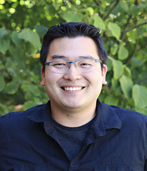 Adrian KC Lee合作论文数Department of Speech & Hearing Sciences, University of Washington3
 Armin Lak合作论文数Department of Physiology, Anatomy & Genetics, University of Oxford3
 Martin Booth合作论文数Department of Engineering Science, University of Oxford;St Hugh's College, University of Oxford3
 Stephen Michael Town合作论文数Department of Zoology, University of Cambridge3
 Vladyslav V. Vyazovskiy合作论文数Department of Physiology, Anatomy & Genetics, University of Oxford3
 Cayla E. Murphy合作论文数2Jones Gareth P合作论文数School of Pharmacy and Biomolecular Sciences, University of Brighton2Ivan P. Lazarte合作论文数1Jennifer L Marchant合作论文数1Heather K. Ortega合作论文数1Danail Stoychev合作论文数1
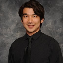 Tai Chaiamarit合作论文数1
 Dalia S Gala合作论文数Department of Biochemistry, University of Oxford1
 Jon Driver合作论文数Wellcome Trust Centre for Neuroimaging, University College London1Peter Zatka-Haas合作论文数Dept Physiol Anat & Genet, Univ Oxford1
 Martin Hailstone合作论文数University of Oxford;1
 Matthias Fritsche合作论文数Dept Physiol Anat & Genet, Univ Oxford1Hongli Wang合作论文数1
 Lucas Pinto合作论文数Department of Physiology, Feinberg School of Medicine, Northwestern University1Matthew Wincott合作论文数1
 Olaf Blanke合作论文数Geneva University Hospital1
 Jacopo Antonello合作论文数1Stella J Lee合作论文数1
 Jan Schnupp合作论文数Department of Neuroscience, City University of Hong Kong1Prsa Mario合作论文数1Gale Steven合作论文数1Kaliuzhna Mariia合作论文数School of Life Science, École Polytechnique Fédérale de Lausanne1
 Simon J. B. Butt合作论文数Butt Group, Department of Physiology, Anatomy and Genetics, Medical Sciences Division, University of Oxford1
 Andrew King合作论文数Centre for Integrative Neuroscience, Department of Physiology, Anatomy & Genetics, University of Oxford1
 Richard Parton合作论文数1
 Ilan Davis合作论文数Department of Biochemistry, University of Oxford1
 Kerry M. M. Walker合作论文数Department of Physiology, Anatomy & Genetics, University of Oxford1
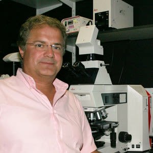 Zoltan Molnar合作论文数Department of Physiology, Anatomy & Genetics, University of Oxford1




