Hydrocephalus, characterized by cerebral ventriculomegaly, is the most common disorder requiring brain surgery in children. Recent studies have implicated SMARCC1, a component of the BRG1-associated factor (BAF) chromatin remodelling complex, as a candidate congenital hydrocephalus gene. However, SMARCC1 variants have not been systematically examined in a large patient cohort or conclusively linked with a human syndrome. Moreover, congenital hydrocephalus-associated SMARCC1 variants have not been functionally validated or mechanistically studied in vivo. Here, we aimed to assess the prevalence of SMARCC1 variants in an expanded patient cohort, describe associated clinical and radiographic phenotypes, and assess the impact of Smarcc1 depletion in a novel Xenopus tropicalis model of congenital hydrocephalus.To do this, we performed a genetic association study using whole-exome sequencing from a cohort consisting of 2697 total ventriculomegalic trios, including patients with neurosurgically-treated congenital hydrocephalus, that total 8091 exomes collected over 7 years (2016-23). A comparison control cohort consisted of 1798 exomes from unaffected siblings of patients with autism spectrum disorder and their unaffected parents were sourced from the Simons Simplex Collection. Enrichment and impact on protein structure were assessed in identified variants. Effects on the human fetal brain transcriptome were examined with RNA-sequencing and Smarcc1 knockdowns were generated in Xenopus and studied using optical coherence tomography imaging, in situ hybridization and immunofluorescence. SMARCC1 surpassed genome-wide significance thresholds, yielding six rare, protein-altering de novo variants localized to highly conserved residues in key functional domains. Patients exhibited hydrocephalus with aqueductal stenosis; corpus callosum abnormalities, developmental delay, and cardiac defects were also common. Xenopus knockdowns recapitulated both aqueductal stenosis and cardiac defects and were rescued by wild-type but not patient-specific variant SMARCC1. Hydrocephalic SMARCC1-variant human fetal brain and Smarcc1-variant Xenopus brain exhibited a similarly altered expression of key genes linked to midgestational neurogenesis, including the transcription factors NEUROD2 and MAB21L2. These results suggest de novo variants in SMARCC1 cause a novel human BAFopathy we term 'SMARCC1-associated developmental dysgenesis syndrome', characterized by variable presence of cerebral ventriculomegaly, aqueductal stenosis, developmental delay and a variety of structural brain or cardiac defects. These data underscore the importance of SMARCC1 and the BAF chromatin remodelling complex for human brain morphogenesis and provide evidence for a 'neural stem cell' paradigm of congenital hydrocephalus pathogenesis.These results highlight utility of trio-based whole-exome sequencing for identifying pathogenic variants in sporadic congenital structural brain disorders and suggest whole-exome sequencing may be a valuable adjunct in clinical management of congenital hydrocephalus patients. Hydrocephalus is the most common disease requiring neurosurgery in children. Singh et al. show an association between mutations in the chromatin remodelling gene SMARCC1 and congenital hydrocephalus with a variety of syndromic features, and perform functional studies to explore the underlying mechanisms.
ABSTRACTImportanceHydrocephalus, characterized by cerebral ventriculomegaly, is the most common disorder requiring brain surgery. A few familial forms of congenital hydrocephalus (CH) have been identified, but the cause of most sporadic cases of CH remains elusive. Recent studies have implicatedSMARCC1, a component of theBRG1-associated factor (BAF) chromatin remodeling complex, as a candidate CH gene. However,SMARCC1variants have not been systematically examined in a large patient cohort or conclusively linked with a human syndrome. Moreover, CH-associatedSMARCC1variants have not been functionally validated or mechanistically studiedin vivo.ObjectivesThe aims of this study are to (i) assess the extent to which rare, damagingde novomutations (DNMs) inSMARCC1are associated with cerebral ventriculomegaly; (ii) describe the clinical and radiographic phenotypes ofSMARCC1-mutated patients; and (iii) assess the pathogenicity and mechanisms of CH-associatedSMARCC1mutationsin vivo.Design, setting, and participantsA genetic association study was conducted using whole-exome sequencing from a cohort consisting of 2,697 ventriculomegalic trios, including patients with neurosurgically-treated CH, totaling 8,091 exomes collected over 5 years (2016-2021). Data were analyzed in 2023. A comparison control cohort consisted of 1,798 exomes from unaffected siblings of patients with autism spectrum disorder and their unaffected parents sourced from the Simons simplex consortium.Main outcomes and measuresGene variants were identified and filtered using stringent, validated criteria. Enrichment tests assessed gene-level variant burden.In silicobiophysical modeling estimated the likelihood and extent of the variant impact on protein structure. The effect of a CH-associatedSMARCC1mutation on the human fetal brain transcriptome was assessed by analyzing RNA-sequencing data.Smarcc1knockdowns and a patient-specificSmarcc1variant were tested inXenopusand studied using optical coherence tomography imaging,in situhybridization, and immunofluorescence microscopy.ResultsSMARCC1surpassed genome-wide significance thresholds in DNM enrichment tests. Six rare protein-altering DNMs, including four loss-of-function mutations and one recurrent canonical splice site mutation (c.1571+1G>A) were detected in unrelated patients. DNMs localized to the highly conserved DNA-interacting SWIRM, Myb-DNA binding, Glu-rich, and Chromo domains ofSMARCC1. Patients exhibited developmental delay (DD), aqueductal stenosis, and other structural brain and heart defects. G0 and G1Smarcc1 Xenopusmutants exhibited aqueductal stenosis and cardiac defects and were rescued by human wild-typeSMARCC1but not a patient-specificSMARCC1mutant. HydrocephalicSMARCC1-mutant human fetal brain andSmarcc1-mutantXenopusbrain exhibited a similarly altered expression of key genes linked to midgestational neurogenesis, including the transcription factorsNEUROD2andMAB21L2.ConclusionsSMARCC1is abona fideCH risk gene. DNMs inSMARCC1cause a novel human BAFopathy we term “SMARCC1-associatedDevelopmentalDysgenesisSyndrome (SaDDS)”, characterized by cerebral ventriculomegaly, aqueductal stenosis, DD, and a variety of structural brain or cardiac defects. These data underscore the importance of SMARCC1 and the BAF chromatin remodeling complex for human brain morphogenesis and provide evidence for a “neural stem cell” paradigm of human CH pathogenesis. These results highlight the utility of trio-based WES for identifying risk genes for congenital structural brain disorders and suggest WES may be a valuable adjunct in the clinical management of CH patients.KEY POINTSQuestionWhat is the role ofSMARCC1, a core component of theBRG1-associated factor (BAF) chromatin remodeling complex, in brain morphogenesis and congenital hydrocephalus (CH)?FindingsSMARCC1harbored an exome-wide significant burden of rare, protein-damagingde novomutations (DNMs) (p = 5.83 × 10⁻⁹) in the largest ascertained cohort to date of patients with cerebral ventriculomegaly, including treated CH (2,697 parent-proband trios).SMARCC1contained four loss-of-function DNMs and two identical canonical splice site DNMs in a total of six unrelated patients. Patients exhibited developmental delay, aqueductal stenosis, and other structural brain and cardiac defects.Xenopus Smarcc1mutants recapitulated core human phenotypes and were rescued by the expression of human wild-type but not patient-mutantSMARCC1. HydrocephalicSMARCC1-mutant human brain andSmarcc1-mutantXenopusbrain exhibited similar alterationsin the expression of key transcription factors that regulate neural progenitor cell proliferation.MeaningSMARCC1is essential for human brain morphogenesis and is abona fideCH risk gene.SMARCC1mutations cause a novel human BAFopathy we term “SMARCC1-associatedDevelopmentalDysgenesisSyndrome (SaDDS)”. These data implicate epigenetic dysregulation of fetal neural progenitors in the pathogenesis of hydrocephalus, with diagnostic and prognostic implications for patients and caregivers.
Cerebral arachnoid cysts (ACs) are one of the most common and poorly understood types of developmental brain lesion. To begin to elucidate AC pathogenesis, we performed an integrated analysis of 617 patient–parent (trio) exomes, 152,898 human brain and mouse meningeal single-cell RNA sequencing transcriptomes and natural language processing data of patient medical records. We found that damaging de novo variants (DNVs) were highly enriched in patients with ACs compared with healthy individuals ( P = 1.57 × 10 −33 ). Seven genes harbored an exome-wide significant DNV burden. AC-associated genes were enriched for chromatin modifiers and converged in midgestational transcription networks essential for neural and meningeal development. Unsupervised clustering of patient phenotypes identified four AC subtypes and clinical severity correlated with the presence of a damaging DNV. These data provide insights into the coordinated regulation of brain and meningeal development and implicate epigenomic dysregulation due to DNVs in AC pathogenesis. Our results provide a preliminary indication that, in the appropriate clinical context, ACs may be considered radiographic harbingers of neurodevelopmental pathology warranting genetic testing and neurobehavioral follow-up. These data highlight the utility of a systems-level, multiomics approach to elucidate sporadic structural brain disease.
In the past forty years, clinician-educators have become indispensable to academic medicine. Numerous clinician-educator-training programs exist within graduate medical education (GME) as clinician-educator tracks (CETs). However, there is a call for the clinician-educator pipeline to begin earlier. This work aims to identify and characterize clinician-educator track-like programs (CETLs) available in undergraduate medical education (UME). We developed an algorithm of 20 individual keyword queries to search the website of each U.S. allopathic medical school for CETLs. We performed the web search between March to April 2021 and repeated the search between July and September 2021. The search identified CETLs for 79 (51%) of the 155 U.S. allopathic medical schools. The identified CETLs commonly address the clinician-educator competency of educational theory (86%, 68/79), are formally organized as concentrations or analogous structures (52%, 41/79), and span all four years of medical school (37%, 29/79). The prevalence of CETLs varies with geography and medical school ranking. We provide an overview of the current state of CETLs as assessed from institutional websites. To create a future with a sustainable output of skilled clinician-educators, UME must continue to increase the number and quality of CETLs.
INTRODUCTION: Arachnoid cysts (ACs) are the most common congenital intracranial lesions. While familial cases implicate genetic factors, their ultimate cause remains unknown. Herein, we describe a multi-omics approach to disease state characterization including traditional whole exome sequencing, integrative genomics with analytics of large bulk-RNA and single cell RNA sequencing (scRNAseq) data, and a novel phenomics technique using natural language processing and Human Phenotypic Ontological (HPO) language clustering. METHODS: Whole exome sequencing was performed on 1560 individuals (520 parent-offspring "trios"). Genes with likely pathogenic mutations were defined as those with >1 de novo variant (DNV) reaching threshold for exome-wide significance (p<2.5 x 10⁻⁶) and having high pLI score (>0.9). This high-confidence pathogenic gene set and a secondary probable pathogenic gene set (>1 DNV, regardless of exome-wide significance or pLI score) were analyzed in bulk-RNA and scRNAseq datasets to assess spatiotemporal and cell-type relationships of gene expression. Phenomic analysis was conducted by subjecting patient medical records to a natural-language-processing (NLP) algorithm. Phenotypes were catalogued with the Human Phenotype Ontology (HPO) language tree and mapped with uniform manifold approximation and projection (UMAP) clustering. RESULTS: 7 genes reached our pathogenic threshold, ADNP, ARIDB1, KDM5C, PURA, FOXP1, MAP2K1, and SCN2A. Strikingly, all but SCN2A play critical roles in epigenetic regulation of gene expression in the developing brain. Bulk-RNA datasets reveal that these genes converge in networks essential for chromatin remodeling and mRNA processing. scRNAseq datasets revealed an association with excitatory and inter-neurons known to play a role in developmental delay. HPO clustering revealed four major phenotypic clusters defined by neurodevelopmental delay (NDD), seizures, both or asymptomatic. In a subgroup of traditionally phenotyped patients, there was statistically significant enrichment of neuraxial phenotypes - including NDD - among those harboring DNVs. CONCLUSION: This novel association between ACs and multiple epigenetic regulators of gene expression provides the first insight into intracranial AC pathobiology. HPO clustering reveals clinically relevant subsets of ACs and implies that traditionally symptomatic or asymptomatic ACs may be harbingers of genetically-driven aberrant neurodevelopment.
Lambdoid craniosynostosis (CS) is a congenital anomaly resulting from premature fusion of the cranial suture between the parietal and occipital bones. Predominantly sporadic, it is the rarest form of CS and its genetic etiology is largely unexplored. Exome sequencing of 25 kindreds, including 18 parent–offspring trios with sporadic lambdoid CS, revealed a marked excess of damaging (predominantly missense) de novo mutations that account for 40
Trigeminal neuralgia (TN) is a common, debilitating neuropathic face pain syndrome often resistant to therapy. The familial clustering of TN cases suggests that genetic factors play a role in disease pathogenesis. However, no unbiased, large-scale genomic study of TN has been performed to date. Analysis of 290 whole exome-sequenced TN probands, including 20 multiplex kindreds and 70 parent-offspring trios, revealed enrichment of rare, damaging variants in GABA receptor-binding genes in cases. Mice engineered with a TN-associated de novo mutation (p.Cys188Trp) in the GABAA receptor Cl- channel γ-1 subunit (GABRG1) exhibited trigeminal mechanical allodynia and face pain behavior. Other TN probands harbored rare damaging variants in Na+ and Ca+ channels, including a significant variant burden in the α-1H subunit of the voltage-gated Ca2+ channel Cav3.2 (CACNA1H). These results provide exome-level insight into TN and implicate genetically encoded impairment of GABA signaling and neuronal ion transport in TN pathogenesis.
Allocco, August A BS; Jin, Sheng C; Dong, Weilai BS; Kundishora, Adam MD; Elsamadicy, Aladine A; Dunbar, Ashley; Reeves, Benjamin; Panchagnula, Shreyas; Nelson-Williams, Carol; Lifton, Richard P MD, PhD; Kahle, Kristopher T MD, PhD Author Information
Panchagnula, Shreyas; Jin, Sheng C; Dong, Weilai BS; Kundishora, Adam MD; Moreno-De-Luca, Andres; Furey, Charuta G BA; Allocco, August A BS; Walker, Rebecca; Nelson-Williams, Carol; Smith, Hannah; Dunbar, Ashley; Conine, Sierra B BS; Lu, Qiongshi; Zen, Xue; Sierant, Michael; Knight, James; Sullivan, William; Phan, Duy BS; DeSpenza, Tyrone BA; Reeves, Benjamin; Karimy, Jason K MS; Marlier, Arnaud; Castaldi, Christopher; Tikhonova, Irina; Li, Boyang; Peña;, Helena; Broach, James; Kabachelor, Edith M; Ssenyonga, Peter; Hehnly, Christine; Ge, Li; Keren, Boris; Timberlake, Andrew T; Goto, June; Mangano, Francesco T DO; Johnston, James M MD; Butler, William MD; Warf, Benjamin C MD; Smith, Edward R MD; Schiff, Steven J MD, PhD; Limbrick, David D MD, PhD; Heuer, Gregory G MD, PhD; Jackson, Eric M MD; Iskandar, Bermans J MD; Mane, Shrikant; Haider, Shozeb PhD; Guclu, Bulent MD; Bayri, Yasar MD; Sahin, Yener; Duncan, Charles C MD; Apuzzo, Michael L.J MD; DiLuna, Michael L MD; Hoffman, Ellen; Sestan, Nenad; Ment, Laura; Alper, Seth; Bilguvar, Kaya; Geschwind, Daniel; Günel, Murat; Lifton, Richard P MD, PhD; Kahle, Kristopher T MD, PhD Author Information
Allocco, August A BS; Kundishora, Adam MD; Viviano, Stephen; Deniz, Engin; Kahle, Kristopher T MD, PhD Author Information
Pediatric midline tumors are devastating high-grade lesions with a dismal prognosis and no curative surgical options. Here, the authors report the clinical presentation, surgical management, whole-exome sequencing (WES), and clonality analysis of a patient with a radically resected H3K27M-mutant pineal parenchymal tumor (PPT) and spine metastases consistent with PPT of intermediate differentiation (PPTID). They identified somatic mutations in H3F3A (H3K27M), FGFR1, and NF1 both in the original PPT and in the PPTID metastases. They also found 12q amplification containing CDK4/MDM2 and chromosome 17 loss of heterozygosity overlapping with NF1 that resulted in biallelic NF1 loss. They noted a hypermutated phenotype with increased C>T transitions within the PPTID metastases and 2p amplification overlapping with the MYCN locus. Clonality analysis detected three founder clones maintained during progression and metastasis. Tumor clones present within the PPTID metastases but not the pineal midline tumor harbored mutations in APC and TIMP2.While the majority of H3K27M mutations are found in pediatric midline gliomas, it is increasingly recognized that this mutation is present in a wider range of lesions with a varied morphological appearance. The present case appears to be the first description of H3K27M mutation in PPTID. Somatic mutations in H3F3A, FGFR1, and NF1 have been suggested to be driver mutations in pediatric midline gliomas. Their clonality and presence in over 80% of tumor cells in our patient’s PPTID are consistent with similarly crucial roles in early tumorigenesis, with progression mediated by copy number variations and chromosomal aberrations involving known oncogenes and tumor suppressors. The roles of APC and TIMP2 mutations in progression and metastasis remain to be investigated.
Congenital hydrocephalus (CH), characterized by enlarged brain ventricles, is considered a disease of excessive cerebrospinal fluid (CSF) accumulation and thereby treated with neurosurgical CSF diversion with high morbidity and failure rates. The poor neurodevelopmental outcomes and persistence of ventriculomegaly in some post-surgical patients highlight our limited knowledge of disease mechanisms. Through whole-exome sequencing of 381 patients (232 trios) with sporadic, neurosurgically treated CH, we found that damaging de novo mutations account for >17% of cases, with five different genes exhibiting a significant de novo mutation burden. In all, rare, damaging mutations with large effect contributed to ~22% of sporadic CH cases. Multiple CH genes are key regulators of neural stem cell biology and converge in human transcriptional networks and cell types pertinent for fetal neuro-gliogenesis. These data implicate genetic disruption of early brain development, not impaired CSF dynamics, as the primary pathomechanism of a significant number of patients with sporadic CH.
Background Congenital hydrocephalus (CH) is a highly morbid disease that features enlarged brain ventricles and impaired cerebrospinal fluid homeostasis. Although early linkage or targeted sequencing studies in large multigenerational families have localized several genes for CH, the etiology of most CH cases remains unclear. Recent advances in whole exome sequencing (WES) have identified five new bona fide CH genes, implicating impaired regulation of neural stem cell fate in CH pathogenesis. Nonetheless, in the majority of CH cases, the pathological etiology remains unknown, suggesting more genes await discovery. Methods WES of family members of a sporadic and familial form of severe L1CAM mutation-negative CH associated with aqueductal stenosis was performed. Rare genetic variants were analyzed, prioritized, and validated. De novo copy number variants (CNVs) were identified using the XHMM algorithm and validated using qPCR. Xenopus oocyte experiments were performed to access mutation impact on protein function and expression. Results A novel inherited protein-damaging mutation (p.Pro605Leu) in SLC12A6, encoding the K+-Cl- cotransporter KCC3, was identified in both affected members of multiplex kindred CHYD110. p.Pro605 is conserved in KCC3 orthologs and among all human KCC paralogs. The p.Pro605Leu mutation maps to the ion-transporting domain, and significantly reduces KCC3-dependent K+ transport. A novel de novo CNV (deletion) was identified in SLC12A7, encoding the KCC3 paralog and binding partner KCC4, in another family (CHYD130) with sporadic CH. Conclusion These findings identify two novel, related genes associated with CH, and implicate genetically encoded impairments in ion transport for the first time in CH pathogenesis.
INTRODUCTION: Congenital Hydrocephalus (CH) affects 1 in 1000 live births and is treated with lifelong surgical cerebrospinal fluid (CSF) diversion with substantial morbidity. Our group recently sequenced 180 probands with CH and identified 4 novel CH genes, all of which regulate neural stem cell (NSC) fate. Nonetheless, these genes account for just 10% of studied cases. METHODS: We doubled the size of our cohort through robust recruitment and exome-sequenced 361 CH probands, including 216 case-parent trios. To study the effect of novel mutations on brain development, we developed a Xenopus tropicalis model of CH using CRISPR/Cas9. Multiple, nonoverlapping CRISPR guide RNAs targeted against candidate genes were designed and injected into fertilized single cell Xenopus embryos via microinjection. Ventricular morphology and CSF flow dynamics were analyzed via optical coherence tomography (OCT) imaging. Histological sections from WT and mutant Xenopus embryos were subjected to whole-mount in Situ hybridization to assess expression of various markers of NSC growth and proliferation. RESULTS: We identified new causative mutations in both previously identified and novel genes, all of which regulate ventricular zone NSC fate. Of these, enrichment in rare, damaging variants was highest in the SWI/SNF chromatin remodeling complex genes SMARCC1 and ARID1B (P = 1.83 × 10⁻⁹). Xenopus tadpoles harboring mutations in SMARCC1 exhibited marked ventriculomegaly, aqueductal stenosis, and impaired CSF flow. In Situ hybridization of SMARCC1 knockout brain sections demonstrated decreased expression of genes regulating NSC growth and proliferation. Notably, injection of wild type SMARCC1 RNA restored normal brain development in SMARCC1 KO embryos. CONCLUSION: These findings implicate SWI/SNF complex genes in CH and validate the use of Xenopus for investigating additional candidate genes. These findings highlight the importance of NSC dysregulation in CH pathology, and suggest that in a subset of patients the risk of adverse neurodevelopmental outcomes may be unaltered whether or not shunting is performed.
Background ATP1A3 encodes the α3 subunit of the Na+/K+ ATPase, a fundamental ion-transporting enzyme. Primarily expressed in neurons, ATP1A3 is mutated in several autosomal dominant neurological diseases. To our knowledge, damaging recessive genotypes in ATP1A3 have never been associated with any human disease. Atp1a3 deficiency in zebrafish results in hydrocephalus; however, no known association exists between ATP1A3 and human congenital hydrocephalus (CH). Methods We utilized whole-exome sequencing (WES), bioinformatics, and computational modeling to identify and characterize novel ATP1A3 mutations in a patient with CH. We performed immunohistochemical studies using mouse embryonic brain tissues to characterize Atp1a3 expression during brain development. Results We identified two germline mutations in ATP1A3 (p. Arg19Cys and p.Arg463Cys), each of which was inherited from one of the patient’s unaffected parents, in a single patient with severe obstructive CH due to aqueductal stenosis, along with open schizencephaly, type 1 Chiari malformation, and dysgenesis of the corpus callosum. Both mutations are predicted to be highly deleterious and impair protein stability. Immunohistochemical studies demonstrate robust Atp1a3 expression in neural stem cells (NSCs), differentiated neurons, and choroid plexus of the mouse embryonic brain. Conclusion These data provide the first evidence of a recessive human phenotype associated with mutations in ATP1A3, and implicate impaired Na+/K+ ATPase function in the pathogenesis of CH.
INTRODUCTION: Vein of Galen malformations (VOGMs) are morbid arteriovenous malformations, with poorly described genetis. 1 Despite improvement in endovascular treatment, VOGM mortality remains high. 2 VOGM has been reported as a rare finding in Capillary Malformation-Arteriovenous Malformation Syndrome (RASA1; OMIM #605384) and Hereditary Hemorrhagic Telangiectasia (ENG, ACVRL1; OMIM #187300, #600376). 3-4 Our previous work has identified EPH receptor tyrosine kinase as also playing a role in VOGM pathogenesis. 5 Here, we report a larger cohort of probands and identify a new gene in the same pathway as EPHB4 as playing a role in VOGM. METHODS: Germline DNA was isolated from 84 unrelated probands harboring radiographically confirmed VOGMs Both parents were available for 69/84 probands. Exome capture and paired-end WES was performed on DNA samples from participating individuals (n = 237). Data was bioinformatically analyzed to identify rare de-novo and transmitted mutations. Binomial analysis tested for exome-wide significance of mutational burden. RESULTS: Only 3/75 patients harbored mutations in previously reported VOGM-associated genes (2.3%; RASA1 n = 2, 1.1%; ACVRL1 n = 1). Significant enrichment of rare damaging mutations was found for a member of the EPH receptor tyrosine kinase family (EPHB4, n = 5; 6.0%; P = 3.31 × 10⁻⁷, 36.62-fold enrichment). Entirely novel mutations in the integrin family were also identified (n = 2; 1.1%, P = 6.03 × 10⁻⁵, 179.6-fold enrichment). Both of these mutations are located in the c-terminal domain and disrupt a binding motif. Furthermore, this integrin protein mutation is involved in the same pathway as EPHB4, although whether or not they directly interact remains unknown. CONCLUSION: This work represents an expansion upon the largest phenotyped, exome-sequenced VOGM cohort in the world. Having discovered a new gene in the same pathway as EPHB4 strongly implicates said pathway in VOMG development. We are currently pioneering tissue sampling from endovascular instruments used during treatment to explore potential somatic mutations. Our findings continue to uncover genetic determinants of VOGM pathogenesis, providing novel insight into vascular developmental biology.
Normal vascular development includes the formation and specification of arteries, veins, and intervening capillaries. Vein of Galen malformations (VOGMs) are among the most common and severe neonatal brain arterio-venous malformations, shunting arterial blood into the brain's deep venous system through aberrant direct connections. Exome sequencing of 55 VOGM probands, including 52 parent-offspring trios, revealed enrichment of rare damaging de novo mutations in chromatin modifier genes that play essential roles in brain and vascular development. Other VOGM probands harbored rare inherited damaging mutations in Ephrin signaling genes, including a genome-wide significant mutation burden in EPHB4. Inherited mutations showed incomplete penetrance and variable expressivity, with mutation carriers often exhibiting cutaneous vascular abnormalities, suggesting a two-hit mechanism. The identified mutations collectively account for ∼30% of studied VOGM cases. These findings provide insight into disease biology and may have clinical implications for risk assessment.
 Kristopher Kahle合作论文数Department of Neurosurgery, School of Medicine, Yale University;Division of Pediatric Neurosurgery, Yale University22
 Jin Sheng Chih合作论文数Department of Psychiatry, Washington University School of Medicine19
 Richard P. Lifton合作论文数Laboratory of Human Genetics and Genomics, The Rockefeller University17
 Carol Nelson-Williams合作论文数16
 Charuta Furey合作论文数School of Medicine, Yale University15
 Murat Gunel合作论文数Computational Biology and Bioinformatics, Biological & Biomedical Sciences, Yale University12
 Xue Zeng合作论文数Dept Genet, Yale Univ11Dong Wei-Lai合作论文数College of Life Sciences, Zhejiang University11
 Adam J Kundishora合作论文数Dept Neurosurg, Yale Univ10
 Duy Phan Q合作论文数Department of Neuroscience, The Ohio State University College of Medicine10Despenza Tyrone合作论文数Department of Physiology and Neurobiology, Geisel School of Medicine at Dartmouth Lebanon10
 Michael DiLuna合作论文数Department of Neurosurgery, Yale School of Medicine, Yale University9
 Haider Shozeb合作论文数Department of Pharmaceutical and Biological Chemistry, School of Pharmacy, Faculty of Life Sciences, University College London9
 Seth Leo Alper合作论文数Molecular and Vascular Medicine Unit, Beth Israel Deaconess Medical Center9Andrew Timberlake合作论文数Dept Genet, Yale Univ8Ashley Dunbar合作论文数7Sierra Conine合作论文数7
 Moreno-De-Luca Andres合作论文数Genomic Medicine Institute, Geisinger Health System7Karimy Jason K合作论文数Department of Neurosurgery, University of Maryland School of Medicine7
 Hannah Smith合作论文数Faculty of History, University of Oxford7Shrikant M. Mane合作论文数7Shreyas Panchagnula合作论文数6
 Emre Kiziltug合作论文数Dept Neurosurg, Yale Univ6Christopher Castaldi合作论文数6Jonathan Gaillard合作论文数6Benjamin C Reeves合作论文数Sch Med, Yale Univ6
 Qiongshi Lu合作论文数Department of Biostatistics & Medical Informatics, School of Medicine and Public Health, University of Wisconsin–Madison6Knight James D R合作论文数Mount Sinai Hospital, Lunenfeld-Tanenbaum Research Institute6Edward R Smith合作论文数5
 Kaya Bilguvar合作论文数School of Medicine, Yale University5
 Jungmin Choi合作论文数School of Medicine, Yale University5
 Garrett Allington合作论文数Dept Pathol, Yale Univ4Daniel Duran合作论文数Yale Sch Med, Dept Neurosurg, New Haven, CT USA4
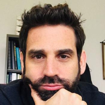 Eric S. Jackson合作论文数Department of Communicative Sciences and Disorders, Steinhardt School of Culture, Education, and Human Development, New York University4
 Engin Deniz合作论文数Department of Pediatrics, Yale School of Medicine, Yale University4
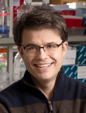 Nenad Sestan合作论文数School of Medicine, Yale University;Genome Editing Center, Yale University4
 Francesc López-Giráldez合作论文数Genètica de la Conservació Animal, Institut de Recerca i Tecnologia Agroalimentàries4Erin Loring合作论文数4Irina R. Tikhonova合作论文数3Stephen McGee合作论文数3Kedous Y Mekbib合作论文数3Stephen Viviano合作论文数3John Shohfi合作论文数3
 Michael Sierant合作论文数Rock, efeller University3
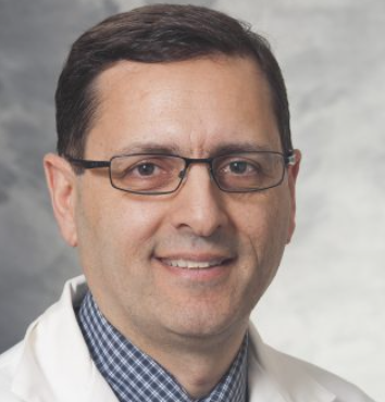 Bermans Iskandar合作论文数Department of Neurological Surgery, School of Medicine and Public Health, University of Wisconsin-Madison3Michael L J Apuzzo合作论文数3
 Shaojie Ma (马少捷)合作论文数Center for Excellence in Brain Science and Intelligent Technology, Chinese Academy of Sciences3Yener Sahin合作论文数School of Medicine, Marmara University3
 Charles Duncan合作论文数Department of Neurosurgery, Yale School of Medicine, Yale University3




