While conventional computer vision emphasizes pixel-level and feature-based objectives, medical image analysis of intricate biological structures necessitates explicit representation of their complex topological properties. Despite their successes, deep learning models often struggle to accurately capture the connectivity and continuity of fine, sometimes pixel-thin, yet critical structures due to their reliance on implicit learning from data. To address this challenge, we introduce Conformable Convolution, a novel convolutional layer designed to explicitly impose topological consistency. Conformable Convolution learns adaptive kernel offsets that focus on regions of high topological significance within an image. This prioritization is guided by our proposed Topological Posterior Generator (TPG) module, which leverages persistent homology. The TPG module identifies key topological features and guides the convolutional layers by applying persistent homology to feature maps transformed into cubical complexes. Unlike existing approaches that are merely aware of topology, our method explicitly constrains the learning process to ensure topological correctness. The proposed modules are architecture-agnostic, enabling them to be integrated seamlessly into various architectures. We showcase the effectiveness of our framework in the segmentation task, where preserving the interconnectedness of structures is critical. The results on three diverse datasets demonstrate that our framework effectively preserves the topology both quantitatively and qualitatively.
Semantic scene editing in the surgical domain presents unique challenges due to the need to preserve anatomical fidelity while altering specific scene elements. In this paper, we propose a novel image editing framework for cholecystectomy surgery using diffusion models. Our approach enables targeted modifications of surgical scenes—such as tool removal, relocation, rotation, and replacement—while maintaining a coherent representation of the operative field. By leveraging the conditional control capabilities of the diffusion model, our model semantically understands the surgical context and performs realistic inpainting to generate high-fidelity edited images. Our comprehensive quantitative and qualitative evaluations on the Cholec dataset demonstrate the proposed model’s superiority and effectiveness in preserving structural details and ensuring visual consistency in the scene editing task.
Surgical data science (SDS) is a field that analyzes patient data before, during, and after surgery to improve surgical outcomes and skills. However, surgical data is scarce, heterogeneous, and complex, which limits the applicability of existing machine learning methods. In this work, we introduce the novel task of future video generation in laparoscopic surgery. This task can augment and enrich the existing surgical data and enable various applications, such as simulation, analysis, and robot-aided surgery. Ultimately, it involves not only understanding the current state of the operation but also accurately predicting the dynamic and often unpredictable nature of surgical procedures. Our proposed method, VISAGE (VIdeo Synthesis using Action Graphs for Surgery), leverages the power of action scene graphs to capture the sequential nature of laparoscopic procedures and utilizes diffusion models to synthesize temporally coherent video sequences. VISAGE predicts the future frames given only a single initial frame, and the action graph triplets. By incorporating domain-specific knowledge through the action graph, VISAGE ensures the generated videos adhere to the expected visual and motion patterns observed in real laparoscopic procedures. The results of our experiments demonstrate high-fidelity video generation for laparoscopy procedures, which enables various applications in SDS.
Identifying causative genes from patient phenotypes remains a significant challenge in precision medicine, with important implications for the diagnosis and treatment of genetic disorders. We propose a novel graph-based approach for predicting causative genes from patient phenotypes, with or without an available list of candidate genes, by integrating a rare disease knowledge graph (KG). Our model, combining graph neural networks and transformers, achieves substantial improvements over the current state-of-the-art. On the real-world MyGene2 dataset, it attains a mean reciprocal rank (MRR) of 24.64% and nDCG@100 of 33.64%, surpassing the best baseline (SHEPHERD) at 19.02% MRR and 30.54% nDCG@100. We perform extensive ablation studies to validate the contribution of each model component. Notably, the approach generalizes to cases where only phenotypic data are available, addressing key challenges in clinical decision support when genomic information is incomplete.
Scaling by training on large datasets has been shown to enhance the quality and fidelity of image generation and manipulation with diffusion models; however, such large datasets are not always accessible in medical imaging due to cost and privacy issues, which contradicts one of the main applications of such models to produce synthetic samples where real data is scarce. Also, fine-tuning on pretrained general models has been a challenge due to the distribution shift between the medical domain and the pretrained models. Here, we propose Latent Drift (LD) for diffusion models that can be adopted for any fine-tuning method to mitigate the issues faced by the distribution shift or employed in inference time as a condition. Latent Drifting enables diffusion models to be conditioned for medical images fitted for the complex task of counterfactual image generation, which is crucial to investigate how parameters such as gender, age, and adding or removing diseases in a patient would alter the medical images. We evaluate our method on three public longitudinal benchmark datasets of brain MRI and chest X-rays for counterfactual image generation. Our results demonstrate significant performance gains in various scenarios when combined with different fine-tuning schemes.
Realistic surgical video synthesis is vital for simulation, training, and preoperative planning, yet existing methods often fall short in capturing the intricate motions intrinsic to surgical procedures. In this paper, we introduce a novel framework that generates high-fidelity surgical videos by integrating keypoints extracted via a zero-shot keypoint prediction module with the motion guidance of video diffusion models. Our approach decouples spatial content from temporal dynamics: critical keypoints representing anatomical landmarks and instrument positions are first identified in a zero-shot manner and then used to steer the diffusion process, ensuring that generated video frames adhere to plausible and consistent motion patterns. Extensive experiments on curated surgical video datasets demonstrate that our method produces temporally coherent videos with enhanced motion accuracy and visual realism compared to baseline models. The results suggest that our framework can substantially contribute to surgical training and simulation by generating dynamic and realistic surgical scenarios that faithfully reflect complex procedural movements. | .
This paper introduces Stress-Aware Learning, a resilient neural training paradigm in which deep neural networks dynamically adjust their optimization behavior - whether under stable training regimes or in settings with uncertain dynamics - based on the concept of Temporary (Elastic) and Permanent (Plastic) Deformation, inspired by structural fatigue in materials science. To instantiate this concept, we propose Plastic Deformation Optimizer, a stress-aware mechanism that injects adaptive noise into model parameters whenever an internal stress signal - reflecting stagnation in training loss and accuracy - indicates persistent optimization difficulty. This enables the model to escape sharp minima and converge toward flatter, more generalizable regions of the loss landscape. Experiments across six architectures, four optimizers, and seven vision benchmarks demonstrate improved robustness and generalization with minimal computational overhead. The code and 3D visuals will be available on GitHub: https://github.com/Stress-Aware-Learning/SAL.
While Whole Slide Imaging (WSI) scanners remain the gold standard for digitizing pathology samples, their high cost limits accessibility in many healthcare settings. Other low-cost solutions also face critical limitations: automated microscopes struggle with consistent focus across varying tissue morphology, traditional auto-focus methods require time-consuming focal stacks, and existing deep-learning approaches either need multiple input images or lack generalization capability across tissue types and staining protocols. We introduce a novel automated microscopic system powered by DeepAf, a novel auto-focus framework that uniquely combines spatial and spectral features through a hybrid architecture for single-shot focus prediction. The proposed network automatically regresses the distance to the optimal focal point using the extracted spatiospectral features and adjusts the control parameters for optimal image outcomes. Our system transforms conventional microscopes into efficient slide scanners, reducing focusing time by 80 m on same-lab samples—matching the performance of dual-image methods (0.19 m) with half the input requirements. DeepAf demonstrates robust cross-lab generalization with only 0.72 4× magnification, a significant achievement at lower magnification than typical 20 × WSI scans. This results in a comprehensive hardware-software design enabling accessible, real-time digital pathology in resource-constrained settings while maintaining diagnostic accuracy.
Graph-based holistic scene representations facilitate surgical workflow understanding and have recently demonstrated significant success. However, this task is often hindered by the limited availability of densely annotated surgical scene data. In this work, we introduce an end-to-end framework for the generation and optimization of surgical scene graphs on a downstream task. Our approach leverages the flexibility of graph-based spectral clustering and the generalization capability of foundation models to generate unsupervised scene graphs with learnable properties. We reinforce the initial spatial graph with sparse temporal connections using local matches between consecutive frames to predict temporally consistent clusters across a temporal neighborhood. By jointly optimizing the spatiotemporal relations and node features of the dynamic scene graph with the downstream task of phase segmentation, we address the costly and annotation-burdensome task of semantic scene comprehension and scene graph generation in surgical videos using only weak surgical phase labels. Further, by incorporating effective intermediate scene representation disentanglement steps within the pipeline, our solution outperforms the SOTA on the CATARACTS dataset by 8% accuracy and 10% F1 score in surgical workflow recognition.
Activation functions play a critical role in deep neural networks by shaping gradient flow, optimization stability, and generalization. While ReLU remains widely used due to its simplicity, it suffers from gradient sparsity and dead-neuron issues and offers no adaptivity to input statistics. Smooth alternatives such as Swish and GELU improve gradient propagation but still apply a fixed transformation regardless of the activation distribution. In this paper, we propose VeLU, a Variance-enhanced Learning Unit that introduces variance-aware and distributionally aligned nonlinearity through a principled combination of ArcTan-ArcSin transformations, adaptive scaling, and Wasserstein-2 regularization (Optimal Transport). This design enables VeLU to modulate its response based on local activation variance, mitigate internal covariate shift at the activation level, and improve training stability without adding learnable parameters or architectural overhead. Extensive experiments across six deep neural networks show that VeLU outperforms ReLU, ReLU6, Swish, and GELU on 12 vision benchmarks. The implementation of VeLU is publicly available in GitHub.
Recent advances in generative models for medical imaging have shown promise in representing multiple modalities. However, the variability in modality availability across datasets limits the general applicability of the synthetic data they produce. To address this, we present a novel physics-informed generative model capable of synthesizing a variable number of brain MRI modalities, including those not present in the original dataset. Our approach utilizes latent diffusion models and a two-step generative process: first, unobserved physical tissue property maps are synthesized using a latent diffusion model, and then these maps are combined with a physical signal model to generate the final MRI scan. Our experiments demonstrate the efficacy of this approach in generating unseen MR contrasts and preserving physical plausibility. Furthermore, we validate the distributions of generated tissue properties by comparing them to those measured in real brain tissue.
We propose UTS, a unit-based tissue segmentation framework for histopathology that classifies each fixed-size 32 * 32 tile, rather than each pixel, as the segmentation unit. This approach reduces annotation effort and improves computational efficiency without compromising accuracy. To implement this approach, we introduce a Multi-Level Vision Transformer (L-ViT), which benefits the multi-level feature representation to capture both fine-grained morphology and global tissue context. Trained to segment breast tissue into three categories (infiltrating tumor, non-neoplastic stroma, and fat), UTS supports clinically relevant tasks such as tumor-stroma quantification and surgical margin assessment. Evaluated on 386,371 tiles from 459 H E-stained regions, it outperforms U-Net variants and transformer-based baselines. Code and Dataset will be available at GitHub.
Surgical Video Synthesis has emerged as a promising research direction following the success of diffusion models in general-domain video generation. Although existing approaches achieve high-quality video generation, most are unconditional and fail to maintain consistency with surgical actions and phases, lacking the surgical understanding and fine-grained guidance necessary for factual simulation. We address these challenges by proposing HieraSurg, a hierarchy-aware surgical video generation framework consisting of two specialized diffusion models. Given a surgical phase and an initial frame, HieraSurg first predicts future coarse-grained semantic changes through a segmentation prediction model. The final video is then generated by a second-stage model that augments these temporal segmentation maps with fine-grained visual features, leading to effective texture rendering and integration of semantic information in the video space. Our approach leverages surgical information at multiple levels of abstraction, including surgical phase, action triplets, and panoptic segmentation maps. The experimental results on Cholecystectomy Surgical Video Generation demonstrate that the model significantly outperforms prior work both quantitatively and qualitatively, showing strong generalization capabilities and the ability to generate higher frame-rate videos. The model exhibits particularly fine-grained adherence when provided with existing segmentation maps, suggesting its potential for practical surgical applications. Project Webpage: diegobiagini.github.io/HieraSurg/ .
Despite significant progress in 3D medical image segmentation using deep learning, manual annotation remains a labor-intensive bottleneck. Self-supervised mask propagation (SMP) methods have emerged to alleviate this challenge, allowing intra-volume segmentation with just a single slice annotation. However, the previous SMP methods often rely on 2D information and ignore volumetric contexts. While our previous work, called Vol2Flow, attempts to address this concern, it exhibits limitations, including not focusing enough on local (i.e., slice-pair) information, neglecting global information (i.e., volumetric contexts) in the objective function, and error accumulation during slice-to-slice reconstruction. This paper introduces Flow2Mask, a novel SMP method, developed to overcome the limitations of previous SMP approaches, particularly Vol2Flow. During training, Flow2Mask proposes the Local-to-Global (L2G) loss to learn inter-slice flow fields among all consecutive slices within a volume in an unsupervised manner. This dynamic loss is based on curriculum learning to gradually learn information within a volume from local to global contexts. Additionally, the Inter-Slice Smoothness (ISS) loss is introduced as a regularization term to encourage changes between the slices occur consistently and continuously. During inference, Flow2Mask leverages these 3D flow fields for inter-slice mask propagation in a 3D image, spreading annotation from a single annotated slice to the entire volume. Moreover, we propose an automatic strategy to select the most representative slice as initial annotation in the mask propagation process. Experimental evaluations on different abdominal datasets demonstrate that our proposed SMP method outperforms previous approaches and improves the overall mean DSC of Vol2Flow by +2.1%, +8.2%, and +4.0% for the Sliver, CHAOS, and 3D-IRCAD datasets, respectively. Furthermore, Flow2Mask even exhibits substantial improvements in weakly-supervised and self-supervised few-shot segmentation methods when applied as a mask completion tool. The code and model for Flow2Mask are available at https://github.com/AdelehBitarafan/Flow2Mask, providing a valuable contribution to the field of medical image segmentation.
Rare genetic disease diagnosis faces critical challenges: insufficient patient data, inaccessible full genome sequencing, and the immense number of possible causative genes. These limitations cause prolonged diagnostic journeys, inappropriate treatments, and critical delays, disproportionately affecting patients in resource-limited settings where diagnostic tools are scarce. We propose RareNet, a subgraph-based Graph Neural Network that requires only patient phenotypes to identify the most likely causal gene and retrieve focused patient subgraphs for targeted clinical investigation. RareNet can function as a standalone method or serve as a pre-processing or post-processing filter for other candidate gene prioritization methods, consistently enhancing their performance while potentially enabling explainable insights. Through comprehensive evaluation on two biomedical datasets, we demonstrate competitive and robust causal gene prediction and significant performance gains when integrated with other frameworks. By requiring only phenotypic data, which is readily available in any clinical setting, RareNet democratizes access to sophisticated genetic analysis, offering particular value for underserved populations lacking advanced genomic infrastructure.
Scene graphs have emerged as accurate descriptive priors for image generation and manipulation tasks, however, their complexity and diversity of the shapes and relations of objects in data make it challenging to incorporate them into the models and generate high-quality results. To address these challenges, we propose PRISM, a novel progressive multi-head image manipulation approach to improve the accuracy and quality of the manipulated regions in the scene. Our image manipulation framework is trained using an end-to-end denoising masked reconstruction proxy task, where the masked regions are progressively unmasked from the outer regions to the inner part. We take advantage of the outer part of the masked area as they have a direct correlation with the context of the scene. Moreover, our multi-head architecture simultaneously generates detailed object-specific regions in addition to the entire image to produce higher-quality images. Our model outperforms the state-of-the-art methods in the semantic image manipulation task on the CLEVR and Visual Genome datasets. Our results demonstrate the potential of our approach for enhancing the quality and precision of scene graph-based image manipulation.
Nuclei semantic segmentation is a key component for advancing machine learning and deep learning applications in digital pathology. However, most existing segmentation models are trained and tested on high-quality data acquired with expensive equipment, such as whole slide scanners, which are not accessible to most pathologists in developing countries. These pathologists rely on low-resource data acquired with low-precision microscopes, smartphones, or digital cameras, which have different characteristics and challenges than high-resource data. Therefore, there is a gap between the state-of-the-art segmentation models and the real-world needs of low-resource settings. This work aims to bridge this gap by presenting the first fully annotated African multi-organ dataset for histopathology nuclei semantic segmentation acquired with a low-precision microscope. We also evaluate state-of-the-art segmentation models, including spectral feature extraction encoder and vision transformer-based models, and stain normalization techniques for color normalization of Hematoxylin and Eosin-stained histopathology slides. Our results provide important insights for future research on nuclei histopathology segmentation with low-resource data. Code and dataset: https://github.com/zerouaoui/AMONUSEG.
Although the preservation of shape continuity and physiological anatomy is a natural assumption in the segmentation of medical images, it is often neglected by deep learning methods that mostly aim for the statistical modeling of input data as pixels rather than interconnected structures. In biological structures, however, organs are not separate entities; for example, in reality, a severed vessel is an indication of an underlying problem, but traditional segmentation models are not designed to strictly enforce the continuity of anatomy, potentially leading to inaccurate medical diagnoses. To address this issue, we propose a graph-based approach that enforces the continuity and connectivity of anatomical topology in medical images. Our method encodes the continuity of shapes as a graph constraint, ensuring that the network's predictions maintain this continuity. We evaluate our method on three public benchmarks of retinal vessel segmentation and one neuronal structure segmentation benchmark, showing significant improvements in connectivity metrics compared to previous works while getting better or on-par performance on segmentation metrics.
While conventional computer vision emphasizes pixel-level and feature-based objectives, medical image analysis of intricate biological structures necessitates explicit representation of their complex topological properties. Despite their successes, deep learning models often struggle to accurately capture the connectivity and continuity of fine, sometimes pixel-thin, yet critical structures due to their reliance on implicit learning from data. Such shortcomings can significantly impact the reliability of analysis results and hinder clinical decision-making. To address this challenge, we introduce Conformable Convolution, a novel convolutional layer designed to explicitly enforce topological consistency. Conformable Convolution learns adaptive kernel offsets that preferentially focus on regions of high topological significance within an image. This prioritization is guided by our proposed Topological Posterior Generator (TPG) module, which leverages persistent homology. The TPG module identifies key topological features and guides the convolutional layers by applying persistent homology to feature maps transformed into cubical complexes. Our proposed modules are architecture-agnostic, enabling them to be integrated seamlessly into various architectures. We showcase the effectiveness of our framework in the segmentation task, where preserving the interconnectedness of structures is critical. Experimental results on three diverse datasets demonstrate that our framework effectively preserves the topology in the segmentation downstream task, both quantitatively and qualitatively.
Meta learning or learning to learn has been an attractive topic of research in the past years. Different methods in this area have been proposed to solve existing problems in the machine learning world. One of the common problems in machine learning that has received much attention in recent years is few-shot learning. Meta learning has been the natural solution to many few-shot learning problems. In this chapter, we introduce some background in meta learning. Then, we provide some examples of its applications in different areas, especially in medical imaging.
 Nassir Navab合作论文数School of Computation, Information and Technology, Technische Universitat Munchen;Department of Computer Science, Technical University of Munich39
 Yousef Yeganeh合作论文数Tech Univ Munich25
 Ehsan Adeli合作论文数Department of Psychiatry and Behavioral Sciences, School of Medicine, Stanford University7
 Helisa Dhamo合作论文数Apple4
 Stefano Ghidoni合作论文数Department of Computer Engineering, University of Padova3
 Federico Tombari合作论文数Google;Faculty of Computer Science, Technical University Munich;Open Perception3
 Peter Weinberger合作论文数2Amr Abuzer合作论文数2Maximilian Frantzen合作论文数2Johann Boschmann合作论文数2
 Richard Gaus合作论文数Fac Med, Ludwig Maximilians Univ Munchen2
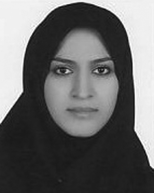 Adeleh Bitarafan合作论文数2
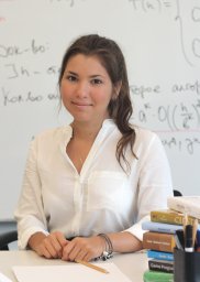 Sabrina Musatian合作论文数2
 Mohammad Farid Azampour合作论文数Chair Comp Aided Med Procedures & Augmented Real, Tech Univ Munich2
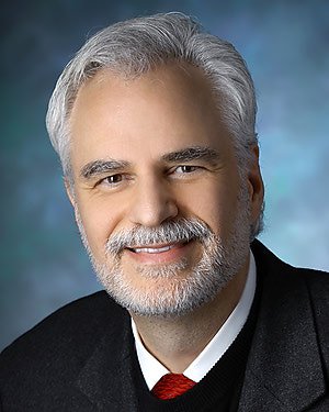 Peter Louis Gehlbach合作论文数Department of Otolaryngology, School of Medicine, Johns Hopkins University2
 Mahdieh Soleymani Baghshah合作论文数Department of Computer Science and Engineering, Sharif University of Technology2
 Björn Ommer合作论文数Computer Vision & Learning Group, Institute of Computer Science, Ludwig Maximilian University of Munich2
 Vasileios Belagiannis合作论文数School of Engineering, Democritus University of Thrace2
 Seyed-Ahmad Ahmadi合作论文数NVIDIA2Martin Hartenberger合作论文数1
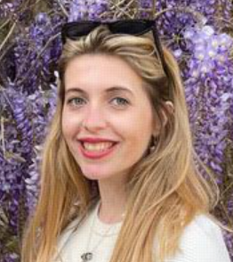 Marta Hasny合作论文数Institute for Computational Imaging and AI in Medicine, Technical University of Munich;Institute of Machine Learning for Biomedical Imaging, Helmholtz Munich1Ioannis Charisiadis合作论文数1
 Yang Zhang合作论文数National University of Singapore;Pixo AI1
 Yu Chi合作论文数Technical University of Munich;Visual Computing and Artificial Intelligence Lab, MPI-INF1Chengzhi Shen合作论文数1
 Sunday Abraham Musa合作论文数1
 Goktug Guvercin合作论文数1
 Hasnae Zerouaoui合作论文数1Felix Holm合作论文数1
 Lorenzo Nicole合作论文数Dept Med DIMED, Univ Padua1
 Pavel Jahoda合作论文数Czech Tech Univ, Fac Informat Technol, Prague, Czech Republic1
 Ghazal Ghazaei合作论文数Sch Engn, Newcastle Univ1
 Shayan Siddiqui合作论文数Department of Child and Adolescent Psychiatry, LMU München1Yawei Li合作论文数1
 Sarthak Garg合作论文数Carnegie Mellon University1
 Karima Echihabi合作论文数College of Computing, Mohammed VI Polytechnic University1
 Shadi Albarqouni合作论文数Klinik für Diagnostische und Interventionelle Radiologie, University of Bonn;Helmholtz AI;Technical University of Munich1
 Salarmehr, R.合作论文数1
 Seong Tae Kim合作论文数Sch Elect Engn, Korea Adv Inst Sci & Technol1
 Guha Roy Abhijit合作论文数1
 Christian Rupprecht合作论文数Magdalen College, University of Oxford;Department of Computer Science, University of Oxford;Yellow1
 Ashkan Khakzar合作论文数Technische Universitat Munchen1
 Gregory D. Hager合作论文数Malone Center for Engineering in Healthcare, The Johns Hopkins University;Department of Computer Science, The Johns Hopkins University1
 Iro Laina合作论文数Department of Engineering Science, University of Oxford1
 Ahmad A. Kardan合作论文数Adv E Learning Technol Lab, Amirkabir Univ Technol1
 Peter Schüffler合作论文数Department of Informatics, Technical University of Munich;Munich Data Science Institute, Technical University of Munich;School of Medicine and Health, Technical University of Munich;School of Computation, Information and Technology, Technical University of Munich1
 Christian Wachinger合作论文数Lab for Artificial Intelligence in Medical Imaging, Ludwig-Maximilians-Universität München;Technical University of Munich;Department of Child and Adolescent Psychiatry, University Hospital Munich1
 Fabio Galasso合作论文数Perception and Intelligence Lab, Dept. of Computer Science, Sapienza University of Rome;ItalAI1




