Aim: This study compared the expression of virulence genes and the ability to form biofilm in Enterococcus faecalis isolated from patients undergoing different endodontic clinical cases. Methods: For the biofilm-forming capacity, a reference strain (E. faecalis ATCC 29212) and four clinically isolated E. faecalis strains were analyzed by counting the colony-forming units, scanning electron microscopy, and optical density measurement. The gene expression of the collagen-binding protein (ace), aggregation substance (asa), endocarditis antigen (efaA), cytolysin (cylA), protein surface (esp), gelatinase (gelE), and histidine kinase (fsrC) was evaluated by a real-time polymerase chain reaction. Results: All strains were capable of forming biofilms in vitro; however, the reference strain exhibited significantly lower biofilm formation than the clinical isolates. The majority of virulence genes were expressed across all strains, with no significant differences between the clinical groups. Notably, expression of the asa gene was detected only in isolates obtained from root canals associated with apical periodontitis. Conclusion: In conclusion, all E. faecalis strains showed biofilm-forming capacity, but analysis of virulence genes showed the expression of asa only in strains isolated from patients with apical periodontitis.
AIM:Metals are essential for metabolism and play a crucial role in various biological processes. Therefore, the aim of this study was to characterize and compare the metallomic profiles of periapical lesions and healthy alveolar bone in rats using complementary analytical techniques. METHODOLOGY:This study included 76 lower first molars (from 38 Wistar rats) where induced periapical lesions and controls (sham) were compared. Periapical lesion induction was performed by pulp exposure of these teeth, allowing infection development. After 40 days, the animals were reweighed, euthanised and their hemimandibles analysed by periapical radiography, histological analysis, micro-computed tomography (μ-CT), X-ray fluorescence microscopy (μ-XRF), scanning electron microscopy with energy dispersive spectroscopy (SEM/EDS), inductively coupled plasma optical emission spectrometry (ICP-OES) and inductively coupled plasma mass spectrometry (ICP-MS). Ten essential metals for metabolism were analysed (sodium, potassium, magnesium, calcium, iron, manganese, cobalt, copper, zinc and molybdenum). The analyses observed a significance level of 5%. RESULTS:Radiographic analysis confirmed the induction of periapical lesions, without difference in animal weight between the conditions (p > .05). Histologically, the periapical lesions showed intense inflammatory infiltrate, cells with bluish cytoplasmic granules, alveolar resorption and scores ranging from moderate to intense. The μ-CT analysis of the induced lesion revealed a significant difference in the periapical region volume (12.74 mm3, p = .0017). SEM/EDS showed limited sensitivity for the investigated chemical elements; however, μ-XRF identified diminished intensities for calcium and zinc and increased intensities for iron. ICP-MS and ICP-OES identified increased concentrations of sodium (p = .0137), potassium (p = .0005), calcium (0.0059), magnesium (p = .0004), iron (p < .001), 56iron (p = .0078), 57iron (p = .0315) and manganese (p < .001) within the induced periapical lesion, suggesting a direct impact on mineral homeostasis following this pathology. CONCLUSIONS:This study demonstrated differences in the levels of various essential elements between conditions with periapical lesions and healthy controls.
Objectives:This study aimed to evaluate whether the agitation protocols using ultrasonic inserts or the XP-endo Finisher R file improved the removal of two different endodontic sealer remnants and the bond strength of fiberglass posts to dentin. Methods:Seventy-two human teeth were selected. The canals were prepared with Reciproc 50 and Easy ProDesign 30/.10 and root filled according to the endodontic sealer groups: AH Plus or EndoSequence BC Sealer HiFlow. The samples were kept at 37ºC and 95% humidity for 28 days. During the post space preparation, the obturation was removed with Largo burs, and the groups were divided according to the irrigant agitation protocols (n = 12): no agitation, agitation with R1-Clearsonic associated with E1-Irrisonic ultrasonic inserts, or agitation with XP-endo Finisher R file. The fiberglass posts were cemented with RelyX ARC. The roots were sectioned into slices and submitted to the push-out test. Micro-computed tomography analysis was used to check the effectiveness of irrigating solution agitation in the elimination of remnants. Results:The cleaning protocols with agitation were more effective in increasing the bond strength of posts to dentin for both sealer groups compared to non-agitation (p < 0.05). There was no difference between the same cleaning protocols for the different sealers. Among the different thirds, there was no statistical difference for the same sealer in the different cleaning protocols (p > 0.05). Conclusions:Both agitation protocols effectively clean root-filled canals sealed with resin-based and calcium silicate-based sealers during fiberglass post space preparation. These protocols result in improved bond strength compared to non-agitation methods.
Este estudo teve como objetivo avaliar as características superficiais e composição química de arcos ativados termicamente utilizados em sistemas autoligados. Arcos de NiTi e CuNiTi com diâmetro 0,014 (Ormco, Aditek, Orthometric, Morelli e Smart4Y) foram submetidos à condições variáveis utilizando segmentos de 10 mm divididos em 3 grupos de acordo com a imersão: não submetidos à imersão, saliva artificial (Kin Hidrat) e solução ácida (pH 4,3) utilizando 2 mL de solução e mantidos a 37ºC por 28 dias. Em seguida, as amostras foram avaliadas em microscopia eletrônica de varredura Personal SEM© eXpress™ (Aspex Corporation, Oregon, EUA). A análise semi-quantitativa realizada no modo de espectroscopia por energia dispersiva de raios X avaliou a composição química da superfície. O fio CuNiTi (Ormco) apresentou maior rugosidade, enquanto o fio termicamente ativado (Morelli), o menor. Nos fios imersos em saliva artificial a alteração superficial foi evidente. Na análise da composição química, os fios CuNiTi apresentaram porcentagens de Cu próximas a 6%. Baixas porcentagens (<0,3%) de Cu foram detectadas nos demais fios avaliados. Para todos os grupos houve predomínio dos íons Ni e Ti. Nos fios imersos em saliva artificial íons Na, Cl e K foram detectados, sugerindo componentes de deposição de saliva na superfície dos fios. Arcos CuNiTi apresentaram características de rugosidade superficial mais irregular em comparação aos demais fios avaliados. A imersão em saliva artificial alterou a superfície dos fios com deposição de íons.
Aim: This study aimed to assess the efficacy of two molecular methods, namely PCR assay and checkerboard DNA-DNA hybridization, for the detection of specific endodontic pathogens in the root canals of deciduous teeth with primary endodontic infection. It also aimed to investigate the most prevalent species detected regardless of the method used. Methods: Samples were taken from 44 cases of deciduous teeth with primary endodontic infection. DNA was extracted and the presence of microorganisms was checked by using PCR assay and checkerboard DNA-DNA hybridization assay. Fisher´s exact test was used to observe the possible difference in the prevalence of each pathogen concerning the molecular methods. Sensitivity, specificity, and accuracy tests were performed between both methods. The statistical significance level was 5%. Results: In our study, PCR and checkerboard DNA-DNA hybridization had specificity higher than sensitivity, demonstrating that these assays can identify true-negative samples. Both methods showed a similar capacity to detect specific endodontic pathogens in root canals of deciduous teeth with primary endodontic infection. PCR and checkerboard showed no statistically significant differences for most species detected, except four species: P. micra and P. nigrescens, which were more prevalent by PCR, and F. alocis and P. intermedia, which were more prevalent by checkerboard DNA-DNA hybridization. Conclusions: In conclusion, both methods are efficient in detecting endodontic pathogens and one should complement the other. Taking together the results from both methods, A. naeslundii and F. nucleatum were the most prevalent species.
This study aimed to evaluate the in vitro cytocompatibility and bioactivity of the newly launched calcium silicate-based sealer BioRoot Flow, compared with other calcium silicate-based sealers (AH Plus Bioceramic Sealer and BioRoot RCS) and the epoxy resin-based AH Plus Jet. NIH/3 T3 fibroblasts were used to assess cell viability, adhesion, and migration. Material characterisation was performed using SEM, EDS, and XRD. BioRoot RCS exhibited the highest cell viability, while BioRoot Flow showed fewer adherent cells and reduced migration at 72 h. EDS revealed zirconium content in BioRoot Flow, and XRD confirmed calcite presence in all calcium silicate-based sealers. Variations in cell behaviour may be related to differences in chemical composition and surface properties, highlighting the need for further investigation.
Introduction: Enterococcus faecalis is the most common enterococcal species associated with infective endocarditis and 1 of the most commonly detected bacteria in cases of secondary/persistent endodontic infection (SPEI). Antimicrobial resistance is a global public health concern. This review aimed to answer the following research question: "Is there a change in the antibiotic resistance profile in clinical strains of E. faecalis over the years?". P (population) - patients with SPEI, I (intervention) -endodontic retreatment, C (comparison) -not included, O (outcome) - profile of Enterococci resistance and susceptibility to systemic antibiotics used. Methods: Two authors independently performed study selection, data extraction, and risk of bias assessment. The literature search was conducted using the following electronic databases: PubMed, Scopus, EMBASE, Web of Science, and Medline. Clinical studies in which Enterococci strains were isolated to assess their antimicrobial resistance were included. Results: Eleven clinical trials were included. Overall, E. faecalis isolated from teeth with SPEI presented an intermediate resistance to 16 antibiotics. In recent years, E. faecalis showed a little resistance to amoxicillin (without clavulanate) and benzylpenicillin. Erythromycin and rifampicin presented an increase in the intermediateresistance status between the first and the last studies. E. faecium presented intermediateresistance results. Conclusion: The most effective drugs remain the combination of amoxicillin and clavulanate, followed by amoxicillin and benzylpenicillin. In patients allergic to penicillin derivatives, moxifloxacin and azithromycin may be indicated with caution. The antibiotics with the highest pattern of resistance against E. faecalis are clindamycin, gentamicin, metronidazole, and rifampicin and are therefore, contraindicated in cases of SPEI. Very few clinical studies using a microbiological approach in teeth with endodontic failure have been carried out to improve the efficacy of prophylactic regimens. However, as bacteria periodically develop resistance to the main drugs used, regular studies should be carried out on the action of these drugs in infection control. (J Endod 2024;50:299-309.)
This study evaluated the glass fibre post (GFP) removal using three different protocols with ultrasonic tips. Thirty-six extracted single-rooted teeth were endodontically treated and cemented with a 10-mm GFP. Three protocols were used for GFP removal: 20 s activated with cooling, 20 s activated without cooling and 40 s activated without cooling. The removal time and temperature increase on the external root surface were recorded. The amount of residual material and the volume of dentin loss were assessed using micro-computed tomography. Data were analysed using ANOVA with a 5% significance level. The removal time and the amount of residual material were not affected by the protocol. There was no difference in temperature increase between the 20-s groups. The 40s activated without cooling produced the highest temperatures, especially in the apical region, and greater dentin loss. It is not advisable to use the ultrasonic insert continuously for more than 20 s.
This review aimed to determine the prevalence of species of yellow, purple and green microbial complexes in root canals (RC) and periodontal pockets (PP) of teeth with endodontic-periodontal lesions. For this purpose, two reviewers searched the literature up to January 2022. Studies reporting the prevalence of species of the yellow, purple and green microbial complexes in teeth diagnosed with endodontic-periodontal lesions were included. The risk of bias of the included studies was assessed using the 14 criteria from the NIH Quality Assessment Tool. Of 1,611 references identified in the initial search, only four studies were eligible and included in the qualitative analysis. The profile and prevalence rates of bacterial species in RC and PP varied among the included studies: levels of Agregatibacter actinomycetemcomitans (12% RC, 58% PP), Capnocytophaga granulosa (10% RC, 35% PP), Capnocytophaga sputigena (15-70% RC, 0-30% PP), Streptococcus mitis (30% RC, 35% PP), Streptococcus sanguinis (30% RC, 35% PP), and Veillonella parvula (70% RC, 50% PP) were identified. The high methodological heterogeneity prevented grouping and quantitative analysis of data. The risk of bias was considered 'moderate' for all studies. The included studies identified the presence of seven bacterial species belonging to the yellow, purple, and green microbial complexes in RC and PP, but with different prevalence rates. Future clinical studies are encouraged to investigate the presence and role of these species in the occurrence and development of endodontic-periodontal lesions.
AIM:The aim of this study is to investigate the expression of inflammatory biomarkers (TNF-α, IL-10, IL-1β) and the pulpitis-associated miRNA (miR-30a-5p and miR-128-3p) in pulp tissue samples from unrestored teeth with a vital normal pulp (NP), teeth with symptomatic irreversible pulpitis (IP) and in unrestored teeth with periodontal disease, unresponsive to periodontal therapy, and a vital pulp (EP). METHODOLOGY:Thirty patients were included in this observational study (10 teeth with NP, 10 teeth with IP, 10 teeth with EP). Dental pulp tissues samples were collected from patients during root canal treatment (RCT). RNA was extracted and qRT-PCR of target genes (tumour necrosis factor [TNF]-α, interleukin [IL]-1β, IL-10) and miRNAs (has-miR-30a-5p, has-miR-128-3p) performed to assess the expression profile. Fold-change in expression was calculated using the formula 2-(ΔCt(Exp)-ΔCt(Ctrl)). One-way anova with post-hoc Tukey's was used to determine significant differences between groups. The significance level was set at 5% (p < .05). All teeth were also followed up clinically for 1 year and evaluated for a range of endodontic and periodontal-related outcomes. RESULTS:All investigated genes significantly increased in expression and miRNAs significantly decreased in expression in the IP and EP groups compared with the NP group (p < .05). With regards to TNF-α and IL-1β there were no significant differences in expression between the IP and EP groups (p > .05), whereas IL-10 expression levels were significantly reduced in the EP compared with the IP group (p < .05). Both miR-30a-5p and miR-128-3p showed significantly reduced expression in both IP and EP lesions, compared with NP (p < .05); however, no significant differences in miRNA expression were observed between IP and EP groups (p > .05). One year after root canal treatment and periodontal maintenance, tooth mobility and probing depth were significantly reduced in the EP group (p < .05). CONCLUSION:Pulp tissues from teeth with IP and EP presented similar levels of altered inflammatory markers compared with NP. TNF-α, IL-10, IL-1β cytokines and miRNAs (miR-30a-5p and miR-128-3p) are potential objective biomarkers to indicate pulpal inflammatory status, aiding diagnosis and directing clinical decision-making. RCT may be beneficial to improve stage III periodontitis unresponsive to non-surgical periodontal treatment, but further research is required.
BACKGROUND/AIM:This study assessed the epidemiology of luxation injuries with or without dental fractures in patients attending the outpatient clinic of a Brazilian dental school over the past decade. MATERIAL AND METHODS:We reviewed clinical records from a specialized center for dental trauma care in Brazil, focusing on patients who experienced at least one traumatic dental injury (TDI) in a permanent tooth between 2012 and 2022. The extracted data included sex, age, etiology, time between trauma occurrence and the search for initial care, TDI classification, and the need for endodontic treatment. The statistical analysis involved Pearson's chi-squared and Fisher's exact tests at a 5% significance level. RESULTS:The 366 analyzed clinical records included 166 patients (350 teeth) with luxation injuries. Men (n = 102) showed a higher prevalence of luxation injuries than women (n = 64). Extrusive luxation prevailed (n = 99 patients and 208 teeth). Patients with luxation injuries sought care promptly after dental trauma incidents (p = .02) and demonstrated a higher incidence of endodontic treatment (p < .0001) than those without luxation injuries. Lateral luxation was notably associated with traffic accidents (p < .0001). The combination of luxation injuries and tooth fractures did not correlate with a higher need for endodontic treatment (p > .05). CONCLUSIONS:Age and trauma etiology seemed to have influenced the epidemiological profile of luxation injuries. Additionally, these injuries affected the time to seek initial care and the need for endodontic treatment.
This in vitro study aimed to verify the influence of chlorhexidine (CHX) (gel and solution) in association with different activation protocols on disinfection of root canals contaminated with Enterococcus faecalis. In total, 120 lower premolar roots were selected, contaminated and divided into 12 groups according to irrigation technique and substance. Samples were collected before and after each irrigation technique and analysed for colony-forming units (CFU). Three dentin discs were obtained for quantification of viable intratubular cells by using confocal laser scanning microscopy (CLSM). CFU results showed that sonic and ultrasonic were more effective than conventional irrigation. Ultrasonic activation was more effective than sonic for CHX gel (p < 0.05). CLSM showed that either activation was better than conventional in all groups and root canal thirds (p < 0.05). It was concluded that ultrasonic/sonic activation was more effective than conventional techniques and CHX enhanced intratubular dentin decontamination across all irrigation methods.
Introduction: In this study, we used metatranscriptomics for the first time to investigate microbial composition, functional signatures, and antimicrobial resistance gene expression in endodontic infections. Methods: Root canal samples were collected from ten teeth, including five primary and five persistent/secondary endodontic infections. RNA from endodontic samples was extracted, and RNA sequencing was performed on a NovaSeq6000 system (Illumina). Taxonomic analysis was performed using the Kraken2 bacterial database. Then, sequences with a taxonomic classification were annotated against the Universal Protein Knowledgebase for functional annotation and the Comprehensive Antibiotic Resistance Database for AR-like gene identification. Results: Proteobacteria, Bacteroidetes, Firmicutes, and Actinobacteria represented the dominant phyla, whereas Fusobacteria, Spirochetes, and Synergistetes were among the nondominant phyla. The top ten species were mainly represented by obligate (or quasiobligate) anaerobes, including Gram-negative (eg, Capnocytophaga sp. oral taxon 323, Fusobacterium nucleatum, , Prevotela l a intermedia, , Prevotella oris, , Tannerella forsythia, and Tannerela l a sp. oral taxon HOT2286) and Gram-positive species (eg, Olsenella uli and Parvimonas micra). ). Transcripts encoding moonlighting proteins (eg, glycolytic proteins, translational elongation factors, chaperonin, and heat shock proteins) were highly expressed, potentially affecting bacterial adhesion, biofilm formation, host defense evasion, and inflammation induction. Endodontic bacteria expressed genes conferring resistance to antibiotic classes commonly used in dentistry, with a high prevalence and expression of tetracycline and lincosamide resistance genes. Antibiotic efflux and antibiotic target alteration/protection were the main resistance mechanisms. Conclusions: Metatranscriptomics revealed the activity of potential endodontic pathogens, which expressed putative virulence factors and a wide diversity of genes potentially involved in AR. (J Endod 2024;50:1059-1072.)
Endocarditis is a severe infection affecting the heart’s inner layer, the endocardium. Its pathophysiology may involve heart valve damage, bacteria adhesion and biofilm formation, potentially leading to fatal complications. Bacteria from various sources, including from endodontic diseases and its treatments may enter the bloodstream provoking this condition. This systematic review aimed to explore the influence of endodontic factors on endocarditis. Searches across PubMed, Embase, Cochrane Library and manual sources yielded 14 relevant articles from 1562 screened studies. Assessment platforms from JBI Critical Appraisal Tools evaluated studies biases. Findings mainly focused on transient bacteraemia as a key indicator of risk correlating bacterial virulence and counts with endocarditis development. Worryingly, multi-species bacteraemia post-endodontic treatment was noted including the genera Enterococcus, Parvimonas, Streptococcus and Staphylococcus. Conclusive validation of the incidence and association between endodontic patients and endocarditis was limited due to a lack of robust longitudinal investigations, such as randomized controlled trials. This emphasizes the need for further research with well-designed methodologies to provide a full understanding of the causative bacterial population and its pathological mechanisms. A current guideline (2023 European Society of Cardiology) was developed to support healthcare professionals in diagnosing and managing infective endocarditis; this 2023 version is introducing a new diagnostic algorithm to aid in patient classification aiming to improve outcomes for this challenging disease. The study was a priori registered on PROSPERO (CRD42023407736).
Introdução: Enterococcus faecalis é uma espécie bacteriana Gram-positiva anaeróbia facultativa, e Fusobacterium nucleatum é uma espécie bacteriana Gram-negativa anaeróbia estrita, sendo ambas frequentemente encontradas em dentes com insucesso do tratamento endodôntico.Objetivo: O objetivo deste estudo foi verificar as prevalências de E. faecalis e F. nucleatum em lesões periapicais de dentes em que houve insucesso do tratamento endodôntico e associá-las com as características clínicas e radiográficas observadas nos casos.Metodologia: Dez lesões periapicais foram coletadas por meio de cirurgia parendodôntica seguindo todos os cuidados com antissepsia.O procedimento foi realizado em dentes com insucesso do retratamento endodôntico e em alguns casos específicos de insucesso do tratamento endodôntico primário, como uma alternativa ao retratamento
Aim: To compare the effect of different sodium hypochlorite (NaOCl) agitation techniques on an ex vivo oral multispecies biofilm during passive disinfection of simulated immature roots. Methodology: Extracted human teeth were prepared to simulate immature roots. They were infected with a dental plaque-derived multispecies biofilm and cultured for 14 days. The roots were randomly designated into four groups: (1) negative control (PBS), (2) 1.5% NaOCl (CNI), (3) CNI + Ultrasonic activation (UA), (4) CNI + EasyClean agitation (ECA), (5) CNI + XP-endo finisher agitation (XPF), and (6) positive control (6% NaOCl). Biofilm samples were collected from the root canals and used to determine the number of viable cells (colony-forming units), scanning electron microscopy, and 16S rRNA gene sequencing. The mean colony-forming units per mL (CFU/mL) were analysed using One-way anova. 16S rRNA sequencing data were analysed for alpha (observed OTUs, Shannon index, and Chao1) and beta diversity (Bray-Curtis dissimilarities). The LEfSe analysis was used to determine the effect of treatment procedures on the abundance of root canal microbiota. The significance was set at .05. Results: PBS and CNI samples had significantly higher CFU/mL counts than UA, ECA, XPF, and 6% NaOCl samples (p < .05). The pre-treatment, PBS, and CNI groups had significantly greater alpha diversity than the UA, ECA, XPF, and 6% NaOCl groups (p < .05). NaOCl agitation groups and the 6% NaOCl group achieved a more pronounced reduction in bacteria from the genera Fusobacterium, Actinomyces, Porphyromonas, and Capnocytophaga. Conclusions: The effectiveness of passive disinfection protocols was enhanced by NaOCl agitation techniques, suggesting that this supplementary method can improve the outcome of revitalization procedures.
OBJECTIVE:To evaluate the effect of different agitation methods on apical extrusion of 1.5% sodium hypochlorite (NaOCl) in an ex vivo model of immature teeth. METHODS:Sixty extracted human inferior incisors were prepared to simulate immature teeth and embedded in an artificial root socket made of silicone impression material. The teeth were then divided into four groups: Conventional needle irrigation (CNI) alone, CNI supplemented with Ultrasonic Irrigant Activation (UIA), EasyClean (EC), or XP-endo Finisher (XPF). Extruded NaOCl was collected, reacted with m-cresol purple, and its absorbance values were measured. The data were statistically analyzed using One-way analysis of variance with a significance level of 5%. RESULTS:All groups showed apically extruded irrigating solution, and the mean volumes of extruded NaOCl did not differ significantly between any of the test groups (p⟩0.05). CONCLUSION:The activation of 1.5% NaOCL by UIA, EC, or XPF as supplementary to CNI does not promote greater apical extrusion when compared to CNI alone in simulated immature teeth.
Introduction: Nonsurgical root canal retreatment (NS-RCRT) becomes necessary when primary endodontic procedures fail. This study evaluates the efficacy of NS-RCRT using 2% chlorhexidine gel and foraminal enlargement techniques, aiming to assess whether these approaches enhance periapical healing outcomes and success rates compared to traditional NS-RCRT techniques reported in the literature. Methods: This retrospective cohort study analyzed 120 teeth diagnosed with persistent apical periodontitis, from 80 patients who underwent NS-RCRT between January 2014 and December 2018 at a specialist's private practice. Data were collected following the Preferred Reporting Items for Observational Studies in Endodontics 2023 guidelines. Periapical healing was evaluated using digital periapical radiographs by three calibrated examiners. The outcome of the treatment was analyzed through descriptive statistics and bivariate analyses, including the Chi-Square and Fisher's Exact tests. Treatment outcomes were deemed successful if they showed complete or incomplete repair and unsuccessful if no repair was observed. Results: The average followup period was 30 months. Under loose criteria, 92.50% (n = 111) of the teeth were categorized as successful, and 7.5% (n = 9) as unsuccessful. Bivariate analysis indicated that the radiographic restoration of apical transportation was the only factor that significantly influenced the outcome. Conclusions: NS-RCRT performed in a single visit using the foraminal enlargement technique and 2% chlorhexidine gel demonstrated high success rates and may be an effective alternative to tooth extraction. This method promoted periapical healing and could significantly improve NS-RCRT protocols. Further prospective studies are recommended to corroborate these findings. (J Endod 2024;50:1551-1559.)
Aim: The aim of the present study was to evaluate the solubility, flow and radiopacity of Calen (SS White Artigos Dentários Ltd, Rio de Janeiro, Brazil), Calen with zinc oxide (Calen/ZnO), calcium hydroxide + zinc oxide + 2% chlorhexidine (CH/ZnO/CHX), Calplus and Ultracal XS. Methods: 28 bovine incisor specimens were standardized, instrumented and filled with pastes with the aid of a centrix syringe. To assess solubility, the root canals of acrylic teeth (N = 10) were filled with the previously mentioned pastes and scanned by using micro–computed tomography before (initial) and after 7 days of immersion in ultrapure water. The solubility of each specimen was the difference between the initial and final volume. Flow properties were analysed according to the ISO 6876/2012 standard. Radiopacity was radiographically analysed by using Image J software. Data were submitted to analysis of variance and Tukey’s test (alpha 5%). Results: CH/ZnO/CHX and Calplus presented similar values regarding radiopacity. The group composed of Calen paste showed higher flow values. As for solubility, CH/ZnO/ CHX showed lower volume loss, but there was no statistical difference compared to other medications. Conclusion: The medication composed of calcium hydroxide, zinc oxide and 2% chlorhexidine is a suitable alternative for use in traumatized teeth.
 Alexandre Augusto Zaia合作论文数Piracicaba Dental School, State University of Campinas142
 Caio Cezar Randi Ferraz合作论文数Faculdade de Odontologia de Piracicaba, Universidade Estadual de Campinas113
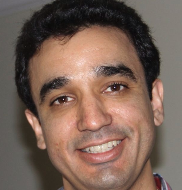 José Flávio Affonso De Almeida合作论文数Departamento of Odontologia Restauradora, Universidade Estadual de Campinas81
 Adriana De Jesus Soares合作论文数69
 Francisco Jose de Souza-Filho合作论文数48
 CCR Ferraz合作论文数47FJ Souza合作论文数42FB Teixeira合作论文数38
 Ericka T Pinheiro合作论文数Department of Dentistry, School of Dentistry, University of São Paulo/ Clinic of Conservative and Preventive Dentistry, Center of Dental Medicine, University of Zurich32
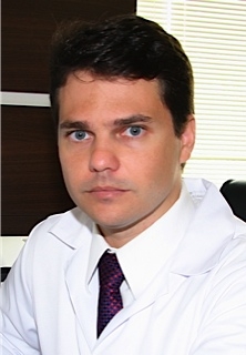 Rogerio de Castilho Jacinto合作论文数Departamento de Odontologia Preventiva e Restauradora, Faculdade de Odontologia, Sao Paulo State University32
 Marciano Marina Angélica合作论文数Bauru Dental School, University of São Paulo32Rodrigo Arruda-Vasconcelos合作论文数Div Endodont, State Univ Campinas UNICAMP31
 Martinho Frederico C合作论文数Piracicaba Dental School, State University of Campinas30
 Maira do Prado合作论文数Department of Restorative Dentistry, State University of Campinas-UNICAMP29
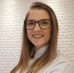 Lidiane Mendes Louzada合作论文数Piracicaba Dental School, State University of Campinas - UNICAMP27
 Francisco Montagner合作论文数Piracicaba Dental School, University of Campinas25
 Herrera Daniel Rodrigo合作论文数Dept Restorat Dent, Univ Campinas Unicamp23Juliana D. Bronzato合作论文数Ctr Nat & Human Sci, Fed Univ ABC17Francisco J. Souza-Filho合作论文数16
 Emmanuel João Nogueira Leal da Silva合作论文数Escola de Ciências da Saúde, Grande Rio University15
 Renata A. Simao合作论文数PEMM/COPPE, Universidade Federal do Rio de Janeiro14Marlos Barbosa Ribeiro合作论文数13Esdras Gabriel Alves-Silva合作论文数Piracicaba Dental School, State University of Campinas - UNICAMP13
 Thais Mageste Duque合作论文数Piracicaba Dental School, State University of Campinas13
 Endo Marcos S合作论文数São José dos Campos Dental School, State University of São Paulo13Francisco Jose De Souza Filho合作论文数13CR Gade-Neto合作论文数12ME Vianna合作论文数12
 Lauter Eston Pelepenko合作论文数Sch Dent Piracicaba, Univ Estadual Campinas12Ariane Cassia Salustiano Marinho合作论文数12Andrea Cardoso Pereira合作论文数11Db Drucker合作论文数11A Zaia合作论文数11
 Fábio Renato Manzolli Leite合作论文数Department of Dentistry and Oral Health, Aarhus University11
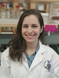 Priscila Amanda Francisco合作论文数Piracicaba Dent Sch, Univ Estadual Campinas10C Ferraz合作论文数10
 Walbert de Andrade Vieira合作论文数State University of Campinas10
 Vanessa Galego Pecorari合作论文数10Iadasa De Quadros合作论文数10ELR Sousa合作论文数9Nagata Juliana Yuri合作论文数9
 Augusto Rodrigues Lima合作论文数9
 Dametto Fabio Roberto合作论文数9
 Pedro Rosalen合作论文数Piracicaba Dental School, University of Campinas (UNICAMP);Avenida;Piracicaba;Piracicaba Dental School, University of Campinas9Hans-Peter Horz合作论文数9
 Georg Conrads合作论文数Institute of Medical Microbiology, University of Aachen [RWTH]9
 Fernanda G C Signoretti合作论文数8Marina A. Marciano合作论文数8A de-Jesus-Soares合作论文数8




