OBJECTIVE:The efficacy of nucleic acid-based vaccines against SARS-CoV-2 varies across individuals, partly due to genetic factors influencing neutralizing antibody production. In patients with systemic autoimmune diseases (SADs), this response may be further altered by immune dysregulation. METHODS:We conducted a genome-wide association study (GWAS) to identify genetic variants associated with postvaccination anti-SARS-CoV-2 IgG antibody levels and to assess whether these associations differ between patients with SAD and healthy individuals. RESULTS:The study included 165 participants (138 with SADs, 27 healthy controls), all of whom received nucleic acid-based vaccines. Antibody levels targeting the spike protein receptor-binding domain (RBD) and nucleocapsid were measured between 1 and 12 months after vaccination. GWAS results were metaanalyzed with data from a previously published GWAS with 1076 healthy individuals. We identified a novel association near RACGAP1 (rs706785; βmeta = -0.30, P meta = 3.85 × 10⁻⁸) and replicated a known association at HLA-DRB1 position 71 (βmeta = -0.23, P meta = 1.94 × 10⁻¹¹). No significant interactions were observed between genotype and disease status. CONCLUSION:This study highlights both MHC and non-MHC genetic contributions to SARS-CoV-2 vaccine responses and suggests these effects are consistent across patients with SADs and healthy individuals, supporting standard vaccination strategies for individuals with systemic autoimmune conditions.
Head and neck cancer imposes a substantial disease burden, with approximately 65,000 new cases annually in the United States and 500,000 cases worldwide. Radiation therapy remains a primary treatment modality for many patients with head and neck cancer. Despite advances aimed at limiting exposure of normal tissues within the field of radiation, salivary gland injury is a common and often unavoidable consequence of treatment. Although the mechanisms underlying radiation-induced salivary gland dysfunction remain incompletely understood, the resulting reduction in salivary flow is clinically evident and can cause substantial morbidity, including increased risk of oral infections, oral mucositis, dysphagia, and difficulty eating and speaking. Current therapeutic options are largely palliative, and durable, effective, and patient-acceptable treatments remain limited. Gene therapy offers a potential strategy for sustained functional improvement in irradiated salivary glands. The purpose of this work is to review the historical development of modified viral vectors for delivery of aquaporin-1, a water channel, to the salivary glands. We first summarize early studies using adenoviral vectors for gene delivery, which provided the basis for a Phase I clinical trial of adenoviral aquaporin-1 transfer for radiation-induced salivary gland dysfunction. We then describe the justification for our subsequent clinical trial using modified adeno-associated virus vectors for aquaporin-1 gene transfer. Finally, we outline the design of this trial, including subject recruitment, study procedures, and outcome measures.
Identifying cell types and states remains a time-consuming, error-prone challenge for spatial biology. While deep learning increasingly plays a role, it is difficult to generalize due to variability at the level of cells, neighborhoods, and niches in health and disease. To address this, we develop TACIT, an unsupervised algorithm for cell annotation using predefined signatures that operates without training data. TACIT uses unbiased thresholding to distinguish positive cells from background, focusing on relevant markers to identify ambiguous cells in multiomic assays. Using five datasets (5,000,000 cells; 51 cell types) from three niches (brain, intestine, gland), TACIT outperforms existing unsupervised methods in accuracy and scalability. Integrating TACIT-identified cell types reveals new phenotypes in two inflammatory gland diseases. Finally, using combined spatial transcriptomics and proteomics, we discover under- and overrepresented immune cell types and states in regions of interest, suggesting multimodality is essential for translating spatial biology to clinical applications.
Introduction Individuals with Fanconi anemia (FA) have an exceptionally high risk of squamous cell carcinoma (SCC) and predominantly affects the tongue and oral mucosa. FA is caused by biallelic germline pathogenic variants in one of >22 genes (e.g., FANCA, FANCD1) involved in the FA/BRCA DNA repair pathway; dysfunction in this pathway portends a 20-50-fold increased risk of SCC. Hematopoietic cell transplantation for bone marrow failure has extended the survival of FA patients by ∼20 years with patients living into their 40s. Therefore, regular monitoring for SCC is recommended for FA starting at a young age. Early detection and treatment of cancers can improve outcomes and increase the chances of survival. Materials and Methods We designed a longitudinal study (NCT00027274) to understand the natural history of oral premalignant lesions (OPL) and facilitate interception of SCC in individuals with FA. Individuals (N = 200; >8 years of age) will be enrolled over 5 years and followed prospectively for 10 years. Subjects will receive annual comprehensive cancer screening including digital intraoral scanning and oral brush biopsy to examine for evidence of oral epithelial dysplasia and oral SCC, and excisional biopsy of OPLs when warranted. Prospective cytopathological findings will be correlated with changes in DNA ploidy and histopathological grade longitudinally to determine the sensitivity and specificity of oral brush cytology and DNA cytometry in predicting clinically actionable dysplasia and SCC. Results The primary outcome measure of the study will define the success of oral screening in characterizing the natural history of OPLs. Secondary exploratory outcome measures will help to identify predictive biomarkers of oral SCC development and possibly help risk stratify patients who may require invasive, oftentimes repeated, biopsy procedures. Conclusions This is longitudinal natural history study will help us to determine the utility of brush biopsy to identify oral dysplasia and SCC in FA patients.
Lysosome-associated membrane protein 3 (LAMP3) is a unique lysosomal membrane protein specifically expressed in mature dendritic cells and type II pneumocytes. Its ectopic expression in salivary gland epithelial cells (SGECs) is induced by type I interferon (IFN) signaling and is further amplified through Toll-like receptor 7 (TLR7) activation, which is implicated in the pathogenesis of Sjögren disease (SjD). This aberrant up-regulation disrupts glandular function by promoting endolysosomal degradation of aquaporin 5 (AQP5) and sodium-potassium-chloride cotransporter 1, leading to impaired fluid secretion and associated clinical sequelae (eg, dry mouth). Additionally, LAMP3-mediated lysosomal exocytosis of DAMPs enhances monocytic bone morphogenetic protein 6 (BMP6) production, which in turn suppresses AQP5 transcription. Moreover, LAMP3 drives the extracellular vesicle-mediated release of autoantigens, which further amplifies autoimmunity, and lysosome-dependent cell death in SGECs contributes to tissue damage. These findings establish LAMP3 as a pivotal regulatory hinge in SjD pathogenesis, linking IFN/TLR7 signaling, lysosomal dysfunction, and glandular hypofunction. Its central role makes it a compelling therapeutic target, with strategies including IFN and TLR7 pathway inhibitors to limit its induction, restoration of lysosomal function, BMP6 inhibition to preserve AQP5 expression, and AQP gene therapy to improve fluid secretion.
Case A 56y male report sudden severe oral dryness 2 weeks post initiation of pembrolizumab therapy for metastatic papillary thyroid cancer (mPTC, pT2N1bMx); diagnosis and treatment for mPTC 20 months prior). The oral dryness significantly reduced his quality-of-life (QoL). No other immune-related adverse events (irAEs; i.e., ocular) were reported. Oral examination revealed erythematous mucosae and hyperlobulated depapillated tongue. Whole unstimulated saliva flow (WUSF) was 0 mL/5 minutes. Schirmer test showed reduced tear production. Laboratory testing showed elevated ACE(50.6 U/L), ANA+(1:160), and anti-SSA antibodies. Labial salivary gland biopsy (LSGB) demonstrated multiple small non-necrotizing granulomas not typical of sarcoid granulomas; microorganisms and polarizable foreign material were not identified (Focus Score[FS]:7). Lymphocytes were concentrated in granulomas and consisted mainly of T cells; CD4 = CD8 cells in the infiltrates. T cells were PD-1+; PD-L1 marked histiocytes and epithelial cells. Pembrolizumab was halted and a 5wk, 60mg prednisone taper initiated. At 5wk follow-up, the patient-reported increased saliva and restored oral QoL. WUSF was 1.8 mL/5 minutes. Schirmer test showed improved tear flow. LSGB displayed improvement in the granulomas (e.g., smaller in number and size) and infiltrating lymphocytes (FS:3). T cells (CD4 > CD8) and small clusters of B cells were associated with the granulomas. Single-cell RNA-sequencing (scRNAseq) of the LSGB confirmed changes in the immune infiltrates before/after prednisone taper. A 2-fold loss of seromucous acinar cells (SMACs) persisted after prednisone therapy; expansion of fibroblasts was observed suggestive of scarring. Differential gene expression showed activation of T-cells (GZMB, IL1B, IFNG, MKI67) after pembrolizumab and normalization after prednisone relative to healthy gland captures; whereas SMACs exhibited partial restoration salivary-specific gene expression. Conclusion To our knowledge, this is the first report of sarcoid-like granulomas involving the salivary glands in a patient treated with pembrolizumab. scRNAseq resolves the effector cells driving tissue damage illustrate response to therapy. Our data provide direct tissue evidence of irAEs and extend our mechanistic understanding of autoimmune/autoinflammatory diseases affecting the salivary glands.
Sjögren's Disease (SjD) is a systemic autoimmune disease without a clear etiology or effective therapy. Utilizing unbiased single-cell and spatial transcriptomics to analyze human minor salivary glands in health and disease we developed a comprehensive understanding of the cellular landscape of healthy salivary glands and how that landscape changes in SjD patients. We identified novel seromucous acinar cell types and identified a population of PRR4+CST3+WFDC2- seromucous acinar cells that are particularly targeted in SjD. Notably, GZMK+CD8 T cells, enriched in SjD, exhibited a cytotoxic phenotype and were physically associated with immune-engaged epithelial cells in disease. These findings shed light on the immune response's impact on transitioning acinar cells with high levels of secretion and explain the loss of this specific cell population in SjD. This study explores the complex interplay of varied cell types in the salivary glands and their role in the pathology of Sjögren's Disease.
OBJECTIVES:In patients with dermatomyositis with anti-Mi2 autoantibodies, autoantibodies can enter muscle cells, leading to the aberrant expression of genes normally repressed by the Mi2/nucleosome remodelling and deacetylation (NuRD) complex. However, the mechanism by which autoantibodies interfere with Mi2/NuRD function remains unclear. This study aimed to identify additional autoantibodies in anti-Mi2-positive patients and the epitopes recognised by these autoantibodies. METHODS:Phage immunoprecipitation sequencing (PhIP-Seq) was used to screen sera from patients with anti-Mi2 autoantibody-positive myositis. Enzyme-linked immunosorbent assays (ELISAs) and luciferase immunoprecipitation system (LIPS) immunoassays were used to detect autoantibodies in sera from healthy controls, patients with myositis, and those with other autoimmune diseases. RESULTS:PhIP-Seq identified autoantibodies recognising the autoimmune regulator (AIRE) in sera from anti-Mi2 autoantibody-positive patients. Both anti-AIRE and anti-Mi2 autoantibodies predominantly recognised a homologous region containing the plant homeodomain zinc finger type I (PHD1), which is critical for AIRE and Mi2/NuRD function. ELISA and LIPS showed that anti-Mi2 autoantibody-positive patients were positive for anti-AIRE autoantibodies, whereas AIRE reactivity was largely absent in healthy comparators, anti-Mi2 autoantibody-negative myositis, and other autoimmune diseases. Affinity-purified anti-Mi2 autoantibodies recognised both Mi2 and AIRE by ELISA, whereas anti-Mi2-depleted fractions did not recognise either protein. CONCLUSIONS:Autoantibodies targeting Mi2 recognise AIRE at a shared PHD1 epitope - a conserved motif found in numerous transcriptional regulators. These findings support a model in which anti-Mi2 autoantibodies disrupt the Mi2/NuRD complex, and potentially other PHD1-containing proteins, by interfering with chromatin binding, although further studies are needed to directly demonstrate this mechanism in vivo.
Objectives:In dermatomyositis patients with anti-Mi2 autoantibodies, autoantibodies can enter muscle cells, leading to the aberrant expression of genes normally repressed by the Mi2/nucleosome remodeling and deacetylation (NuRD) complex. However, the mechanism by which autoantibodies interfere with Mi2/NuRD function remains unclear. This study aimed to identify additional autoantibodies in anti-Mi2-positive patients as well as the specific epitopes recognized by anti-Mi2 and any novel autoantibodies. Methods:Phage ImmunoPrecipitation Sequencing (PhIP-Seq) was used to screen serum samples from anti-Mi2-positive myositis patients for autoantibodies. Enzyme-linked immunosorbent assays (ELISA) and luciferase immunoprecipitation system (LIPS) immunoassays were used to detect autoantibodies in serum samples from myositis patients and healthy controls. Results:PhIP-Seq identified autoantibodies recognizing the autoimmune regulator (AIRE) in sera from anti-Mi2 autoantibody-positive patients. Both anti-AIRE and anti-Mi2 autoantibodies predominantly recognized a homologous region of the plant homeodomain zinc finger type I (PHD1), which is critical for AIRE and Mi2/NuRD function. ELISA and LIPS testing showed that anti-Mi2 autoantibody-positive patients were positive for anti-AIRE autoantibodies, while AIRE reactivity was largely absent in healthy comparators, anti-Mi2 autoantibody-negative-myositis, and other autoimmune diseases. Affinity-purified anti-Mi2 autoantibodies recognized both Mi2 and AIRE by ELISA, whereas anti-Mi2-depleted immunoglobulin fractions did not recognize either protein. Conclusions:Autoantibodies recognizing Mi2 also recognize AIRE at a homologous PHD1 finger. This region is required by the Mi2/NuRD complex to anchor the nucleosome and consequently repress gene expression. Our findings suggest that anti-Mi2 autoantibodies disrupt NuRD complex function by binding to the PHD1 domain. Further studies are needed to determine if anti-Mi2 autoantibodies bind other PHD1-containing proteins and their functional implications.
Objectives Plant homeodomain (PHD) fingers are present in many chromatin-binding proteins. We recently discovered that anti-Mi2 autoantibodies recognize PHD fingers in Mi2 and AIRE. The purpose of this study was to characterize anti-Mi2 autoantibody recognition of PHD fingers in SP140L and TIF1γ as well as to explore recognition of TIF1γ by both anti-TIF1γ and anti-Mi2 autoantibodies. Methods Luciferase immunoprecipitation system (LIPS) assays were performed to detect autoantibodies against full-length and protein fragments of SP140L and TIF1γ in serum samples from myositis patients, disease controls, and healthy controls. Results Anti-Mi2 autoantibodies recognized SP140L. When a 49 amino acid fragment of the PHD finger of SP140L was used as the target, the specificity for selectively detecting anti-Mi2 autoantibodies increased. Additionally, anti-Mi2 autoantibodies weakly bound TIF1γ compared with anti-TIF1γ autoantibodies. Excluding the TIF1γ PHD finger from the TIF1γ target autoantigen eliminated cross-reactivity with anti-Mi2 autoantibodies, confirming that anti-Mi2 autoantibodies specifically target the PHD finger of TIF1γ. Switching two amino acids in the TIF1γ PHD finger to resemble those in AIRE markedly enhanced anti-Mi2 autoantibody immunoreactivity. Anti-TIF1γ autoantibodies primarily recognized the N-terminal fragment outside of the PHD finger, indicating this region contains the immunodominant epitopes. Conclusions Anti-Mi2 autoantibodies recognize the PHD fingers of SP140L and TIF1γ. TIF1γ is recognized by two different myositis-specific autoantibodies: anti-Mi2 autoantibodies bind the C-terminal PHD domain and anti-TIF1γ autoantibodies predominantly bind the N-terminal region. Removing the PHD finger from the anti-TIF1γ target autoantigen can improve the specificity of anti-TIF1γ autoantibody assays by reducing cross-reactivity with anti-Mi2 autoantibodies.
The immunoregulatory architecture of human oral tissues remains poorly defined despite their central role as barrier interfaces. We present the first integrated single cell and dual platform spatial proteotranscriptomic atlas of oral tissues, profiling >250,000 single-cell transcriptomes and >4 million spatially-resolved cells across 13 niches. Using our AI enabled AstroSuite (TACIT, Constellation, STARComm, hist2omics), we defined tissue cellular neighborhoods (TCNs) and multicellular interaction modules (MCIMs) in health, revealing peri-epithelial fibroblast-centered hubs enriched for effector cytokines. We harmonized eight fibroblast subtypes (universal, immune, peri-epithelial, peri-vascular, peri-neural, APC like, stress-responsive, and myofibroblasts) with stress-responsive subtypes partitioning between mucosae (Type I) and glands (Type II). Spatial multiomics mapped receptor ligand circuits and showed mucosal stress-responsive fibroblasts as immunoregulatory hubs. In chronic periodontitis, niche-aware integration of healthy and diseased datasets revealed rewiring of fibroblast phenotypes and ligand::receptor networks into interdigitated inflammatory and reparative niches. Disease neighborhoods exhibited fragmentation, expansion of MHC_I, MHC_II;, and PDL1; fibroblasts, and predicted spatial engagement with T cells at ectopic lymphoid structures. Drug2Cell analysis highlighted druggable stromal::immune networks. Together, this proteotranscriptomic atlas positions fibroblasts as central architects of structural immunity in human oral tissues and establishes a scalable framework for precision targeting of stromal::immune ecosystems across other barrier organs in health and chronic disease.
Fine mapping and bioinformatic analysis of the DDX6-CXCR5 genetic risk association in Sjögren's Disease (SjD) and Systemic Lupus Erythematosus (SLE) identified five common SNPs with functional evidence in immune cell types: rs4938573, rs57494551, rs4938572, rs4936443, rs7117261. Functional interrogation of nuclear protein binding affinity, enhancer/promoter regulatory activity, and chromatin-chromatin interactions in immune, salivary gland epithelial, and kidney epithelial cells revealed cell type-specific allelic effects for all five SNPs that expanded regulation beyond effects on DDX6 and CXCR5 expression. Mapping the local chromatin regulatory network revealed several additional genes of interest, including lnc-PHLDB1-1. Collectively, functional characterization implicated the risk alleles of these SNPs as modulators of promoter and/or enhancer activities that regulate cell type-specific expression of DDX6, CXCR5, and lnc-PHLDB1-1, among others. Further, these findings emphasize the importance of exploring the functional significance of SNPs in the context of complex chromatin architecture in disease-relevant cell types and tissues.
Aim/Introduction:We aimed to achieve direct quantitative measurement of activated and therapeutically actionable pathways (e.g., Type-I interferon) in target organs of autoimmune disease using flow cytometry of human salivary glands. Sjögren's Disease (SjD) is a systemic autoimmune disorder characterized by lymphocytic inflammation and dysfunction of the lacrimal and salivary glands. Minor salivary glands are routinely biopsied and are used for the histopathological diagnosis of SjD. In this study, we optimized the dissociation, permeabilization, antibody panel, and analytical parameters to characterize both the immune and epithelial cells in the glands, and the activation status of a specified pathway by measuring intracellular phosphorylated proteins. Methods:Fresh human MSG biopsies were dissociated into single-cell suspensions and permeabilized under optimized conditions. MSG suspensions were stained for cell surface and intracellular markers then analyzed using nine-color conventional flow cytometry, including two intracellular markers. Results:Our optimized dissociation and permeabilization protocols for human MSG preserved key cell surface markers. Our flow cytometry panel identified major immune cell populations and distinguished epithelial cells via cytokeratin-18. We demonstrate the protocol's utility showing differential interferon pathway activity in SjD vs. healthy MSG leukocytes and epithelial cells. We provide guidance on panel selection, analytical capabilities, and the impact of cell yield on resolution using conventional flow cytometry. Conclusion:Our optimized protocol enables high-resolution characterization of immune and epithelial cell populations in human MSG, preserving key markers and capturing interferon pathway activity. Our protocol provides a robust framework for the direct study of immune heterogeneity and signaling dynamics in SjD at single cell resolution.
Objectives:Plant homeodomain (PHD) fingers are present in many chromatin-binding proteins. We recently discovered that anti-Mi2 autoantibodies recognize PHD fingers in Mi2 and AIRE. The purpose of this study was to characterize anti-Mi2 autoantibody recognition of PHD fingers in SP140L and TIF1γ as well as to explore recognition of TIF1γ by both anti-TIF1γ and anti-Mi2 autoantibodies. Methods:Luciferase immunoprecipitation system (LIPS) assays were performed to detect autoantibodies against full-length and protein fragments of SP140L and TIF1γ in serum samples from myositis patients, disease controls, and healthy controls. Results:Anti-Mi2 autoantibodies recognized SP140L. When a 49 amino acid fragment of the PHD finger of SP140L was used as the target, the specificity for selectively detecting anti-Mi2 autoantibodies increased. Additionally, anti-Mi2 autoantibodies weakly bound TIF1γ compared to anti-TIF1γ autoantibodies. Excluding the TIF1γ PHD finger from the TIF1γ target autoantigen eliminated cross-reactivity with anti-Mi2 autoantibodies, confirming that anti-Mi2 autoantibodies specifically target the PHD finger of TIF1γ. Switching two amino acids in the TIF1γ PHD finger to resemble those in AIRE markedly enhanced anti-Mi2 autoantibody immunoreactivity. Anti-TIF1γ autoantibodies primarily recognized the N-terminal fragment outside of the PHD finger, indicating this region contains the immunodominant epitopes. Conclusions:Anti-Mi2 autoantibodies recognize the PHD fingers of SP140L and TIF1γ. TIF1γ is recognized by two different myositis-specific autoantibodies: anti-Mi2 autoantibodies bind the C-terminal PHD domain and anti-TIF1γ autoantibodies predominantly bind the N-terminal region. Removing the PHD finger from the anti-TIF1γ target autoantigen can improve the specificity of anti-TIF1γ autoantibody assays by reducing cross-reactivity with anti-Mi2 autoantibodies.
Saliva contains antimicrobial peptides considered integral components of host innate immunity, and crucial for protection against colonizing microbial species. Most notable is histatin-5 which is exclusively produced in salivary glands with uniquely potent antifungal activity against the opportunistic pathogen Candida albicans. Recently, SARS-CoV-2 was shown to replicate in salivary gland acinar cells eliciting local immune cell activation. In this study, we performed mechanistic and clinical studies to investigate the implications of SARS-CoV-2 infection on salivary histatin-5 production and Candida colonization. Bulk RNA-sequencing of parotid salivary glands from COVID-19 autopsies demonstrated statistically significant decreased expression of histatin genes. In situ hybridization, coupled with immunofluorescence for co-localization of SARS-CoV-2 spike and histatin in salivary gland cells, showed that histatin was absent or minimally present in acinar cells with replicating viruses. To investigate the clinical implications of these findings, salivary histatin-5 levels and oral Candida burden in saliva samples from three independent cohorts of mild and severe COVID-19 patients and matched healthy controls were evaluated. Results revealed significantly reduced histatin-5 in SARS-CoV-2 infected subjects, concomitant with enhanced prevalence of C. albicans. Analysis of prospectively recovered samples indicated that the decrease in histatin-5 is likely reversible in mild-moderate disease as concentrations tended to increase during the post-acute phase. Importantly, salivary cytokine profiling demonstrated correlations between activation of the Th17 inflammatory pathway, changes in histatin-5 concentrations, and subsequent clearance of C. albicans in a heavily colonized subject. The importance of salivary histatin-5 in controlling the proliferation of C. albicans was demonstrated using an ex vivo assay where C. albicans was able to proliferate in COVID-19 saliva with low histatin-5, but not with high histatin-5. Taken together, the findings from this study provide direct evidence implicating SARS-CoV-2 infection of salivary glands with compromised oral innate immunity, and potential predisposition to oral candidiasis.
Abstract Sjögren's Disease (SjD) is a prevalent autoimmune disorder characterized by dysfunctional lacrimal and salivary glands (SG) and lacks an effective therapy. Despite unknown pathogenic mechanisms, elevated interferon (IFN) expression is observed in SjD patients. Recent findings indicate activation of the cyclic GMP-AMP synthase (cGAS)-stimulator of interferon genes (STING) pathway in systemic lupus, prompting investigation into its role in SjD. We hypothesize cGAS-STING pathway activation in SjD contributing IFN dysregulation and symptoms. Bulk and single-cell RNA sequencing of SjD SG shows elevated IFN-related genes in epithelial and inflammatory cells. Investigation of the affected human SG by flow cytometry confirms upregulated phosphorylation status of cGAS-STING proteins in epithelial and immune cells; and epithelial cGAS-STING pathway activation directly correlates with the level of immune cell infiltration. Treatment of patient’s SG and peripheral blood mononuclear cells (PBMC) with a STING antagonist ex vivo reduces phosphorylated protein levels without cytotoxicity. These findings support our hypothesis that targeting the cGAS-STING pathway holds promise for mitigating salivary gland inflammation in SjD, preventing chronic inflammation's secondary effects and resulting tissue destruction.
Objectives: Inflammatory cytokines that signal through the JAK- STAT pathway, especially interferons (IFNs), are implicated in Sjogrens Disease (SjD). Although inhibition of JAKs is effective in other autoimmune diseases, a systematic investigation of IFN-JAK-STAT signaling and effect of JAK inhibitor (JAKi) therapy in SjD-affected human tissues has not been reported. Methods: Human minor salivary glands (MSGs) and peripheral blood mononuclear cells (PBMCs) were investigated using bulk or single cell (sc) RNA sequencing (RNAseq), immunofluorescence microscopy (IF), and flow cytometry. Ex vivo culture assays on PBMCs and primary salivary gland epithelial cell (pSGEC) lines were performed to model changes in target tissues before and after JAKi. Results: RNAseq and IF showed activated JAK-STAT pathway in SjD MSGs. Elevated IFN-stimulated gene (ISGs) expression associated with clinical variables (e.g., focus scores, anti-SSA positivity). scRNAseq of MSGs exhibited cell-type specific upregulation of JAK-STAT and ISGs; PBMCs showed similar trends, including markedly upregulated ISGs in monocytes. Ex vivo studies showed elevated basal pSTAT levels in SjD MSGs and PBMCs that were corrected with JAKi. SjD-derived pSGECs exhibited higher basal ISG expressions and exaggerated responses to IFN{beta}, which were normalized by JAKi without cytotoxicity. Conclusions: SjD patients tissues exhibit increased expression of ISGs and activation of the JAK-STAT pathway in a cell type-dependent manner. JAKi normalizes this aberrant signaling at the tissue level and in PBMCs, suggesting a putative viable therapy for SjD, targeting both glandular and extraglandular symptoms. Predicated on these data, a Phase Ib/IIa randomized controlled trial to treat SjD with tofacitinib was initiated.
IntroductionProstate-specific membrane antigen (PSMA) is present in high amounts in salivary glands, but it is unclear whether labeled binders of PSMA are excreted in the saliva.MethodsTen patients with prostate cancer underwent whole-body [18F]DCFPyL PET/CT (NCT03181867), and saliva samples were collected between 0-120 minutes post-injection. [18F]DCFPyL salivary excretion was measured over 120 minutes and expressed as %ID/g. Protein-associated binding was estimated by the percentage of [18F]DCFPyL versus parent radiotracer.ResultsAll PET scans of 10 patients (69 ± 8 years) with histologically confirmed prostate cancer (PSA= 2.4 ± 2.4, and Gleason Grade = 6-9) showed high uptake of [18F]-DCFPyL in salivary glands while 8 patients demonstrated high uptake in the saliva at 45 minutes. The intact [18F]-DCFPyL (98%) was also confirmed in the saliva samples at 120 min with increasing salivary radioactivity between 30-120 min.ConclusionSystemically injected [18F]DCFPyL shows salivary gland uptake, an increasing amount of which is secreted in saliva over time and is not maximized by 120 minutes post-injection. Although probably insignificant for diagnostic studies, patients undergoing PSMA-targeted therapies should be aware of radioactivity in saliva.
 John A. Chiorini合作论文数38
 Paola Jacqueline Perez合作论文数National Institute of Dental and Craniofacial Research, National Institutes of Health26
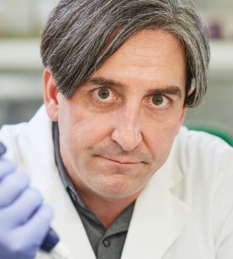 Christopher J. Lessard合作论文数Department of Pathology, University of Oklahoma Health Sciences Center;Center for Autoimmune Disease Research, Universidad del Rosario19
 David E. Kleiner合作论文数Laboratory of Pathology, Center for Cancer Research, National Cancer Institute17Margaret Beach合作论文数16
 Thomas Pranzatelli合作论文数AAV Biol Sect, NIH16
 Christopher M. Weghorst合作论文数College of Public Health, The Ohio State University16Eileen Pelayo合作论文数15
 Thomas J Knobloch合作论文数15
 Astrid Rasmussen合作论文数Department of Neurogenetics and Molecular Biology, Instituto Nacional de Neurología y Neurocirugía Manuel Velasco Suárez15Bhuwan Khatri合作论文数Genes and Human Disease Research Program, Oklahoma Medical Research Foundation14Burbelo Peter D合作论文数Lab Sensory Biol, NIH14
 A. Darise Farris合作论文数14Scofield R H合作论文数14Casto Bruce C合作论文数13
 Judith A. James合作论文数Oklahoma Medical Research Foundation;University of Oklahoma Health Sciences Center13
 Torsten Witte合作论文数13
 Maureen Rischmueller合作论文数University College London Hospital13
 Xavier Mariette合作论文数Department of Immuno-Rheumatology, Bicêtre Hospital;Immunology of Viral, Autoimmune, Haematological and Bacterial Diseases Laboratory, Université Paris-Saclay13Kevin M. Byrd合作论文数12
 Omdal Roald合作论文数Clin Immunol Unit, Stavanger Univ Hosp12
 Roland Jonsson合作论文数Department of Clinical Science, University of Bergen12
 Gunnel Nordmark合作论文数Department of Medical Sciences, Uppsala University12
 Hiroyuki Nakamura合作论文数12
 Ilias Alevizos合作论文数National Institute of Dental and Craniofacial Research, National Institutes of Health12Lars Ronnblom合作论文数12Tsutomu Tanaka合作论文数11Radfar Lida合作论文数11
 Tatsuya Atsumi合作论文数Dept Rheumatol Endocrinol & Nephrol, Hokkaido Univ11Khavandgar Zohreh合作论文数11
 Masayuki Noguchi (野口雅之)合作论文数Tsukuba Human Tissue Diagnostic Center, University of Tsukuba;Faculty of Medicine, University of Tsukuba;University Hospital, University of Tsukuba11
 Patrick M Gaffney合作论文数College of Marine Studies, University of Delaware11Shyh-Ing Jang合作论文数11
 Simon J Bowman合作论文数Institute of Inflammation and Ageing, College of Medical and Dental Sciences, University of Birmingham11Marie Wahren-Herlenius合作论文数Department of Medicine, Karolinska Hospital11Alan Baer合作论文数10
 Changyu Zheng合作论文数10Joachims Michelle L合作论文数9
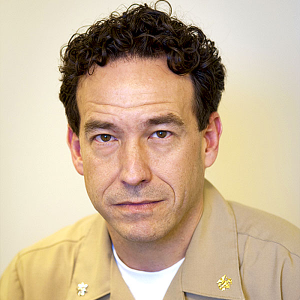 Stephen M. Hewitt合作论文数Laboratory of Pathology, Center for Cancer Research, National Cancer Institute, National Institutes of Health8
 Iago Pinal-Fernandez合作论文数National Institute of Arthritis and Musculoskeletal and Skin Disease8
 Marta E Alarcón Riquelme合作论文数Centro Pfizer-Universidad de Granada-Junta de Andalucía de Genómica e Investigación Oncológica8Sandra Afione合作论文数8Sydney Stein合作论文数7W-F Ng合作论文数7Rider Lisa G合作论文数7Chertow Daniel S合作论文数7Mammen Andrew L合作论文数7
 Peter L. Choyke合作论文数National Cancer Institute, National Institutes of Health7Betty P. Tsao合作论文数7




