Abstract Objectives Many hospitals use pneumatic tube systems (PTS) for transport of diagnostic samples. Continuous monitoring of PTS and evaluation prior to clinical use is recommended. Data loggers with specifically developed algorithms have been suggested as an additional tool in PTS evaluation. We compared two different data loggers. Methods Transport types – courier, conventional (cPTS) and innovative PTS (iPTS) – were monitored using two data loggers (MSR145® logger, CiK Solutions GmbH, Karlsruhe, Germany, and a prototype developed at the University Medicine Greifswald). Data loggers differ in algorithm, recording frequencies and limit of acceleration detection. Samples from apparently healthy volunteers were split among the transport types and results for 37 laboratory measurands were compared. Results For each logger specific arbitrary units were calculated. Area-under-the-curve (AUC)-values (MSR145®) were lowest for courier and highest for iPTS and increased with increasing recording frequencies. Stress (St)-values (prototype logger) were obtained in kmsu (1,000*mechanical stress unit) and were highest for iPTS as well. Statistical differences between laboratory measurement results of transport types were observed for three measurands sensitive for hemolysis. Conclusions The statistical, but not clinical, differences in the results for hemolysis sensitive measurands may be regarded as an early sign of preanalytical impairment. Both data loggers record this important interval of beginning mechanical stress with a high resolution indicating their potential to facilitate early detection of preanalytical impairment. Further studies should identify suitable recording frequencies. Currently, evaluation and monitoring of diagnostic sample transport should not only rely on data loggers but also include diagnostic samples.
Biopreservation and BiobankingAhead of Print Isber CornerFree AccessISBER 2021 Annual MeetingAmanda J. Moors, Benjamin Otto, Debra L. Garcia, and Marianne K. HendersonAmanda J. MoorsAddress correspondence to: Amanda J. Moors, BS, NIST Biorepository, Chemical Sciences Division, National Institute of Standards and Technology, 331 Fort Johnson Road, Charleston, SC 29412, USA E-mail Address: amanda.moors@nist.govNIST Biorepository, Chemical Sciences Division, National Institute of Standards and Technology, Charleston, South Carolina, USA.Search for more papers by this author, Benjamin OttoUniversity Medical Center Hamburg, Eppendorf, Hamburg, Germany.Search for more papers by this author, Debra L. GarciaPrivate Consultant, San Mateo, California, USA.Search for more papers by this author, and Marianne K. HendersonNational Cancer Institute, NIH, DHHS, Bethesda, MD, USA.Search for more papers by this authorPublished Online:19 Aug 2021https://doi.org/10.1089/bio.2021.29092.mjhAboutSectionsPDF/EPUB ToolsAdd to favoritesDownload CitationsTrack Citations Back To Publication ShareShare onFacebookTwitterLinked InRedditEmail The International Society for Biological and Environmental Repositories (ISBER) held its first completely virtual annual meeting from May 10 to 14, 2021. The virtual platform gave ISBER an opportunity to present, engage, and connect with the global biobanking community on current issues.This year's theme, “Connect and Collaborate through Biobanking: Powering Innovation and Discovery,” was well received and appropriate as 2020 was certainly spent seeking creative ways to connect and collaborate for everyone. While ISBER has always connected and collaborated with fellow biobankers, the past year has only punctuated the importance of bringing together experts to share and exchange their knowledge and experience with ISBER members and the global biobanking community.ISBER 2021 hosted a wide range of experts collaborating in different disciplines: academia, hospitals, biological & life sciences, clinical diagnostics, pharma, and for the first time standards in food sciences! Experts shared their experiences and research, and discussed how closer collaboration can lead to more successful future research and development and address the needs to facilitate such collaboration.The 4-day program included a Keynote address by Dr. Jim Vaught, round table discussions, 11 symposia sessions, 4 contributed article sessions, 6 educational workshops, and opportunities to network with ISBER members and learn from our corporate sponsors during Sponsor Showcases, corporate workshops, and dedicated exhibit hall time. We were thrilled to have >80 abstract submissions for our annual meeting! In addition, virtual poster sessions were available to attendees that included a range of biorepository topics of interest.The Biobanking 101 workshop was held for a 2-day period with two outstanding sessions before the annual meeting. The goal of the workshop was to provide education and interaction with newer biobankers or those that are expanding their biobank scopes. The first workshop consisted of two sections. Brent Schacter presented an update on the Qualifications in Biorepository Science Program and Helen Morrin and Rebecca Pugh presented “Success is the Result of Preparation: Emergency Planning for Biobanking.” This workshop covered preparation for emergencies that may be more frequent (e.g., power outages) to less frequent (e.g., earthquakes). The second workshop, “Basic Business Planning for BioBanking,” was presented by Marianne Henderson, Daniel Simeon-Dubach, and Kirstin Goldring provided attendees with guidance on how to fulfill their banks mission by formulating a business plan that will satisfy stakeholders through the use of Specific, Measurable, Achievable, Relevant and Time-based (SMART) performance metrics and audit points. Both of the workshop days were recorded for future use and featured polling questions to the participants, questions and answer periods, and white board interactive activities. The feedback from the participants was overwhelmingly positive, due to the interactive nature of both days' lessons.Each year, ISBER honors well-deserving members with awards and this year was no exception. ISBER paid tribute to the late Dr. Andrew Brooks by honoring him posthumously with the Outstanding Achievement in Biobanking Award. As described by Rohit Gupta “Dr. Brooks helped spawn a new generation of biobankers, inspiring them to become more involved in the discovery process, enabling new technologies and build infrastructure for emerging markets.” Dr. Fay Betsou was the recipient of the Pioneer Award for her role in developing standards and practical methods to enhance quality in biobanking. The Distinguished Leadership award was presented to Ms. Nicole Sieffert. Throughout Nicole's involvement with ISBER, she has been devoted to promoting and improving ISBER's role in the biobanking community through her exemplary work on the board of directors and her leadership of the governance committee. ISBER Special Service Awards were presented to Clare Allocca, Birendra Kumar Yadav, Billy Scheif, Karolin Bergenstrahle, Heidi Wagner, and Anusha Hettiaratchi. The ISBER Special Service Award recognizes individuals who have made exceptional contributions toward the goals of the society through the performance of a special service or act on behalf of the organization.This year, ISBER brought back the Poster Awards. The 2021 Poster Award winner was Poster PF-03: ‘Driving Innovation and Discovery Through Collaborations to Address Global Challenges at UHN Biospecimen Services’ presented by Ana Bolanos from the University Health Network. The Honorable Mentions in the Poster Awards were Poster PC-01: ‘Genetic Assessment of Cattle in the Western Gauteng Region of South Africa’ presented by Pranisha Soma from the Agricultural Research Council; Poster PJ-08: ‘Optimizing Tissue Acquisition for Biobanking through Patient Prescreening: Lessons Learned at a Tertiary Cancer Center Biorepository’ presented by Arvind Rishi from Northwell Health; and Poster PJ-10: ‘Emergent Transformation of Cancer Center Tissue Biobank to a COVID-19 Biospecimen Processing Facility: Creation of Shared Institution Resource’ presented by Sharon Fox from Northwell Health Cancer Institute.ISBER 2021 would not have had such amazing success without our invited speakers, members, workshop presenters, and the amazing work of ISBER's head office staff. A large benefit of this year's virtual format is that the meeting does not need to end; presentations, workshops, and posters are still accessible using the conference platform. For more information about ISBER and the 2021 annual meeting, please visit the website, or reach out to the authors who can point you in the right direction.Author Disclosure StatementThe authors declare that no competing financial interests exist.FiguresReferencesRelatedDetails Volume 0Issue 0 InformationCopyright 2021, Mary Ann Liebert, Inc., publishersTo cite this article:Amanda J. Moors, Benjamin Otto, Debra L. Garcia, and Marianne K. Henderson.Biopreservation and Biobanking.ahead of printhttp://doi.org/10.1089/bio.2021.29092.mjhOnline Ahead of Print:August 19, 2021PDF download
Background: Interpretation of postmortem fentanyl concentrations after transdermal application remains a challenge. There are indications that fentanyl shows relevant postmortem redistribution. The aim of this study was to investigate the time course of these changes and to develop recommendations for toxicological case work. Material and method: Blood specimens were collected from palliative care patients who were treated with fentanyl transdermal patches. Antemortem reference samples (ethylenediaminetetraacetic acid (EDTA) and serum specimens) were collected at stable dose rates. Postmortem femoral venous blood specimens were collected at four postmortem time-points: 2 hpm (hours postmortem), 6-8 hpm, 11-16 hpm and approximately 24 hpm. Liquid chromatography tandem mass spectrometry was applied to quantify fentanyl and norfentanyl. Results: Ten patients were included in the study (8 men, 2 women). Fentanyl patches with delivery rates of 12-150 mu g/h were applied. Antemortem fentanyl levels in EDTA samples varied between 0.19 and 4.64 mu g/L. At 6 to 8 hpm, blood concentrations of fentanyl were already significantly (p =0.05) higher in postmortem samples compared to the paired antemortem reference. On average, the antemortem concentration (range: 0.19-4.64 mu g/L) increased 3-fold within 6-8 hpm (range: 0.4-14.9 mu g/L), and 5.5-fold within 24 hpm (range: 0.39-21.88 mu g/L). Norfentanyl concentrations increased significantly (p = 0.01) within 6-8 hpm, too. In half of the patients, norfentanyl concentrations were below fentanyl concentrations, antemortem as well as postmortem. Conclusion: Postmortem fentanyl concentrations increased quickly. As early as 6-8 h after death, postmortem concentrations differ significantly from antemortem ones. Our results strongly indicate that postmortem blood concentrations of fentanyl after transdermal application should be interpreted carefully. (C) 2019 Elsevier B.V. All rights reserved.
We investigated the functional role of CEACAM1 in a spontaneous metastasis xenograft model of human melanoma in scid mice using BRAF wildtype MeWo cells with and without RNAi mediated knockdown of CEACAM1. Tumors from the xenograft model were subjected to whole genome expression analysis and metastasis was quantified histologically. Results and identified markers were verified using tissue samples of over 100 melanoma patients. Knockdown of CEACAM1 prolonged the animals' survival by significantly reducing subcutaneous growth of MeWo tumors and spontaneous lung metastasis. Microarray analysis revealed a strong influence of CEACAM1 knockdown on the network of EMT associated genes in the xenograft tumors (e.g. downregulation of BRAF, FOSL1, NRAS and TWIST). IGFBP7 and Latexin (highest up- and downregulated expression in microarray analysis) were found to be associated with longer and shorter survival, respectively, of melanoma patients. High FOSL1 and altered TWIST1 expression were found to be correlated with shortened survival in the cohort of melanoma patients. After a stepwise selection procedure combining above markers, multivariate analysis revealed IGFBP7, Latexin and altered TWIST to be prognostic markers for death. CEACAM1 could be a target for melanoma therapy as an alternative to (or in combination with) immune checkpoint and BRAF inhibitors.
Driven by genetic and epigenetic alterations, progression, therapy resistance and metastasis are frequent events in colorectal cancer (CRC). Although often speculated, the function of cell-cell contact for radiochemosensitivity, particularly associated with E-cadherin/catenin complex, warrants further clarification. In this study, we investigated the role of the E-cadherin/catenin complex proteins under more physiological three-dimensional (3D) cell culture conditions in a panel of CRC cell lines. In contrast to floating spheroids and growth in the laminin-rich matrix, collagen type 1 induced the formation of two distinct growth phenotypes, i.e., cell groups and single cells, in 5 out of the 8 CRC cell lines. Further characterization of these subpopulations revealed that, intriguingly, cell-cell contact proteins are important for invasion, but negligible for radiochemosensitivity, proliferation and adhesion. Despite the generation of genomic and transcriptomic data, we were unable to elucidate the mechanisms through which α-catenin affects collagen type 1 invasion. In a retrospective analysis of patients with rectal carcinoma, a low α-catenin expression trended with overall survival, as well as locoregional and distant control. Our results suggest that the E-cadherin/catenin complex proteins forming cell-cell contacts are mainly involved in the invasion, rather than the radiochemosensitivity of 3D grown CRC cells. Further studies are warranted in order to provide a better understanding of the molecular mechanisms controlling cell-cell adhesion in the context of radiochemoresistance.
While hepatitis E virus (HEV) infections have become a relevant topic in Europe, knowledge about HEV in the U.S. and South-America is still limited as no study or meta-analysis has yet compared the anti-HEV rate in the Americas. Therefore, the aim of this study was to develop a clearer understanding of anti-HEV IgG seroprevalence in the Americas identifying risk factors associated with anti-HEV seroprevalence.
The pathogenesis of the progressive liver disease, primary sclerosing cholangitis (PSC), remains largely elusive. The strong genetic association with HLA loci suggests that T cell-dependent, adaptive immune reactions could contribute to disease pathogenesis. Recent studies have indicated that PSC is also associated with polymorphisms in the locus encoding for proapoptotic Bim (BCL2L11). Bim is crucial for the maintenance of immunologic tolerance through induction of apoptosis in activated T cells. Of interest with regard to PSC is the finding that BCL2L11-deficient mice develop periductular infiltrates. We, therefore, investigated, whether defective apoptosis of T cells might contribute to the phenotype of PSC. Thus, we induced apoptosis of T cells from patients with PSC and controls by repeated T cell receptor (TCR) stimulation or cytokine withdrawal. We found that CD4(+) T cells, but not CD8(+) T cells, from patients with PSC exhibited significantly reduced apoptosis in response to both, TCR restimulation or cytokine withdrawal. This increased apoptosis resistance was associated with significantly reduced upregulation of proapoptotic Bim in T cells from patients with PSC. However, T cell apoptosis did not seem to be influenced by the previously described BCL2L11 polymorphisms. Reduced CD4(+) T cell apoptosis in patients with PSC was not due to reduced cell activation, as indicated by a similar surface expression of the activation markers CD69, CD25, and CD28 in T cells from patients and controls. Thus, decreased apoptosis of activated CD4(+) T cells may be part of the immune dysregulation observed in patients with PSC.
AIM:To identify predictive factors associated with long-term patient and graft survival (> 15 years) in liver transplant recipients.METHODS:Medical charts of all de novo adult liver transplant recipients (n = 140) who were transplanted in Hamburg between 1997 and 1999 were retrospectively reviewed. In total, 155 transplantations were identified in this time period (15 re-transplantations). Twenty-six orthotopic liver transplant (OLT) recipients were early lost to follow-up due to moving to other places within 1 year after transplantation. All remaining 114 patients were included in the analysis. The following recipient factors were analysed: Age, sex, underlying liver disease, pre-OLT body mass index (BMI), and levels of alanine aminotransferase (ALT), bilirubin, creatinine and gamma-glutamyltransferase (gamma-GT), as well as warm and cold ischemia times. Furthermore, the following donor factors were assessed: Age, BMI, cold ischemia time and warm ischemia time. All surviving patients were followed until December 2014. We divided patients into groups according to their underlying diagnosis: (1) hepatocellular carcinoma (n = 5, 4%); (2) alcohol toxic liver disease (n = 25, 22.0%); (3) primary sclerosing cholangitis (n = 6, 5%); (4) autoimmune liver diseases (n = 7, 6%); (5) hepatitis C virus cirrhosis (n = 15, 13%); (6) hepatitis B virus cirrhosis (n = 21, 19%); and (7) other (n = 35, 31%). The group "other" included rare diagnoses, such as acute liver failure, unknown liver failure, stenosis and thrombosis of the arteria hepatica, polycystic liver disease, Morbus Osler and Caroli disease.RESULTS:The majority of patients were male (n = 70, 61%). Age and BMI at the time point of transplantation ranged from 16 years to 69 years (median: 53 years) and from 15 kg/m2 to 33 kg/m2 (median: 24), respectively. Sixty-six OLT recipients (58%) experienced a follow-up of 15 years after transplantation. Recipient's age (P = 0.009) and BMI (P = 0.029) were identified as risk factors for death by χ2-test. Kaplan-Meier analysis confirmed BMI or age above the median as predictors of decreased long-term survival (P = 0.008 and P = 0.020). Hepatitis B as underlying disease showed a trend for improved long-term survival (P = 0.049, χ2-test, P = 0.055; Kaplan-Meier analysis, Log rank). Pre-transplant bilirubin, creatinine, ALT and gamma-GT levels were not associated with survival in these patients of the pre-era of the model of end stage liver disease.CONCLUSION:The recipients' age and BMI were predictors of long-term survival after OLT, as well as hepatitis B as underlying disease. In contrast, donors' age and BMI were not associated with decreased survival. These findings indicate that recipient factors especially have a high impact on long-term outcome after liver transplantation.
Background & Aims: Reduced numbers of regulatory T cells (Treg) have been reported in patients with primary sclerosing cholangitis (PSC); therefore, Treg expansion might serve as a therapeutic approach. Here, we explored whether treatment with IL-2/IL-2 monoclonal antibody complex (IL-2/IL-2Ab complex) could provide in vivo Treg expansion and treatment of experimental sclerosing cholangitis.Methods: Treg were expanded by repeated injection of IL-2/IL2Ab complex in mouse models of cholangitis (Mdr2(-/-), DDC) or colitis (dextran sulfate sodium [DSS]) as control. In vitro suppressive capacity and gene expression were analyzed in isolated hepatic and splenic Treg.Results: In vivo expansion resulted in a 5-fold increase in hepatic Treg, which localized within the inflamed portal tracts. However, although Treg expansion was associated with reduced proinflammatory IL-17 and increased anti-inflammatory IL-10 production by hepatic lymphocytes, the severity of cholangitis was not reduced. In contrast, DSS-induced colitis could be improved by Treg expansion, suggesting a selectively reduced functionality of intrahepatic Treg. Indeed, hepatic Treg manifested reduced Foxp3 expression and reduced suppressive capacity compared to splenic Treg. Hepatic Treg dysfunction could be linked to increased IL-12 signaling due to an upregulation of the IL-12 receptor. Accordingly, IL-12 receptor beta 2 knockout mice (IL12rb2(-/-)) were able to maintain hepatic Treg functionality.Conclusions: Hepatic Treg expanded in vivo failed to improve the course of cholangitis, which was related to the effects of hepatic IL-12 on Treg. Therefore, neutralization of IL-12 should be considered as part of treatment strategies targeting Treg in sclerosing cholangitis.Lay summary: Primary sclerosing cholangitis (PSC) is associated with a paucity of regulatory T cells (Treg) that have a particular ability to control immune responses; therefore, in vivo expansion of Treg might serve as a treatment of cholangitis. However, in a mouse model of PSC, we show that Treg enrichment in the liver was not sufficient to provide effective control of cholangitis, as the suppressive functionality of hepatic Treg was significantly limited by IL-12 signals. Thus, neutralization of IL-12 should be considered as part of treatment strategies to improve the efficacy of Treg-based treatments for liver diseases.
Background: The innovative pneumatic tube system (iPTS) transports one sample at a time without the use of cartridges and allows rapid sending of samples directly into the bulk loader of a laboratory automation system (LAS). We investigated effects of the iPTS on samples and turn-around time (TAT). Methods: During transport, a mini data logger recorded the accelerations in three dimensions and reported them in arbitrary area under the curve (AUC) units. In addition representative quantities of clinical chemistry, hematology and coagulation were measured and compared in 20 blood sample pairs transported by iPTS and courier. Results: Samples transported by iPTS were brought to the laboratory (300 m) within 30 s without adverse effects on the samples. The information retrieved from the data logger showed a median AUC of 7 and 310 arbitrary units for courier and iPTS transport, respectively. This is considerably below the reported limit for noticeable hemolysis of 500 arbitrary units. Conclusions: iPTS reduces TAT by reducing the hands-on time and a fast transport. No differences in the measurement results were found for any of the investigated 36 analytes between courier and iPTS transport. Based on these findings the iPTS was cleared for clinical use in our hospital.
BACKGROUND:Hepatitis C virus (HCV) infection is associated with a particularly poor outcome after liver transplantation. In December 2014, sofosbuvir/ledipasvir (SOF/LDV) fixed-dose combination (FDC) was approved for HCV genotype 1 and 4 in Europe. In orthotopic liver transplantation (OLT) recipients, the interferon-free treatment of HCV re-infection with novel direct-acting antivirals has been demonstrated to be safe and effective in clinical trials, but real-world data are missing. The aim of this study was to investigate the safety and efficacy of SOF/LDV FDC in OLT recipients in the real-life setting.METHODS:All consecutive OLT patients started on SOF/LDV FDC for 12 or 24 weeks at the University Medical Center Hamburg-Eppendorf and Medical School Hannover between October 2014 and August 2015 were retrospectively analyzed (n = 30). The primary efficacy endpoint was sustained virological response (SVR), i.e., absence of viremia 12 weeks after end of treatment (SVR 12). Liver function tests, creatinine, blood count, and HCV RNA (by polymerase chain reaction assay) were determined at each visit.RESULTS:SVR was achieved in 29/30 patients (96.67%) treated with SOF/LDV ± ribavirin (RBV) for 12 (n = 4) or 24 weeks (n = 25). Twenty-five patients (86.2%) received RBV. However, in 15 of the 25 patients, RBV administration had to be discontinued because of severe anemia (57.7%). One RBV-treated patient died of a myocardial infarction during antiviral therapy; this event was most likely not directly related to SOF/LDV. Aside from RBV-associated anemia, no severe side effects of the antiviral regimen were observed.CONCLUSION:Antiviral treatment with SOF/LDV is highly effective, safe, and well tolerated in OLT recipients. The addition of RBV often results in severe anemia, requiring dose reduction or discontinuation.
e23010 Background: E-selectin-mediated adhesion of circulating tumor cells (CTCs) to vascular endothelial cells (ECs) initiates CTC extravasation at a distant site and is hence a critical step of the metastatic cascade. E-selectin expression is induced by a pro-inflammatory milieu caused by the primary tumor, which might depend on disinhibition of NF-kB in the proteasome. Methods: in vivo, ex vivo and in vitro analyses of metastasis formation, dynamic tumor cell adhesion, glycosylation, glycsoyltransferase and glycoprotein expression. Results: We show that the approved proteasome inhibitor bortezomib (BZM) counteracts cytokine-mediated up-regulation of E-selectin on ECs. This can lead to impaired endothelial adhesion of tumor cells in vitro, reducedspontaneous pulmonary metastasis in vivo and decreased adhesion of human CTCs to pulmonary microvessels ex vivo. Surprisingly, efficacy of BZM is crucially determined by the nature of the tumor cell’s E-selectin ligands. BZM-‘sensitive’ tumors (neural crest-derived tumors) are characterized by newly specified, non-canonical, low affinity E-selectin ligands such as poly-N-acetyllactosamine, which require shear stress for E-selectin binding and mediate cell rolling under flow. In contrast, BZM-‘resistant’ tumors (gastrointestinal adenocarcinomas) are characterized by the canonical, high affinity E-selectin ligand sialyl Lewis A (CA 19-9), which does not require shear stress for E-selectin binding and mediates firm adhesion under flow. For both classes of ligands, we exemplarily demonstrate MGAT5B and C2GNT2 as key glycosyltransferases and CD44 and CD24 as glycoprotein carriers on neuroblastoma and colorectal cancer cells, respectively. The identified glycoconjugates are shown to be frequently co-expressed in corresponding clinical samples and experimental metastases. Conclusions: This is the first report proving anti-metastatic efficacy of BZM in a subset of solid human tumors. Clinical studies are needed to investigate whether BZM might be helpful to reduce metastatic spread particularly during transient periods of increased CTC release such as biopsy and surgery and whether serum sLeA (CA 19-9) can be used as a predictor of therapy response.
The transcription factor grainyhead-like 2 (GRHL2) plays a crucial role in various developmental processes. Although GRHL2 recently has attracted considerable interest in that it could be identified as a novel suppressor of the epithelial-to-mesenchymal transition, evidence is emerging that GRHL2 also exhibits tumour-promoting activities. Aim of the present study therefore was to help defining the relevance of GRHL2 for human cancers by performing a comprehensive immunohistochemical analysis of GRHL2 expression in normal (n = 608) and (n = 3,143) tumour tissues using tissue microarrays. Consistent with its accepted role in epithelial morphogenesis, GRHL2 expression preferentially but not exclusively was observed in epithelial cells. Regenerative and proliferating epithelial cells with stem cell features showed a strong GRHL2 expression. Highly complex GRHL2 expression patterns indicative of both reduced and elevated GRHL2 expression in tumours, possibly reflecting potential tumour-suppressing as well as oncogenic functions of GRHL2 in distinct human tumours, were observed. A dysregulation of GRHL2 expression for the first time was found in tumours of non-epithelial origin (e.g., astrocytomas, melanomas). We also report GRHL2 copy number gains which, however, did not necessarily translate into increased GRHL2 expression levels in cancer cells. Results obtained by meta-analysis of gene expression microarray data in conjunction with functional assays demonstrating a direct regulation of HER3 expression further point to a potential therapeutic relevance of GRHL2 in ovarian cancer. Hopefully, the results presented in this study may pave the way for a better understanding of the yet largely unknown function of GRHL2 in the initiation, progression and also therapy of cancers.
BACKGROUND & AIMS:The IL-23/IL-17 axis plays an important role in the pathogenesis of autoimmune diseases and the pathological consequences of infection. We previously showed that immunopathologic mechanisms mediated by inflammatory monocytes underlie the severe focal liver damage induced by the protozoan parasite, Entamoeba histolytica. Here, we analyze the contribution of the IL-23/IL-17 axis to the induction and subsequent recovery from parasite-induced liver damage. METHODS:IL-23p19(-/-), IL-17A/F(-/-), CCR2(-/-), and wild-type (WT) mice were intra-hepatically infected with E. histolytica trophozoites and disease onset and recovery were analyzed by magnetic resonance imaging. Liver-specific gene and protein expression during infection was examined by qPCR, microarray, FACS analysis and immunohistochemistry. Immuno-depletion and substitution experiments were performed in IL-23p19(-/-) and WT mice to investigate the role of IL-13 in disease outcome. RESULTS:Liver damage in infected IL-23p19(-/-), IL-17A/F(-/-), and CCR2(-/-) mice was strongly attenuated compared with that in WT mice. IL-23p19(-/-) mice showed reduced accumulation of IL-17 and CCL2 mRNA and proteins. Increased numbers of IL-13-producing CD11b(+)Ly6C(lo) monocytes were associated with disease attenuation in IL-23p19(-/-) mice. Immuno-depletion of IL-13 in IL-23p19(-/-) mice reversed this attenuation and treatment of infected WT mice with an IL-13/anti-IL-13-mAb complex supported liver recovery. CONCLUSIONS:The IL-23/IL-17 axis plays a critical role in the immunopathology of hepatic amebiasis. IL-13 secreted by CD11b(+)Ly6C(lo) monocytes may be associated with recovery from liver damage. An IL-13/anti-IL13-mAb complex mimics this function, suggesting a novel therapeutic option to support tissue healing after liver damage.
The cytosolic protein Sharpin is a component of the linear ubiquitin chain assembly complex, which regulates NF-kappa B signaling in response to specific ligands, such as TNF-alpha. Its inactivating mutation in chronic proliferative dermatitis mutation (Cpdm) mice causes multiorgan inflammation, yet this phenotype is not transferable into wild-type mice by hematopoietic stem cell transfer. Recent evidence demonstrated that Cpdm mice additionally display low bone mass, and that this osteopenia is corrected by Tnf deletion. Because the cellular mechanism underlying this pathology, however, was still undefined, we performed a thorough skeletal phenotyping of Cpdm mice on the basis of nondecalcified histology and cellular and dynamic histomorphometry. We show that the trabecular and cortical osteopenia in Cpdm mice is solely explained by impaired bone formation, whereas osteoclastogenesis is unaffected. Consistently, Cpdm primary calvarial cells display reduced osteogenic capacity ex vivo, and the same was observed with CD11b(-) bone marrow cells. Unexpectedly, short-term treatment of these cultures with TNF-alpha did not reveal an impaired molecular response in the absence of Sharpin. Instead, genome-wide and gene-specific expression analyses revealed that Cpdm mesenchymal cells display increased responsiveness toward TNF-alpha-induced expression of specific cytokines, such as CXCL5, IL-1 beta, and IL-6. Therefore, our data not only demonstrate that the skeletal defects of Cpdm mice are specifically caused by impaired differentiation of osteoprogenitor cells, they also suggest that increased cytokine expression in mesenchymal bone marrow cells contributes to the inflammatory phenotype of Cpdm mice.
Objectives: To investigate whether the adherens junction protein vascular endothelial cadherin (VE-cadherin) is released during Shiga toxin 2 producing Escherichia coli (STEC) infection with haemolytic uraemic syndrome (HUS) and thus could be used to assist diagnosis.Design: Using data from the large 2011 STEC outbreak in northern Europe, we determined VE-cadherin plasma concentrations in 356 patients distributed over three patient cohorts: patients with STEC infection accompanied by HUS (STEC-HUS), STEC patients without HUS (STEC) and control patients with diarrhoea but without STEC infection. We then looked for associations between VE-cadherin concentrations and disease severity defined by changes in lactate dehydrogenase, haemoglobin, creatinine, platelet count, haptoglobin and neurological symptoms.Setting: This study was conducted at the University Medical Center Hamburg-Eppendorf, Germany.Participants: 79 STEC-HUS patients, 77 STEC patients and 200 control patients were enrolled in the study.Results: We analysed 864 specimens (207 STEC, 449 STEC-HUS and 208 controls) in total. At admission, VE-cadherin concentration tended to be lower in STEC-HUS patients compared to other patients. However, HUS patients later showed an increase in VE-cadherin concentrations with prolonged elevation beyond remission. This pattern clearly differs from that observed in non-HUS patients.Conclusions: VE-cadherin concentrations are elevated in STEC-HUS patients and might be a biomarker reflecting endothelial damage in patients with HUS.
ANCA-associated vasculitis is the most frequent cause of crescentic GN. To define new molecular and/or cellular biomarkers of this disease in the kidney, we performed microarray analyses of renal biopsy samples from patients with ANCA-associated crescentic GN. Expression profiles were correlated with clinical data in a prospective study of patients with renal ANCA disease. CC chemokine ligand 18 (CCL18), acting through CC chemokine receptor 8 (CCR8) on mononuclear cells, was identified as the most upregulated chemotactic cytokine in patients with newly diagnosed ANCA-associated crescentic GN. Macrophages and myeloid dendritic cells in the kidney were detected as CCL18-producing cells. The density of CCL18(+) cells correlated with crescent formation, interstitial inflammation, and impairment of renal function. CCL18 protein levels were higher in sera of patients with renal ANCA disease compared with those in sera of patients with other forms of crescentic GN. CCL18 serum levels were higher in patients who suffered from ANCA-associated renal relapses compared with those in patients who remained in remission. Using a murine model of crescentic GN, we explored the effects of the CCL18 murine functional analog CCL8 and its receptor CCR8 on kidney function and morphology. Compared with wild-type mice, Ccr8(-/-) mice had significantly less infiltration of pathogenic mononuclear phagocytes. Furthermore, Ccr8(-/-) mice maintained renal function better and had reduced renal tissue injury. In summary, our data indicate that CCL18 drives renal inflammation through CCR8-expressing cells and could serve as a biomarker for disease activity and renal relapse in ANCA-associated crescentic GN.
In order to explore the abundance and potential environmental functions of green algal laccases, we screened various algae for extracellular laccase-like activities, characterized basic features of these activities in selected species and exemplarily studied the transformation of environmental pollutants and complex natural compounds by the laccase of Tetracystis aeria. Oxidation of the classical laccase substrate ABTS was found to be widespread in chlorophycean algae. The oxidation activity detected in members of the 'Scenedesmus' clade was caused by an unknown thermostable low-molecular-mass compound. In contrast, species of the Moewusinia, including Chlamydomonas moewusii and T. aeria, excreted putative 'true' laccases. Phenolic substrates were oxidized by these enzymes optimally at neutral to alkaline pH. The Tetracystis laccase efficiently transformed bisphenol A, 17α-ethinylestradiol, nonylphenol and triclosan in the presence of ABTS as redox mediator, while anthracene, veratrylalcohol and adlerol were not attacked. Lignosulfonate and humic acid underwent slight (de)polymerization reactions in the presence of the laccase and mediator(s), probably involving the oxidation of phenolic constituents. Possible natural functions of the enzymes, such as the synthesis of complex polymers or detoxification processes, may assist the survival of the algae in adverse environments. In contaminated surface waters, laccase-producing green algae might contribute to the environmental breakdown of phenolic pollutants.
Neurofibromatosis Type 2 (NF2) is an autosomal disorder caused by mutations of the NF2 gene. More than half of all NF2 patients have unaffected parents and carry de novo mutations, which may be of prezygotic or postzygotic origin. The latter can result in mosaicism, which is relatively common in NF2 patients. Previous studies indicated that, in 50% of patients with mosaic NF2 mutations, the mutant allele is only detectable by Sanger sequencing of PCR products amplified from tumor tissue but not from blood samples. In order to establish a highly sensitive method that has the power to detect low levels of NF2 mutant alleles from blood samples of mosaic NF2 patients, we performed ultra deep sequencing and calculated the percentage of mutant and wildtype NF2 alleles. The mutant allele frequencies detected ranged from 2.6% to 19.7%. In three patients, however, the NF2 mutation previously identified in tumor tissue was not identified in blood samples by means of deep sequencing, suggesting absence of mutant cells in the blood. Remarkably, we observed a correlation between the age at onset of the disease and the mutant allele frequency. Our study indicates that ultra deep sequencing is an effective and highly sensitive method to determine the mutant allele frequency in patients with mosaic NF2 gene mutations, which enables extended phenotype/correlations in these patients. © 2015 Wiley Periodicals, Inc.
 Thomas Streichert合作论文数18Udo Schumacher合作论文数7Christoph Wagener合作论文数7Tolstonog Genrich V合作论文数5Lohse Ansgar W合作论文数4Klaetschke Kristin合作论文数4
 Petersmann Astrid合作论文数4Wolfgang Deppert合作论文数4
 Daniel Wicklein合作论文数Diagnostic Center, University Hospital Hamburg-Eppendorf4Jeschke Anke合作论文数3Matthias Nauck合作论文数Institute of Clinical Chemistry and Laboratory Medicine, University Medicine of Greifswald3
 Ulrich Lehmann合作论文数Institut für Pathologie, Medizinische Hochschule Hannover3
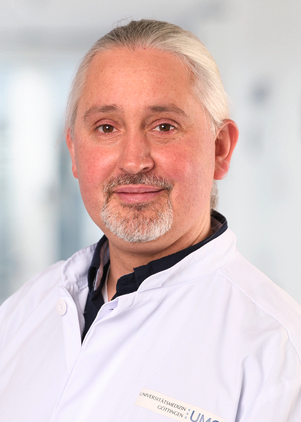 Florian Wegwitz合作论文数Department of Gynecology and Obstetrics, University Medical Center Göttingen3
 Dietmar Schlosser合作论文数3Anne Greiser合作论文数2
 Martin Horstmann合作论文数2Ronald Simon合作论文数2
 Christoph Schramm合作论文数2
 Gerrit Wolters-Eisfeld合作论文数Department of General, Visceral and Thoracic Surgery, University Medical Center Hamburg-Eppendorf2
 Dorothee Schwinge合作论文数2Harry R Dalton合作论文数Royal Cornwall Hospitals NHS Trust2Pischke S合作论文数2Balabanov Stefan合作论文数2
 Pierre Abramowski合作论文数2
 Johannes Hartl合作论文数2Maar Hanna合作论文数2Suchsland Juliane合作论文数2Harder Lena合作论文数2Michael Amling合作论文数2Schoknecht Tanja合作论文数2
 Jakob R. Izbicki合作论文数Department of General, Visceral & Thoracic Surgery, University Medical Center Hamburg-Eppendorf2
 Klaus Pantel合作论文数Institute of Tumor Biology, University Hospital Hamburg-Eppendorf;Center for Experimental Medicine, University Hospital Hamburg-Eppendorf2
 Frank Tacke合作论文数Charité – Universitätsmedizin Berlin2
 Carsten Bokemeyer合作论文数Zentrum Für Onkologie, Universitätsklinikum Hamburg-Eppendorf2A. W. Lohse合作论文数2Johannes Herkel合作论文数2Werner Reisser合作论文数2
 Guido Sauter合作论文数Zentrum für Diagnostik, Universitätsklinikum Hamburg-Eppendorf;Ambulanzzentrum des UKE GmbH, Universitätsklinikum Hamburg-Eppendorf;Fachbereich Pathologie und Gynpathologie, Universitätsklinikum Hamburg-Eppendorf2Ursula Valentiner合作论文数2Martina Sterneck合作论文数2Christina Heinlein合作论文数2S. Pischke合作论文数2Thorsten Schinke合作论文数2Katharina Gruner合作论文数2
 Tobias Lange合作论文数2
 Samuel Huber合作论文数I. Department of Medicine, University Medical Centre Hamburg-Eppendorf2D Westhölter合作论文数1Hauke Stamm合作论文数1Tim H. Brümmendorf合作论文数1




