Purpose: The progressive accumulation of lipofuscin in the retinal pigment epithelium (RPE), correlates with the pathogenesis of age-related macular degeneration (AMD). We seek a better molecular understanding of the sources and consequences of lipofuscin accumulation, including the protein content of lipofuscin. Methods: Human RPE lipofuscin was purified by conventional sucrose density gradient centrifugation methods. Lipofuscin granule purity was evaluated by light, fluorescence, confocal, and electron microscopy. Lipofuscin preparations were extracted with chloroform/methanol then the chloroform insoluble material was extracted with SDS and subjected to SDS-PAGE, gel bands excised and proteins identified by LC MS/MS. Western analysis was used to probe for oxidative protein modifications. Results: Ultrastructural analyses of lipofuscin purified by conventional methods revealed a heterogeneous core structure composed of lipofuscin granules surrounded by substantial extra-granular material. The chloroform insoluble lipofuscin fraction of the conventional preparation exhibited many fuzzy Coomassie blue stained SDS-PAGE bands, suggesting post-translational modifications. Western blot analysis confirmed the presence of abundant carboxyethylpyrrole adducts. Over 160 proteins were identified, ~33% of which exhibited apparent mass additions. Essentially “pure” lipofuscin granules, free of extra-granular material, were obtained by proteolytic digestion of the conventional preparation. Boiling the purified granules in SDS has so far failed to yield SDS-PAGE detectable bands with Coomassie or silver staining. Conclusions: Lipofuscin granules appear to be embedded in a protein “matrix” similar in content to drusen. Proteomic characterization of purified lipofuscin granules is underway.
Purpose: To model lipofuscin–induced and age–related changes to ubiquitin conjugates in the RPE Methods: Confluent ARPE19 cultures were fed isolated lipofuscin granules (~300 per cell) and maintained in basal medium for 7 days. Control cultures lacking lipofuscin were maintained under similar conditions. The medium was replaced with photosensitizer free medium and the cultures either exposed to blue light (409–490nm; 2.8mWcm2) from a sunlight source or maintained in the dark. Cultures were lysed at 0, 2, 4 and 6 h post exposure. Lysates were divided for determination of protein content, carbonyl content and levels of ubiquitin conjugates. Results: There was no significant change in total protein content with any of the treatments. Measurement of carbonyl content demonstrated a significant increase in oxidised membrane proteins in cells exposed to light and lipofuscin compared to either light alone or cultures maintained in the dark. Total carbonyl content was greatest in lipofuscin–fed cells exposed to blue light (3.8 fold compared to dark controls at 4 h) although light alone did show a lesser but significant increase in carbonyl content. There was little change in ubiquitin conjugate levels in samples which were exposed to light but not treated with lipofuscin. In contrast, treatment with lipofuscin and exposure to blue light was associated with a time–dependent increases in endogenous ubiquitin conjugates of 2, 3.3, and 3.5 fold at 2,4,and 6 h respectively compared to control. Discussion: We previously demonstrated that the ubiquitin pathway is associated with protein quality control during aging or upon stress in several systems including RPE. This data confirms that lipofuscin, a known photoinducible generator of reactive oxygen species, is associated with increased accumulation of high mass ubiquitin conjugates in the RPE. The accumulation is due to either enhanced formation or delayed deubiquitination and degradation of the conjugates and may contribute to retinal ageing and age–related macular degeneration.
PURPOSE:Lipofuscin accumulates in human retinal pigment epithelium (RPE) cells with age and may be the main factor responsible for the increasing susceptibility of RPE to photo-oxidation with age. As the composition, absorption, and fluorescence of lipofuscin undergo age-related changes, the purpose of this study was to determine whether photoreactivity of lipofuscin granules also changes with the donor age.METHODS:To determine whether the photoreactivity of lipofuscin itself is age related, lipofuscin granules were isolated from human RPE and pooled into age groups. Photoreactivity was assessed by measuring action spectra of photo-induced oxygen uptake and photogeneration of reactive oxygen species. Separation of chloroform-soluble (ChS) and -insoluble (ChNS) components by Folch's extraction was used to determine the factors responsible for the age-related increase in lipofuscin photoreactivity.RESULTS:The observed rates of photo-induced oxygen uptake and photo-induced accumulation of superoxide-derived spin adducts indicated that when normalized to equal numbers of lipofuscin granules, aerobic photoreactivity of lipofuscin increased with age. Both ChS and ChNS mediated photogeneration of singlet oxygen, superoxide radical anion, and photo-oxidation of added lipids and proteins. Although both ChS and ChNS exhibited substantial photoreactivities, neither exhibited significant age-related changes when normalized to equal dry mass. In contrast, ChNS contents in lipofuscin granules significantly increased with aging.CONCLUSIONS:Aerobic photoreactivity of RPE lipofuscin substantially increases with aging. This effect may be ascribed to the increased content of insoluble components.
Purpose: The photophysical properties - absorption and fluorescence - of RPE melanosomes (MS) and lipofuscin granules (LF) change with age. The aim of this study was to determine whether their photoreactivities also undergo age-related changes. Methods: MS and LF granules were isolated from human RPE pooled into four age groups - below 40, 41-60, 61-80, and above 80 years of age. The concentrations of pigment granules were measured by counting in a hemocytometer. Photoreactivity was assessed by photo-induced oxygen uptake measured by electron spin resonance (ESR) oximetry, and photogeneration of free radicals measured by ESR spin trapping during irradiation of suspension of pigment granules with narrow or broad band blue light. Results: The rates of photo-induced oxygen uptake increased with age of pigment granule donor by a factor of 2.4 and 1.6 for MS and LF, respectively. Also the rates of photo-induced accumulation of spin adducts with superoxide increased with donors' age. Most oxygen was consumed by oxidation of intragranular components and only about 27% and 1 % of oxygen accumulated as hydrogen peroxide in the presence of photoexcited aged MS and LF, respectively. Despite the rate of broad band blue light-induced oxygen uptake mediated by aged MS was lower by a factor of about 3 than that mediated by the same concentration of LF granules, it significantly increased in the presence of 0.2 mM ascorbate and became similar to that of LF. Conclusion: Blue-light induced aerobic photoreactivity of both MS and LF granules increases with age, perhaps providing a source of reactive oxygen species and leading to depletion of vital cellular reductants, which together with increased number of LF granules may contribute to cellular dysfunction.
PURPOSETo determine whether aging is accompanied by changes in aerobic photoreactivity of retinal pigment epithelial (RPE) melanosomes isolated from human donors of different ages, and to compare the photoreactivity of aged melanosomes with that of RPE lipofuscin.METHODSHuman RPE pigment granules were isolated from RPE cells pooled into groups according to the age of the donors. Photoreactivity was determined by blue-light-induced oxygen uptake and photogeneration of reactive oxygen species. Short-lived radical intermediates were detected by spin-trapping, hydrogen peroxide by an oxidase electrode, singlet oxygen by cholesterol assay, and lipid hydroperoxides by iodometric assay.RESULTSBlue-light photoexcitation of melanosomes resulted in age-related increases in both oxygen uptake and the accumulation of superoxide anion spin adducts. The efficiencies of these processes, however, were still significantly lower than that induced by photoexcited lipofuscin. During irradiation of melanosomes, a substantial amount of oxygen was converted into hydrogen peroxide, whereas for lipofuscin, hydrogen peroxide accounted for not more than 3% of oxygen consumed. In contrast to lipofuscin, photoexcited melanosomes did not substantially increase the rate of oxidative reactions in the presence of polyunsaturated lipids or albumin. However, oxygen uptake was significantly elevated in the presence of ascorbate. Thus, the rate of photo-induced oxygen uptake in samples containing both ascorbate and melanosomes approached that observed in lipofuscin samples.CONCLUSIONSBlue-light-induced photoreactivity of melanosomes increases with age, perhaps providing a source of reactive oxygen species and leading to depletion of vital cellular reductants, which, together with lipofuscin, may contribute to cellular dysfunction.
 Malgorzata Rozanowska合作论文数School of Optometry and Vision Sciences, Cardiff University19
 Anna Pawlak合作论文数8Mike Boulton合作论文数7
 Tadeusz Sarna合作论文数Department of Biophysics, Faculty of Biochemistry, Biophysics and Biotechnology, Jagiellonian University7Me Boulton合作论文数6
 Janice Burke合作论文数4
 John Douglas Simon合作论文数Harvard's Belfer Center3
 Mariusz Zareba合作论文数Department of Biochemistry, Medical College of Wisconsin3b czuba pelech合作论文数2Christine M. B. Skumatz合作论文数2Witold Korytowski合作论文数2Stuart G. Jarrett合作论文数2A Cantrell合作论文数1m e boulton合作论文数1P Dayhawbarker合作论文数1Me Rayborn合作论文数1F Shang合作论文数1m gallagher合作论文数1John W Crabb合作论文数Cleveland Clinic1
 Gabriel Bogdan Gugiu合作论文数1
 Joe G. Hollyfield合作论文数Cleveland Clinic1Bigaj J合作论文数1
 Allen Taylor合作论文数Jean Mayer USDA Human Nutrition Research Centre, Friedman School of Nutrition Science and Policy, Tufts University1T. George Truscott合作论文数1Michael E. Boulton合作论文数1
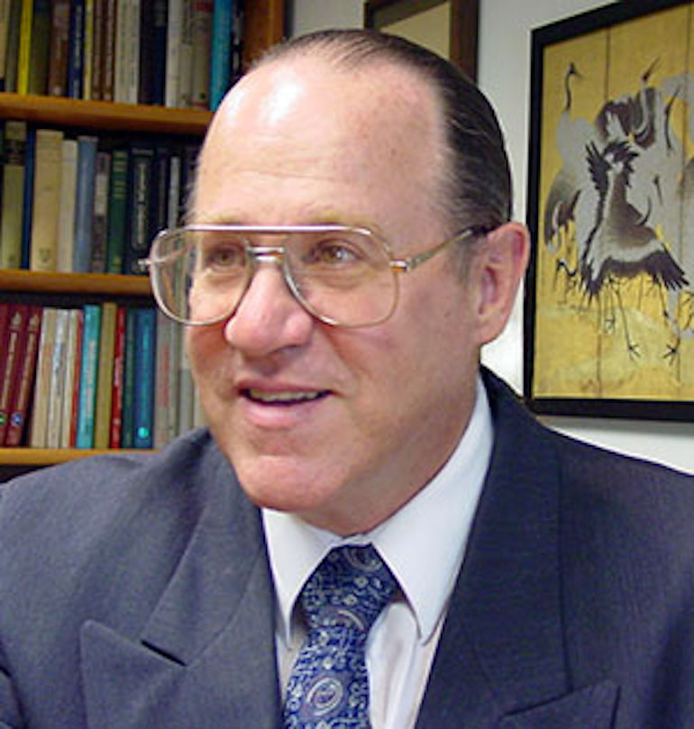 Robert G. Salomon合作论文数Case Comprehensive Cancer Center, Case Western Reserve University;Department of Ophthalmology and Visual Sciences, School of Medicine, Case Western Reserve University1Farrukh A. Shamsi合作论文数1Matthew W. Davies合作论文数1
 Vera L Bonilha合作论文数Cole Eye Institute, Cleveland Clinic Foundation1Edward J. Land合作论文数1John T. Landrum合作论文数1
 Xiaorong Gu合作论文数1




