We propose an alternate minimization algorithm for estimating the point-spread function (PSF) of a confocal laser scanning microscope and the specimen fluorescence distribution. A three-dimensional separable Gaussian model is used to restrict the PSF solution space and a constraint on the specimen is used so as to favor the stabilization and convergence of the algorithm. The results obtained from the simulation show that the PSF can be estimated to a high degree of accuracy, and those on real data show better deconvolution as compared to a full theoretical PSF model.
In order to denoise Poisson count data, we introduce a variance stabilizing transform (VST) applied on a filtered discrete Poisson process, yielding a near Gaussian process with asymptotic constant variance. This new transform, which can be deemed as an extension of the Anscombe transform to filtered data, is simple, fast, and efficient in (very) low-count situations. We combine this VST with the filter banks of wavelets, ridgelets and curvelets, leading to multiscale VSTs (MS-VSTs) and nonlinear decomposition schemes. By doing so, the noise-contaminated coefficients of these MS-VST-modified transforms are asymptotically normally distributed with known variances. A classical hypothesis-testing framework is adopted to detect the significant coefficients, and a sparsity-driven iterative scheme reconstructs properly the final estimate. A range of examples show the power of this MS-VST approach for recovering important structures of various morphologies in (very) low-count images. These results also demonstrate that the MS-VST approach is competitive relative to many existing denoising methods.
Accurate computational localization of single fluorescent particles is of interest to many biophysical studies and underlies recent approaches to high resolution microscopy using photo-switchable fluorophores. The position of individual particles is typically computed by least-squares fitting of a Gaussian intensity profile to the image, whose band-width is either derived from an idealized theoretical model of the point spread function (PSF), or itself fitted to the image. However, the band-width best approximating the actual PSF may differ significantly from its theoretical value, while fitting it is expected to degrade localization accuracy. Here, instead, we measure the real PSF bandwidth using fluorescent beads as calibration probes, and use this new bandwidth in a Gaussian model fitting algorithm. We show on simulated and real images that this simple modification of the standard localization procedure results in significant improvement of the 3D accuracy in the nanometer range.
Optical Sections of biological samples obtained from a fluorescence Confocal Laser Scanning Microscopes (CLSM) are often degraded by out-of-focus blur and photon counting noise. Such physical constraints on the observation are a result of the diffraction-limited nature of the optical system, and the reduced amount of light detected by the photomultiplier respectively. Hence, the image stacks can benefit from postprocessing restoration methods based on deconvolution. The parameters of the acquisition system's Point Spread Function (PSF) may vary during the course of experimentation, and so they have to be estimated directly from the observation data. We describe here an alternate minimization algorithm for the simultaneous blind estimation of the specimen 3D distribution of fluorescent sources and the PSF. Experimental results on real data show that the algorithm provides very good de- convolution results in comparison to theoretical microscope PSF models.
Methods based on hypothesis tests (HTs) in the Haar domain are widely used to denoise Poisson count data. Facing large datasets or real-time applications, Haar-based denoisers have to use the decimated transform to meet limited-memory or computation-time constraints. Unfortunately, for regular underlying intensities, decimation yields discontinuous estimates and strong "staircase" artifacts. In this paper, we propose to combine the HT framework with the decimated biorthogonal Haar (Bi-Haar) transform instead of the classical Haar. The Bi-Haar filter bank is normalized such that the p-values of Bi-Haar coefficients (pBH) provide good approximation to those of Haar (pH) for high-intensity settings or large scales; for low-intensity settings and small scales, we show that pBH are essentially upper-bounded by pH. Thus, we may apply the Haar-based HTs to Bi-Haar coefficients to control a prefixed false positive rate. By doing so, we benefit from the regular Bi-Haar filter bank to gain a smooth estimate while always maintaining a low computational complexity. A Fisher-approximation-based threshold imple- menting the HTs is also established. The efficiency of this method is illustrated on an example of hyperspectral-source-flux estimation.
In this paper, we present a novel object-based statistical colocalization method. Our colocalization relies on multiple hypothesis tests on the distances between all pairs of the (spot-shaped) objects from the two markers. We wish to test among all these pairs how many are significantly close to each other such that they cannot occur just "by chance". Two objects are decided to be co-localized if the test on their distance is significant. For this purpose, we first extract the objects by applying a wavelet-based spot detection approach which fully takes into account the mixed-Poisson-Gaussian noise process of confocal fluorescence images. Then, we build a null hypothesis model in which the distribution of the distance between two independently randomly drawn detections in the cell is estimated by a kernel method. The observed distances are tested against this null model. Our tests control the false discovery rate (FDR) of the co-localizations. Simulations show that this approach has a good specificity. Furthermore, our method has been successfully applied in a real problem of protein colocalization analysis during the endocytic process.
In this paper, we propose a method for the iterative restoration of fluorescence Confocal Laser Scanning Microscopic (CLSM) images and parametric estimation of the acquisition system's Point Spread Function (PSF). The CLSM is an optical fluorescence microscope that scans a specimen in 3D and uses a pinhole to reject most of the out-of-focus light. However, the quality of the images suffers from two basic physical limitations. The diffraction-limited nature of the optical system, and the reduced amount of light detected by the photomultiplier cause blur and photon counting noise respectively. These images can hence benefit from post-processing restoration methods based on deconvolution. An efficient method for parametric blind image deconvolution involves the simultaneous estimation of the specimen 3D distribution of fluorescent sources and the microscope PSF. By using a model for the microscope image acquisition physical process, we reduce the number of free parameters describing the PSF and introduce constraints. The parameters of the PSF may vary during the course of experimentation, and so they have to be estimated directly from the observed data. A priori model of the specimen is further applied to stabilize the alternate minimization algorithm and to converge to the solutions.
Fluorescence microscopy images are contaminated by photon and readout noises, and hence can be described by mixed-Poisson-Gaussian (MPG) processes. In this paper, a new variance stabilizing transform (VST) is designed to convert a filtered MPG process into a near Gaussian process with a constant variance. This VST is then combined with the isotropic undecimated wavelet transform leading to a multiscale VST (MS-VST). We demonstrate the usefulness of MS-VST for image denoising and spot detection in fluorescence microscopy. In the first case, we detect significant Gaussianized wavelet coefficients under the control of a false discovery rate. A sparsity-driven iterative scheme is proposed to properly reconstruct the final estimate. In the second case, we show that the MS-VST can also lead to a fluorescent-spot detector, where the false positive rate of the detection in pure noise can be controlled. Experiments show that the MS-VST approach outperforms the generalized Anscombe transform in denoising, and that the detection scheme allows efficient spot extraction from complex background.
We comprehensively study the least-squares Gaussian approximations of the diffraction-limited 2D-3D paraxial-nonparaxial point-spread functions (PSFs) of the wide field fluorescence microscope (WFFM), the laser scanning confocal microscope (LSCM), and the disk scanning confocal microscope (DSCM). The PSFs are expressed using the Debye integral. Under an L(infinity) constraint imposing peak matching, optimal and near-optimal Gaussian parameters are derived for the PSFs. With an L1 constraint imposing energy conservation, an optimal Gaussian parameter is derived for the 2D paraxial WFFM PSF. We found that (1) the 2D approximations are all very accurate; (2) no accurate Gaussian approximation exists for 3D WFFM PSFs; and (3) with typical pinhole sizes, the 3D approximations are accurate for the DSCM and nearly perfect for the LSCM. All the Gaussian parameters derived in this study are in explicit analytical form, allowing their direct use in practical applications.
Zhang, Fadili, & Starck have recently developed a denoising procedure for Poisson data that offers advantages over other methods of intensity estimation in multiple dimensions. Their procedure, which is nonparametric, is based on thresholding wavelet coefficients. The restoration algorithm applied after thresholding provides good conservation of source flux. We present an investigation of the procedure of Zhang et al. for the detection and characterization of astrophysical sources of high-energy gamma rays, using realistic simulated observations with the Large Area Telescope (LAT). The LAT is to be launched in late 2007 on the Gamma-ray Large Area Space Telescope mission. Source detection in the LAT data is complicated by the low fluxes of point sources relative to the diffuse celestial background, the limited angular resolution, and the tremendous variation of that resolution with energy (from tens of degrees at {approx}30 MeV to 0.1{sup o} at 10 GeV). The algorithm is very fast relative to traditional likelihood model fitting, and permits immediate estimation of spectral properties. Astrophysical sources of gamma rays, especially active galaxies, are typically quite variable, and our current work may lead to a reliable method to quickly characterize the flaring properties of newly-detected sources.
Cell migration is a field of intense current research, where biologists increasingly rely on methods and expertise from physics and engineering. Signal processing approaches can contribute significantly to this research, most notably to help analyze the exploding quantity of imaging data produced with standard and new microscopy techniques. In this article, we first provide a brief background on the importance of understanding cell movements, then review a selection of current efforts on tracking moving cells, with an emphasis on deformable model approaches (some ideas expressed in this article have been previously discussed. We will point out some of the main difficulties posed by cellular imaging, discuss advantages and limitations of different tracking techniques, and suggest a few directions for future advances
We describe a method designed to detect fluorescent rods from 2D microscopy images. It is motivated by the desire to study the dynamics of bacteria such as Shigella. The methodology is adapted from a super-resolution spot detection algorithm and is based on a parametric model of the rods and the microscope PSF. The algorithm consists of two parts: 1) a pre-detection step, based on thresholding a score computed from the product of the mean curvature and the local intensity of the filtered image, 2) an iterative procedure, where a mixture model of blurred segments is fitted to the image, and segments are removed, then added under the control of hypothesis tests. An upper bound is provided for the probability of erroneously detecting rods in noise. We show that the algorithm can reliably detect and accurately localize rods from low SNR images and can distinguish rods separated by subresolution distances. We also illustrate its ability to identify and separate overlapping bacteria on a real image
Despite the availability of rigorous physical models of microscopy point spread functions (PSFs), approximative PSFs, particularly separable Gaussian approximations are widely used in practical microscopic data processing. In fact, compared with a physical PSF model, which usually involves non-trivial terms such as integrals and infinite series, a Gaussian function has the advantage that it is much simpler and can be computed much faster. Moreover, due to its special analytical form, a Gaussian PSF is often preferred to facilitate the analysis of theoretical models such as Fluorescence Recovery After Photobleaching (FRAP) process and of processing algorithms such as EM deconvolution. However, in these works, the selection of Gaussian parameters and the approximation accuracy were rarely investigated. In this paper, we present a comprehensive study of Gaussian approximations for diffraction-limited 2D/3D paraxial/non-paraxial PSFs of Wide Field Fluorescence Microscopy (WFFM), Laser Scanning Confocal Microscopy (LSCM) and Disk Scanning Confocal Microscopy (DSCM) described using the Debye integral. Besides providing an optimal Gaussian parameter for the 2D paraxial WFFM PSF case, we further derive nearly optimal parameters in explicit forms for each of the other cases, based on Maclaurin series matching. Numerical results show that the accuracy of the 2D approximations is very high (Relative Squared Error (RSE) < 2% in WFFM, < 0.3% in LSCM and < 4% in DSCM). For the 3D PSFs, the approximations are average in WFFM (RSE similar or equal to 16 - 20%), accurate in DSCM (RSE similar or equal to 3 - 6%) and nearly perfect in LSCM (RSE similar or equal to 0.3 - 0.5%).
Optical microscopy in 2 or 3 dimensions allows extensive observations of the motility and morphology of living cells, in culture or in tissue. This leads to an exploding accumulation of imaging data and shifts the bottleneck from data acquisition to data analysis. Manual image analysis is often either impossible or exceedingly time-consuming and subject to uncontrollable user bias and errors. Computerized methods promise to ensure fast, accurate and reproducible processing, but the basic image analysis functions available in standard commercial software are generally not adapted to the complexity of biological images. For this reason, we develop methods based on active contours, a powerful and flexible technique to segment and track objects, that has become very popular in computer vision research. Here, we describe the main benefits and limitations of active contours for our application, and our efforts to adapt and improve these methods for the analysis of cellular dynamics.
The study of cell and pathogen motility in biology requires computerized methods to enable objective quantitative analysis of large amounts of data. In this paper, we propose a method to detect and track multiple moving biological objects showing different kind of dynamics in image sequences acquired through fluorescence video microscopy. It enables the extraction and analysis of informations such as number, position, speed and movement phases of, e.g., endosomes and viral particles. The method consists of four stages. After a detection stage performed by an undecimated wavelet transform, we compute, for each detected spot, several predictions of its future state in the next frame. This is accomplished thanks to an Interacting Multiple Model (IMM) algorithm which includes several models corresponding to different movement types. Tracks are constructed thereafter by a data association algorithm based on the maximization of the estimated likelihood of each IMM. The last stage consists in updating the IMM filters in order to compute final estimations for the present image and to improve predictions for the next image. The performance of the method is illustrated on synthetic image data.
This paper presents a method based on geometric active contours to segment and track biological cells in fluorescence videomicroscopy. To achieve robust segmentation of fluorescent cells with fuzzy boundaries, we use the region-based model introduced by Chan and Vese. The level set approach allows fully automatic initialization, detection of dividing cells and of cells entering the field of view during the sequence. However, the standard approach using only one level set function leads to undesired merging of touching cells and failure of tracking. To prevent this, we represent each cell by its own level set function and introduce a coupling constraint that minimizes overlaps of active contours. This technique maintains the separation of contours and allows tracking to proceed even when the boundary between cells is weak or invisible.
A method which allows for the first time to perform automatically the detection and the tracking of microscopic objects directly from three dimensional image data is presented. It enables to analyse biological moving objects in three dimensional fluorescence image sequences coming from biological immunomicroscopy experiments, and get quantitative data such as the number of objects, their position, movement phases and speed. After a detection step is performed through the multiscale analysis of images using a shift-invariant wavelet transform, the tracking is achieved using a Kalman filter and an association which enable the position of the moving objects to be predicted, refined and updated. Trajectories are analysed in terms of different parameters relevant for the motility analysis of biological objects.
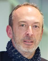 Jean-Christophe Olivo Marin合作论文数Bioimage Analysis Unit, Institut Pasteur;Institut Carnot Pasteur Microbes & Santé13
 Josiane Zerubia合作论文数INRIA5
 Christophe Zimmer合作论文数Station d'Agronomie, INRA5
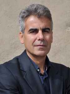 Jean-Luc Starck合作论文数CosmoStat laboratory, Institute of Research into The Fundamental Laws of The Universe, Departement D'Astrophysique, CEA-Saclay4Vannary Meas-Yedid合作论文数Institut Pasteur4Zvi Kam合作论文数3
 Praveen Pankajakshan合作论文数Amrita Vishwa Vidyapeetham;Roc Tranquil Community Lab3
 Laure Blanc-Féraud合作论文数French National Centre for Scientific Research, Laboratoire d'Informatique, Signaux et Systèmes, Côte d'Azur University3
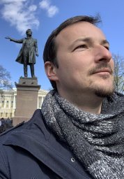 Alexandre Dufour合作论文数Image Mining group, Institut Pasteur Korea2Jalal Fadili合作论文数ENSI Caen2
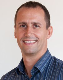 Auguste Genovesio合作论文数Image Mining group, Institut Pasteur Korea2
 Sylvain Berlemont合作论文数2Samantha Blazquez合作论文数1l mcmahon合作论文数1c jacques合作论文数1
 Marcio de Moraes Marim合作论文数1S. W. Digel合作论文数1
 James Chiang合作论文数SLAC National Accelerator Laboratory, Stanford University;Kavli Institute for Particle Astrophysics and Cosmology, Stanford University1Robert Menard合作论文数1A. Thebaud合作论文数1
 Gregory Scherrer合作论文数Department of Cell Biology and Physiology, School of Medicine, The University of North Carolina at Chapel Hil1Alexandre Grassart合作论文数1
 Jost Enninga合作论文数U389 INSERM, Institut Pasteur;28,;Unité de Pathogénie Microbienne Moléculaire, U389 INSERM Institut Pasteur1Angela Giangrande合作论文数1
 Nancy Guillen合作论文数INSERM U786, Paris, France1
 Michel Labouesse合作论文数Institut de Biologie Paris-Seine1Laurent Soustelle合作论文数1
 Nathalie Sauvonnet合作论文数Unité de Biologie des Interactions Cellulaires;Institut Pasteur;Centre National de la Recherche Scientifique1
 Nicolas Chenouard合作论文数Institut Pasteur, CNRS1Seth W. Digel合作论文数1
 Elisabeth Labruyere合作论文数Unite Anal Images Quantitat, Inst Pasteur1
 Friedrich Frischknecht合作论文数Hygiene Institute, Heidelberg University School of Medicine1




