Two cases of trisomy 4 mosaicism are reported including one with molecularly confirmed uniparental disomy (UPD) of chromosome 4. Cytogenetic analysis of a chorionic villus sample (CVS) in Case 1 showed complete trisomy 4 in trophoblast and diploidy in chorionic stroma. Amniotic fluid analysis demonstrated a 46,XX complement. After intrauterine fetal death at 30 weeks, molecular analysis confirmed the presence of trisomy 4 of maternal meiotic origin, while fetal tissues showed maternal UPD for chromosome 4. Cultured CVS in Case 2 revealed trisomy 4 in 2/30 cells analyzed. This pregnancy resulted in a healthy livebirth with biparental inheritance of chromosome 4. Molecularly confirmed UPD4 has not been previously reported, and therefore, although the adverse outcome in Case 1 is likely due to the trisomy 4 in the placenta, an imprinting effect associated with UPD4 cannot be excluded.
Recurrence of trisomy 21 was observed in a family in which both parents had a normal chromosome complement. Mosaic trisomy 21 was found in a blood karyotype of the first child, a second pregnancy ended in spontaneous abortion, and a full trisomy 21 was found at prenatal diagnosis of the third pregnancy of this same couple. Although recurrent trisomy 21 may be due to chance, the possibility of germline mosaicism for trisomy 21 in one of the parents has important implications for recurrence risk. Molecular analysis was therefore undertaken in this family to determine the parental origin and the stage of nondisjunction of the extra chromosome 21 in both cases. Although a maternal origin of both instances of trisomy 21 was observed, the mosaic case showed homozygosity for all markers along the duplicated maternal chromosome. Such a finding would normally suggest a postzygotic origin of the trisomy 21. However, the diploid cell line in this same case showed maternal uniparental disomy 21, implying that it was the result of a trisomic conception. We suggest that a somatic nondisjunction in the maternal germ cells is the most likely explanation for these findings. The apparent meiotic II stage of nondisjunction of the nonmosaic trisomy 21 fetus was consistent with maternal mosaicism. A review of the literature for recurrent trisomy 21 cases studied by molecular means, suggests that mosaicism in germ cells may account for more cases than is detected cytogenetically. These results also show that DNA marker analysis does not provide a valuable tool for patient counseling in case of recurrent trisomy 21.
Paternal uniparental disomy (UPD) for chromosome 15 (UPD15), which is found in ∼2% of Angelman syndrome (AS) patients, is much less frequent than maternal UPD15, which is found in 25% of Prader–Willi syndrome patients. Such a difference cannot be easily accounted for if ‘gamete complementation’ is the main mechanism leading to UPD. If we assume that non‐disjunction of chromosome 15 in male meiosis is relatively rare, then the gain or loss of the paternal chromosome involved in paternal and maternal UPD15, respectively, may be more likely to result from a post‐zygotic rather than a meiotic event. To test this hypothesis, the origin of the extra chromosome 15 was determined in 21 AS patients with paternal UPD15 with a paternal origin of the trisomy. Only 4 of 21 paternal UPD15 cases could be clearly attributed to a meiotic error. Furthermore, significant non‐random X‐chromosome inactivation (XCI) observed in maternal UPD15 patients (p<0.001) provides indirect evidence that a post‐zygotic error is also typically involved in loss of the paternal chromosome. The mean maternal and paternal ages of 33.4 and 39.4 years, respectively, for paternal UPD15 cases are increased as compared with normal controls. This may be simply the consequence of an age association with maternal non‐disjunction leading to nullisomy for chromosome 15 in the oocyte, although the higher paternal age in paternal UPD15 as compared with maternal UPD15 cases is suggestive that paternal age may also play a role in the origin of paternal UPD15.
Non-disjoined chromosomes 15 from 115 cases of uniparental disomy (ascertained through Prader-Willi syndrome) and 13 cases of trisomy of maternal origin were densely typed for microsatellite loci spanning chromosome 15q. Of these 128 cases a total of 97 meiosis I (MI) errors, 19 meiosis II (MII) errors and 12 mitotic errors were identified. The genetic length of a map created from the MI errors was 101 cM, as compared with a maternal length of 137 cM based on CEPH controls. No significant differences were detected in the distribution of recombination events along the chromosome arm and a reduction was seen for most of the chromosome 15 intervals examined. It was estimated that 21% of tetrads leading to MI non-disjunction were achiasmate, which may account for most or all of the reduction in recombination noted. The mean age of mothers of cases involving MI errors which showed no transitions from heterodisomy to isodisomy was significantly lower (32.7) than cases showing one or more observable transitions (36.3) (P < 0.003, t -test). However, even among chiasmate pairs the highest mean maternal age was seen for multiple exchange tetrads. Chromosome-specific differences in maternal age effects may be related to the normal distribution of exchanges (and their individual susceptibilities) for each chromosome. However, they may also reflect the presence of multiple factors which act to ensure normal segregation, each affected by maternal age in a different way and varying in importance for each chromosome.
Fluorescence in situ hybridization (FISH) provides a rapid and accurate method for the detection of chromosomal aneuploidy. We have developed a technique for the use of FISH on single cell suspensions produced from either formalin-fixed or paraffin-embedded tissues. Preparation of such tissues involves sequential rehydration, enzymatic digestion to release single nuclei, and hybridization with a fluorescently labeled chromosome-specific centromeric probe. In a clinical setting formalin-fixed tissue from many tissue types is readily available for additional retrospective study. FISH on formalin-fixed tissues is especially beneficial in follow-up studies of cases involving termination after prenatal diagnosis or patients with a malignant disease where previous routine cytogenetics established the chromosomal aneuploidy. The use of this technique eliminates the biases of cytogenetic analysis due to clonal selection in tissue culture, the low number of cells analyzed, and the restriction to only dividing cell populations. We have demonstrated that this application of interphase cytogenetics to the study of various formalin-fixed tissues is amenable to the detection of chromosomal aneuploidies and has specific advantages over cytogenetic analysis.
 Wendy Robinson合作论文数BC Children's Hospital Research Institute;Department of Medical Genetics, Faculty of Medicine, The University of British Columbia8
 TJ HASSOLD合作论文数3
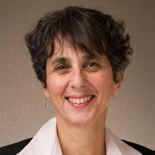 Sylvie Langlois合作论文数Department of Medical Genetics, Faculty of Medicine, The University of British Columbia;BC Children's Hospital Research Institute3Dagmar K. Kalousek合作论文数3
 Simone Schuffenhauer合作论文数3IJ Barrett合作论文数2JL Gair合作论文数2
 Bernhard Horsthemke合作论文数Institute of Human Genetics, University Hospital Essen2
 Ron Charles Michaelis合作论文数2
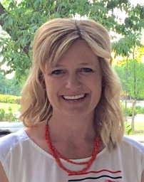 Susan L. Christian合作论文数Department of Human Genetics, The University of Chicago2BL Lomax合作论文数1M S Peñaherrera合作论文数1AA SCHINZEL合作论文数1A. Schinzel合作论文数1
 Helene Bruyère合作论文数Department of Pathology and Laboratory Medicine, Faculty of Medicine, The University of British Columbia;VGH Cytogenetics Laboratory;Vancouver Coastal Health Research Institute1Marc Lalande合作论文数1
 Mary Stephenson合作论文数College of Medicine, University of Illinois1
 Albert Schinzel合作论文数Institute of Medical Genetics, University of Zurich1Sue Malcolm合作论文数1Florence Bernasconi合作论文数1A Schulze合作论文数1
 Rachael Ritchie合作论文数Institute of Molecular Medicine, John Radcliffe Hospital1Rosemarie Rupps合作论文数1
 Jan Friedman合作论文数Department of Medical Genetics, Faculty of Medicine, The University of British Columbia;BC Children's Hospital1
 Michael Bjorn Petersen合作论文数1Karen Brøndum-Nielsen合作论文数1S. L. Yong合作论文数1




