Sleep is a dangerous part of an animal's life.1,2,3 Nonetheless, following sleep loss, mammals and birds sleep longer and deeper, as reflected by increased electroencephalogram (EEG) slow-wave activity (SWA; ≈1-5 Hz spectral power) during non-rapid eye movement (NREM) sleep.4,5 Stimulating a brain region during wakefulness also causes that region to sleep deeper afterwards,6,7,8,9 indicating that NREM sleep is a local, homeostatically regulated process.10,11 Birds and some marine mammals can keep one eye open during NREM sleep,12,13 a behavior associated with lighter sleep or wakefulness in the hemisphere opposite the open eye-states called asymmetric and unihemispheric NREM sleep, respectively.13,14,15,16,17,18,19,20,21,22,23 Closure of both eyes is associated with symmetric NREM or REM sleep. Birds rely on asymmetric and unihemispheric sleep to stay safe.17,24,25 However, as sleeping deeply with only one hemisphere at a time increases the time required for both hemispheres to fulfill their need for NREM sleep, increased sleep pressure might cause birds to engage in symmetric sleep at the expense of asymmetric sleep.26,27 Using high-density EEG recordings of European jackdaws (Coloeus monedula), we investigated intra- and inter-hemispheric asymmetries during normal sleep and following sleep deprivation (SD). The proportion of asymmetric sleep was lower early in the sleep period and following SD-periods of increased sleep pressure. Our findings demonstrate a trade-off between the benefits of sleep and vigilance and indicate that a bird's utilization of asymmetric sleep is constrained by temporal dynamics in their need for sleep.
Citation: Beckers GJL, Huybregts MAC, Everaert MBH and Bolhuis JJ (2024) No evidence for language syntax in songbird vocalizations. Front. Psychol. 15:1393895. doi: 10.3389/fpsyg.2024.1393895
Sleep's role in memory consolidation is widely accepted. However, the role of the different sleep states and accompanying neurophysiological activity is still actively debated. Most theories of sleep-related memory consolidation are based on studies in a few mammalian species. Recent evidence from research in birds, which exhibit sleep states that are in most respects similar to those found in mammals (despite being distantly related) suggests that the way some types of memories are consolidated during sleep might be different in taxa other than mammals. This review will discuss how the recent sleep-related neurophysiological findings in birds inform our understanding of memory consolidation during sleep.
Sleep-related brain activity occurring during non-rapid eye-movement (NREM) sleep is proposed to play a role in processing information acquired during wakefulness. During mammalian NREM sleep, the transfer of information from the hippocampus to the neocortex is thought to be mediated by neocortical slow-waves and their interaction with thalamocortical spindles and hippocampal sharp-wave ripples (SWRs). In birds, brain regions composed of pallial neurons homologous to neocortical (pallial) neurons also generate slow-waves during NREM sleep, but little is known about sleep-related activity in the hippocampus and its possible relationship to activity in other pallial regions. We recorded local field potentials (LFP) and analogue multiunit activity (AMUA) using a 64-channel silicon multi-electrode probe simultaneously inserted into the hippocampus and medial part of the nidopallium (i.e., caudal medial nidopallium; NCM) or separately into the caudolateral nidopallium (NCL) of adult female zebra finches (Taeniopygia guttata) anesthetized with isoflurane, an anesthetic known to induce NREM sleep-like slow-waves. We show that slow-waves in NCM and NCL propagate as waves of neuronal activity. In contrast, the hippocampus does not show slow-waves, nor sharp-wave ripples, but instead displays localized gamma activity. In conclusion, neuronal activity in the avian hippocampus differs from that described in mammals during NREM sleep, suggesting that hippocampal memories are processed differently during sleep in birds and mammals.
Birds exhibit two types of sleep that are in many respects similar to mammalian rapid eye movement (REM) and non-REM (NREM) sleep. As in mammals, several aspects of avian sleep can occur in a local manner within the brain. Electrophysiological evidence of NREM sleep occurring more deeply in one hemisphere, or only in one hemisphere - the latter being a phenomenon most pronounced in dolphins - was actually first described in birds. Such asymmetric or unihemispheric NREM sleep occurs with one eye open, enabling birds to visually monitor their environment for predators. Frigatebirds primarily engage in this form of sleep in flight, perhaps to avoid collisions with other birds. In addition to interhemispheric differences in NREM sleep intensity, the intensity of NREM sleep is homeostatically regulated in a local, use-depended manner within each hemisphere. Furthermore, the intensity and temporo-spatial distribution of NREM sleep-related slow waves varies across layers of the avian hyperpallium - a primary visual area - with the slow waves occurring first in, and propagating through and outward from, thalamic input layers. Slow waves also have the greatest amplitude in these layers. Although most research has focused on NREM sleep, there are also local aspects to avian REM sleep. REM sleep-related reductions in skeletal muscle tone appear largely restricted to muscles involved in maintaining head posture. Other local aspects of sleep manifest as a mixture of features of NREM and REM sleep occurring simultaneously in different parts of the neuroaxis. Like monotreme mammals, ostriches often exhibit brainstem-mediated features of REM sleep (muscle atonia and REMs) while the hyperpallium shows EEG slow waves typical of NREM sleep. Finally, although mice show slow waves in thalamic input layers of primary sensory cortices during REM sleep, this is not the case in the hyperpallium of pigeons, suggesting that this phenomenon is not a universal feature of REM sleep. Collectively, the local aspects of sleep described in birds and mammals reveal that wakefulness, NREM sleep, and REM sleep are not always discrete states.
Propagating slow-waves in electroencephalogram (EEG) or local field potential (LFP) recordings occur during non-rapid eye-movement (NREM) sleep in both mammals and birds. Moreover, in both, input from the thalamus is thought to contribute to the genesis of NREM sleep slow-waves. Interestingly, the general features of slow-waves are also found under isoflurane anesthesia. However, it is unclear to what extent these slow-waves reflect the same processes as those giving rise to NREM sleep slow-waves. Similar slow-wave spatio-temporal properties during NREM sleep and isoflurane anesthesia would suggest that both types of slow-waves are based on related processes. We used a 32-channel silicon probe connected to a transmitter to make intra-cortical recordings of the visual hyperpallium in naturally sleeping and isoflurane anesthetized pigeons (Columba livia) using a within-bird design. Under anesthesia, the amplitude of LFP slow-waves was higher when compared to NREM sleep. Spectral power density across all frequencies (1.5-100 Hz) was also elevated. In addition, slow-wave coherence between electrode sites was higher under anesthesia, indicating higher synchrony when compared to NREM sleep. Nonetheless, the spatial distribution of slow-waves under anesthesia was more comparable to NREM sleep than to wake or REM sleep. Similar to NREM sleep, slow-wave propagation under anesthesia mainly occurred in the thalamic input layers of the hyperpallium, regions which also showed the greatest slow-wave power during both recording conditions. This suggests that the thalamus could be involved in the genesis of slow-waves under both conditions. Taken together, although slow-waves under isoflurane anesthesia are stronger, they share spatio-temporal activity characteristics with slow-waves during NREM sleep.
Artificial grammar learning (AGL) has become an important tool used to understand aspects of human language learning and whether the abilities underlying learning may be unique to humans or found in other species. Successful learning is typically assumed when human or animal participants are able to distinguish stimuli generated by the grammar from those that are not at a level better than chance. However, the question remains as to what subjects actually learn in these experiments. Previous studies of AGL have frequently introduced multiple potential contributors to performance in the training and testing stimuli, but meta-analysis techniques now enable us to consider these multiple information sources for their contribution to learning-enabling intended and unintended structures to be assessed simultaneously. We present a blueprint for meta-analysis approaches to appraise the effect of learning in human and other animal studies for a series of artificial grammar learning experiments, focusing on studies that examine auditory and visual modalities. We identify a series of variables that differ across these studies, focusing on both structural and surface properties of the grammar, and characteristics of training and test regimes, and provide a first step in assessing the relative contribution of these design features of artificial grammars as well as species-specific effects for learning.
Sleep in birds is composed of two distinct sub-states, remarkably similar to mammalian slow-wave sleep (SWS) and rapid eye movement (REM) sleep. However, it is unclear whether all aspects of mammalian sleep are present in birds. We examined whether birds suppress REM sleep in response to changes in sleeping conditions that presumably evoke an increase in perceived predation risk, as observed previously in rodents. Although pigeons sometimes sleep on the ground, they prefer to sleep on elevated perches at night, probably to avoid nocturnal mammalian ground predators. Few studies to date have investigated how roosting sites affect sleep architecture. We compared sleep in captive pigeons on days with and without access to high perches. On the first (baseline) day, low and high perches were available; on the second day, the high perches were removed; and on the third (recovery) day, the high perches were returned. The total time spent sleeping did not vary significantly between conditions; however, the time spent in REM sleep declined on the low-perch night and increased above baseline when the pigeons slept on the high perch during the recovery night. Although the amount of SWS did not vary significantly between conditions, SWS intensity was lower on the low-perch night, particularly early in the night. The similarity of these responses between birds and mammals suggests that REM sleep is influenced by at least some ecological factors in a similar manner in both groups of animals.
The faculty of language is thought to be uniquely human. Recently, it has been claimed that songbirds are able to associate meaning with sound, comparable to the way that humans do. In human language, the meaning of expressions (semantics) is dependent on a mind-internal hierarchical structure (syntax). Meaning is associated with structure through the principle of compositionality, whereby the meaning of a complex expression is a function of the meaning of its constituent parts and the mode of composition. We argue that while recent experimental findings on songbird call sequences offer exciting novel insights into animal communication, despite claims to the contrary, they are quite unlike what we find in human language. There are indeed remarkable behavioral and neural parallels in auditory-vocal imitation learning between songbirds and human infants that are absent in our closest evolutionary relatives, the great apes. But so far, there is no convincing evidence of syntax-determined meaning in nonhuman animals.
Study ObjectivesThe changes in electroencephalogram (EEG) activity that characterize sleep and its sub-states-slow-wave sleep (SWS) and rapid eye movement (REM) sleep-are similar in mammals and birds. SWS is characterized by EEG slow waves resulting from the synchronous alternation of neuronal membrane potentials between hyperpolarized down-states with neuronal quiescence and depolarized up-states associated with action potentials. By contrast, studies of non-avian reptiles report the presence of high-voltage sharp waves (HShW) during sleep. How HShW relate to EEG phenomena occurring during mammalian and avian sleep is unclear. We investigated the spatiotemporal patterns of electrophysiological phenomena in Nile crocodiles (Crocodylus niloticus) anesthetized with isoflurane to determine whether they share similar spatiotemporal patterns to mammalian and avian slow waves.MethodsRecordings of anesthetized crocodiles were made using 64-channel penetrating arrays with electrodes arranged in an 8 × 8 equally spaced grid. The arrays were placed in the dorsal ventricular ridge (DVR), a region implicated in the genesis of HShW. Various aspects of the spatiotemporal distribution of recorded signals were investigated.ResultsRecorded signals revealed the presence of HShW resembling those reported in earlier studies of naturally sleeping reptiles. HShW propagated in complex and variable patterns across the DVR.ConclusionsWe demonstrate that HShW within the DVR propagate in complex patterns similar to those observed for avian slow waves recorded from homologous brain regions. Consequently, sleep with HShW may represent an ancestral form of SWS, characterized by up-states occurring less often and for a shorter duration than in mammals and birds.
Both mammals and birds exhibit two sleep states, slow wave sleep (SWS) and rapid eye movement (REM) sleep. Studying certain aspects of sleep-related electrophysiology in freely behaving animals can present numerous methodological constraints, particularly when even fine body movements interfere with electrophysiological signals. Interestingly, under light general anesthesia, mammals and birds also exhibit slow waves similar to those observed during natural SWS. For these reasons, slow waves occurring under general anesthesia are commonly used in the investigation of sleep-related neurophysiology. However, how spectral properties of slow waves induced by anesthesia correspond to those occurring during natural SWS in birds has yet to be investigated systematically. In this study, we systematically analyzed spectral properties of electroencephalographic (EEG) patterns of pigeons (Columba livia) occurring under two commonly used anesthetics, isoflurane and urethane. These data were compared with EEG patterns during natural sleep. Slow waves occurring during spontaneous SWS, and those induced with isoflurane and urethane all showed greatest absolute power in the slowest frequencies (<3 Hz). Isoflurane and urethane-induced slow waves had near-identical power spectra, and both had higher mean power than that observed during SWS for all frequencies examined (0-25 Hz). Interestingly, burst suppression EEG activity observed under deeper planes of isoflurane anesthesia could occur bihemispherically or unihemispherically. Electrophysiological patterns while under isoflurane and urethane share phenomenological and spectral similarities to those occurring during SWS, notably the generation of high amplitude, slow waves, and peak low-frequency power. These results build upon other studies which suggest that some anesthetics exert their effects by acting on natural sleep pathways. As such, anesthesia-induced slow waves appear to provide an acceptable model for researchers interested in investigating sleep-related slow waves utilizing electrophysiological methods not suitable for use in freely behaving birds.
Several mammalian-based theories propose that the varying patterns of neuronal activity occurring in wakefulness and sleep reflect different modes of information processing. Neocortical slow-waves, hippocampal sharp-wave ripples, and thalamocortical spindles occurring during mammalian non-rapid eye-movement (NREM) sleep are proposed to play a role in systems-level memory consolidation. Birds show similar NREM and REM (rapid eye-movement) sleep stages to mammals; however, it is unclear whether all neurophysiological rhythms implicated in mammalian memory consolidation are also present. Moreover, it is unknown whether the propagation of slow-waves described in the mammalian neocortex occurs in the avian "cortex" during natural NREM sleep. We used a 32-channel silicon probe connected to a transmitter to make intracerebral recordings of the visual hyperpallium and thalamus in naturally sleeping pigeons (Columba livia). As in the mammalian neocortex, slow-waves during NREM sleep propagated through the hyperpallium. Propagation primarily occurred in the thalamic input layers of the hyperpallium, regions that also showed the greatest slow-wave activity (SWA). Spindles were not detected in both the visual hyperpallium, including regions receiving thalamic input, and thalamus, using a recording method that readily detects spindles in mammals. Interestingly, during REM sleep fast gamma bursts in the hyperpallium (when present) were restricted to the thalamic input layers. In addition, unlike mice, the decrease in SWA from NREM to REM sleep was the greatest in these layers. Taken together, these variant and invariant neurophysiological aspects of avian and mammalian sleep suggest that there may be associated mechanistic and functional similarities and differences between avian and mammalian sleep.
In this Formal Comment the authors respond to objections to their previous Essay, reiterating that comparative linguistics is not an easy undertaking.
Artificial grammar learning is a popular paradigm to study syntactic ability in nonhuman animals. Subjects are first trained to recognize strings of tokens that are sequenced according to grammatical rules. Next, to test if recognition depends on grammaticality, subjects are presented with grammar-consistent and grammar-violating test strings, which they should discriminate between. However, simpler cues may underlie discrimination if they are available. Here, we review stimulus design in a sample of studies that use particular sounds as tokens, and that claim or suggest their results demonstrate a form of sequence rule learning. To assess the extent of acoustic similarity between training and test strings, we use four simple measures corresponding to cues that are likely salient. All stimulus sets contain biases in similarity measures such that grammatical test stimuli resemble training stimuli acoustically more than do non-grammatical test stimuli. These biases may contribute to response behaviour, reducing the strength of grammatical explanations. We conclude that acoustic confounds are a blind spot in artificial grammar learning studies in nonhuman animals.
Unlike nonhuman primates, thousands of bird species have articulatory capabilities that equal or surpass those of humans, and they develop their vocalizations through vocal imitation in a way that is very similar to how human infants learn to speak. An understanding of how speech mechanisms have evolved is therefore unlikely to yield key insights into how the human brain is special.
Brain rhythms occurring during sleep are implicated in processing information acquired during wakefulness, but this phenomenon has almost exclusively been studied in mammals. In this review we discuss the potential value of utilizing birds to elucidate the functions and underlying mechanisms of such brain rhythms. Birds are of particular interest from a comparative perspective because even though neurons in the avian brain homologous to mammalian neocortical neurons are arranged in a nuclear, rather than a laminar manner, the avian brain generates mammalian-like sleep-states and associated brain rhythms. Nonetheless, until recently, this nuclear organization also posed technical challenges, as the standard surface EEG recording methods used to study the neocortex provide only a superficial view of the sleeping avian brain. The recent development of high-density multielectrode recording methods now provides access to sleep-related brain activity occurring deep in the avian brain. Finally, we discuss how intracerebral electrical imaging based on this technique can be used to elucidate the systems-level processing of hippocampal-dependent and imprinting memories in birds.
Background: In mammals, the slow-oscillations of neuronal membrane potentials (reflected in the electroencephalogram as high-amplitude, slow-waves), which occur during non-rapid eye movement sleep and anesthesia, propagate across the neocortex largely as two-dimensional traveling waves. However, it remains unknown if the traveling nature of slow-waves is unique to the laminar cytoarchitecture and associated computational properties of the neocortex.Results: We demonstrate that local field potential slow-waves and correlated multiunit activity propagate as complex three-dimensional plumes of neuronal activity through the avian brain, owing to its non-laminar, nuclear neuronal cytoarchitecture.Conclusions: The traveling nature of slow-waves is not dependent upon the laminar organization of the neocortex, and is unlikely to subserve functions unique to this pattern of neuronal organization. Finally, the three-dimensional geometry of propagating plumes may reflect computational properties not found in mammals that contributed to the evolution of nuclear neuronal organization and complex cognition in birds.
 Niels C. Rattenborg合作论文数11
 Johan Bolhuis合作论文数Department of Psychology, Utrecht University;Department of Psychology, University of Cambridge;St Catharine's College, University of Cambridge9
 Jacqueline van der Meij合作论文数8
 John A. Lesku合作论文数Sleep and Flight Group, Max Planck Institute for Ornithology—Seewiesen8Berwick Robert C合作论文数7N. C. Rattenborg合作论文数4
 Ryan Tisdale合作论文数Biosciences Division, SRI International4
 Kazuo Okanoya合作论文数Teikyo University4
 Carel Ten Cate合作论文数Inst Biol, Leiden Univ4Verena R. Ohms合作论文数4
 Dolores Martinez合作论文数3Marinus A C Huybregts合作论文数3
 M.B.H. Everaert合作论文数3
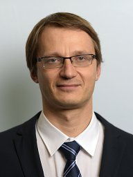 Vyssotski Alexei L合作论文数Inst Neuroinformat, Univ Zurich2Gill Arike合作论文数2van Heijningen Caroline A A合作论文数2Manfred L. Gahr合作论文数2Gianina Ungurean合作论文数1Peter Ch. Snelderwaard合作论文数1Antony S Trotter合作论文数1
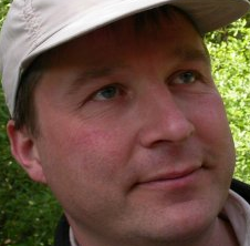 S. Verhulst合作论文数Groningen Institute for Evolutionary Life Sciences, Faculty of Science and Engineering, University of Groningen1
 Morten H. Christiansen合作论文数Department of Psychology, College of Arts and Sciences, Cornell University1
 Padraic Monaghan合作论文数University of Edinburgh1
 Tom V. Smulders合作论文数Newcastle University;School of Biology;School of Biology, Newcastle University1
 Roderick Atkins Suthers合作论文数School of Medicine, Indiana University1
 Peter Meerlo合作论文数Department of Molecular Neurobiology, University of Groningen1
 Buddhamas Kriengwatana合作论文数1




