The structure of the retinal vasculature can indicate various health issues. Quantitatively measuring changes in retinal arteries and veins offers significant potential for disease prevention and management. We propose VAVnets: three variants of a deep-learning network to generate Vessels, Arteries and Veins binary segmentations. Training is conducted in a few-shot and cross-dataset manner using images from four different fundus datasets, collectively comprising 137 images: DRIVE, DUMO, HRF, and LESAV. Training occurs with 3-fold cross-validation. Each fold employs a cross-dataset-build of 40 images (10 from each dataset), with testing on the remaining 97 images. In this article, we discuss our experiments involving architectural choices, transfer-learning, and data augmentation. We assess performances using the dice score as we aim to achieve the best possible pixel-wise segmentations. Our dice scores for each dataset are, for vessels: 0.81, 0.83, 0.81, 0.86; for veins: 0.78, 0.81, 0.78, 0.79; and for arteries: 0.73, 0.78, 0.74, 0.77. To the best of our knowledge, VAVnets demonstrate superior performances compared to existing few-shot methods across these datasets.
The retinal vasculature reveals numerous health conditions, making the quantitative assessment of changes in retinal arteries and veins crucial for disease prevention and management. Quantifying changes in the retinal vasculature requires segmentation to delineate it. Deep-learning techniques demonstrate impressive results for retinal vasculature segmentation in color fundus images. However, even if the generated segmentations are good at the pixel level, they are not coherent at the structural level, (i.e. not anatomically coherent compared to a real retinal vasculature). The vasculature of the retina is composed of two completely connected trees: arteries and veins, whereas segmentations produce several disconnected components. In this article, we propose VNR-AV: a Vasculature Network Retrieval method specifically designed for retinal Arteries and Veins segmentation. The proposed post-processing method achieves two main objectives: it leverages vessels segmentation to enhance the segmentation of arteries and veins by performing reconnection, removal, and detail gathering; and it removes or reconnects segmentation components based on a set of rules developed through an understanding of deep-learning-generated segmentations. VNR-AV retrieve a fully connected thus more anatomically coherent structure of the retinal arteries and veins networks while managing to slightly improve the superposition quality at pixel-level. VNR-AV enable a more coherent assessment of changes in retinal arteries and veins and pave the way for further research in prevention and management of eye-related diseases.
Field of View Nano-CT X-Ray synchrotron imaging is used for acquiring brain neuronal features from Golgi-stained bio-samples. It theoretically requires a large number of acquired radiographs for compensating reconstruction noise reinforced by the brain features sparsity. However reducing the number of radiographs is essential in routine applications but it results to degraded tomograms. In such a case, traditional segmentation methods are no longer able to distinguish neuronal structures from surrounding noise. We investigate several existing deep-learning networks and we define new ones to segment brain features from very degraded tomograms. We demonstrate the superiority of the proposed networks compared to existing ones.
X-ray absorption imaging is used in the medical field since a long time, but recent advance in phase-contrast imaging made it feasible in a clinical setup. X-ray Phase-contrast imaging technique using a Hartmann sensor allows extracting the absorption and phase information in a single acquisition, allowing to extract a phase-shift information with a minimal exposition and deposited dose. An iterative wavefront reconstruction (IR-WF) algorithm is necessary to extract the phase and absorption values from an acquired image. Our method consists of merging the wavefront reconstruction with a computed tomographic iterative reconstruction (IR-CT) to ensure that all images converge to the same result, improving the final 3D volume.
The analysis of fundus images may reflect systemic and cerebral vascular status through a non-invasive, rapid, and cost-effective method. Accurate characterization of the retinal vessels is critical for this status assessment. Medical professionals can perform diagnosis on measurements extracted from the retinal vessels, which are identified through segmentation. Supervised-Learning is used to perform this segmentation task and has been shown to produce higher-quality results compared to traditional methods. However, the Supervised-Learning-based binary method leads to segmentations with multiple Connected Components (CC). Amongst these components, some are disconnected retinal vessels (mentioned as branches), others are artifacts. Artifacts are disconnected miss-classified components resulting from the Supervised-Learning segmentation and that should be removed. Conversely, branches should be kept and further re-connected as they are anatomically supposed to be connected. In this study, we propose a Connected-Components-based post-processing procedure to remove artifacts while preserving the most possible amount of branches. Our methodology involves a relative threshold to cluster the CC based on their areas. We also introduce a useful evaluation metric for the segmentations in the case of measurements extractions on retinal vessels. Over 615 predicted segmentations from six datasets, we improved the dice by a substantial 0.062 leading from 0.782 to 0.844. In conclusion, our method has the potential to significantly enhance the usability and reliability of retinal vessels segmentations, making it a valuable tool for medical professionals in the assessment of systemic and cerebral vascular status. Our work also provides useful insights for future research in this area, especially to address the re-connection of the remaining branches.
Terahertz technology (spanning between 0.1 and 10 THz) is now a well-established tool to achieve contactless sensing and non-destructive testing (NDT). Among the advanced approaches, THz computed tomography (THz CT) is an emerging technique for 3D reconstruction and has been extensively investigated over the last decade. This work focuses on those capabilities for 3D volumetric reconstructions of complex objects through the use of a real-time THz imaging system operating at 2.5 THz. Further work demonstrates that the resulting data are compatible with automated processing for (i) an ad-hoc segmentation, extracting the sample from the background and reconstruction surrounding noise, (ii) a component labelling, and (iii) a skeletonization, providing crucial additional metadata about the sample morphology.
sequences. Sizing, surface and volume rendering can be extracted and compared with targeted dimensions. In this work, we use millimeter wave systems tomographic system (100 and 300 GHz), frequency modulated systems (100 and 300 GHz), and pulse time domain systems (100 GHz to 4 THz) for non destructive characterization of 3D printed additive manufacturing parts. The aim of this talk to to show the advantages and disadvantages of several techniques to define their application aera. This work is associated to a data processing analysis using automated segmentation, extracting of the different volumes of interest (VOI) composing the sample. A mesh is performed for each VOI to numerically calculate the dimensions, surfaces and volume which leads to 3D visualization and dimensional measurements. Overall sequence is implemented onto unique software and validated through different sample analysis.
For standard laboratory microtomography systems, acquired radiographs do not always adhere to the strict geometrical assumptions of the reconstruction algorithm. The consequence of this geometrical inconsistency is that the reconstructed tomogram contains motion artifacts, e.g., blurring, streaking, double-edges. To achieve a motion-artifact-free tomographic reconstruction, one must estimate, and subsequently correct for, the per-radiograph experimental geometry parameters. In this paper, we examine the use of re-projection alignment (RA) to estimate per-radiograph geometry. Our simulations evaluate how the convergence properties of RA vary with: motion-type (smooth versus random), trajectory (helical versus discrete-sampling ‘space-filling’ trajectories) and tomogram resolution. The idealized simulations demonstrate for the space-filling trajectory that RA convergence rate and accuracy is invariant with regard to the motion-type and that the per-projection motions can be estimated to less than 0.25 pixel mean absolute error by performing a single quarter-resolution RA iteration followed by a single half-resolution RA iteration. The direct impact is that, for the space-filling trajectory, one can incorporate RA in an iterative multi-grid reconstruction scheme with only a single RA iteration per multi-grid resolution step. We also find that for either trajectory, slowly varying vertical errors cannot be reliably estimated by employing the RA method alone; such errors are indistinguishable from a trajectory of different pitch. This has minimal effect in practice because RA can be combined with reference frame correction which is effective for correcting low-frequency errors.
We present a new family of X-ray source scanning trajectories for large-angle cone-beam computed tomography. Traditional scanning trajectories are described by continuous paths through space, e.g., circles, saddles, or helices, with a large degree of redundant information in adjacent projection images. Here, we consider discrete trajectories as a set of points that uniformly sample the entire space of possible source positions, i.e., a space-filling trajectory (SFT). We numerically demonstrate the advantageous properties of the SFT when compared with circular and helical trajectories as follows: first, the most isotropic sampling of the data, second, optimal level of mutually independent data, and third, an improved condition number of the tomographic inverse problem. The practical implications of these properties in tomography are also illustrated by simulation. We show that the SFT provides greater data acquisition efficiency, and reduced reconstruction artifacts when compared with helical trajectory. It also possesses an effective preconditioner for fast iterative tomographic reconstruction.
Art painting diagnostic is commonly performed using electromagnetic waves at wavelengths from terahertz to X-ray. These former techniques are essential in conservation and art history research, but they could be also very useful for restoring artwork. While most studies use time domain imaging technique, in this study, a painting has been investigated using both time domain imaging (TDI) and frequency-modulated continuous wave (FMCW) system in the millimeter frequency range. By applying these systems to a painting of the eighteenth century, we detect and analyze the structure of some defects. This study underlines the differences between FMCW and TDI. We present the advantages and disadvantages of each technique on a real artwork.
Seals are part of our cultural heritage but the study of these objects is limited because of their fragility. Terahertz and X-Ray imaging are used to analyze a collection of wax seals from the fourteenth to eighteenth centuries. In this work, both techniques are compared in order to discuss their advantages and limits and their complementarity for conservation state study of the samples. Thanks to 3D analysis and reconstructions, defects and fractures are detected with an estimation of their depth position. The path from the parchment tongue inside the seals is also detected.
Three-dimensional (3D) histology is the next frontier for modern anatomo-pathology. Characterizing abnormal parameters in a tissue is essential to understand the rationale of pathology development. However, there is no analytical technique, in vivo or histological, that is able to discover such abnormal features and provide a 3D distribution at microscopic resolution. Here, we introduce a unique high-throughput infrared (IR) microscopy method that combines automated image correction and subsequent spectral data analysis for 3D-IR image reconstruction. We performed spectral analysis of a complete organ for a small animal model, a mouse brain with an implanted glioma tumor. The 3D-IR image is reconstructed from 370 consecutive tissue sections and corrected using the X-ray tomogram of the organ for an accurate quantitative analysis of the chemical content. A 3D matrix of 89 × 106 IR spectra is generated, allowing us to separate the tumor mass from healthy brain tissues based on various anatomical, chemical, and metabolic parameters. We demonstrate that quantitative metabolic parameters can be extracted from the IR spectra for the characterization of the brain vs. tumor metabolism (assessing the Warburg effect in tumors). Our method can be further exploited by searching for the whole spectral profile, discriminating tumor vs. healthy tissue in a non-supervised manner, which we call 'spectromics'.
IR microscopy was first conceptualized in 1949 and the first commercial system was launched 1983. With the appearance of FPA detectors in the 90's, FTIR microscopy became a technique of choice for histology. Two decades later, the release of QCLs working in the mid-IR range refuels this promise by accelerating tremendously the acquisition of IR images for large tissue areas with high-quality spectra for chemical mapping of parameters of interest. The new QCL-IR imaging system allows a 150× faster spectral data acquisition at equivalent S/N level. The quality of spectral data is comparable while applying spectral curve-fitting treatments, thus showing that laser sources offer reliable signal over a large spectral range. If QCL-IR imaging system seem to offer the opportunity to develop routines for anatomo-pathology, several technological challenges stand in front of us to reach this goal to define the specs of an IR microscope dedicated to hospital.
Achieving sub-micron resolution in lab-based micro-tomography is challenging due to the geometric instability of the imaging hardware (spot drift, stage precision, sample motion). These instabilities manifest themselves as a distortion or motion of the radiographs relative to the expected system geometry. When the hardware instabilities are small (several microns of absolute motion), the radiograph distortions are well approximated by shift and magnification of the image. In this paper we examine the use of re-projection alignment (RA) to estimate per-radiograph motions. Our simulation results evaluate how the convergence properties of RA vary with: motion-type (smooth versus random), trajectory (helical versus space-filling) and resolution. We demonstrate that RA convergence rate and accuracy, for the space-filling trajectory, is invariant with regard to the motion-type. In addition, for the space-filling trajectory, the per-projection motions can be estimated to less than 0.25 pixel mean absolute error by performing a single quarter-resolution RA iteration followed by a single half-resolution RA iteration. The direct impact is that, for the space-filling trajectory, we need only perform one RA iteration per resolution in our iterative multi-grid reconstruction (IMGR). We also give examples of the effectiveness of RA motion correction method applied to real double-helix and space-filling trajectory micro-CT data. For double-helix Katsevich filtered-back-projection reconstruction (approximate to 2500x2500x5000 voxels), we use a multi-resolution RA method as a pre-processing step. For the space-filling iterative reconstruction (approximate to 2000x2000x5400 voxels), RA is applied during the IMGR iterations.
Metal artifact reduction (MAR) is a well-known problem and lots of studies have been performed during the last decades. The common standard methods for MAR consist of synthesizing missing projection data by using an interpolation or in-painting process. However, no method has been yet proposed to solve MAR problem when no sinogram is available. This paper proposes a novel MAR approach using confidence maps to restore an artifacted sinogram computed directly from the reconstructed image.
Additive manufacturing (AM) technology is not only used to make 3D objects but also for rapid prototyping. In industry and laboratories, quality controls for these objects are necessary though difficult to implement compared to classical methods of fabrication because the layer-by-layer printing allows for very complex object manufacturing that is unachievable with standard tools. Furthermore, AM can induce unknown or unexpected defects. Consequently, we demonstrate terahertz (THz) imaging as an innovative method for 2D inspection of polymer materials. Moreover, THz tomography may be considered as an alternative to x-ray tomography and cheaper 3D imaging for routine control. This paper proposes an experimental study of 3D polymer objects obtained by additive manufacturing techniques. This approach allows us to characterize defects and to control dimensions by volumetric measurements on 3D data reconstructed by tomography.
In the context of large-angle cone-beam tomography (CBCT), we present a practical iterative reconstruction (IR) scheme designed for rapid convergence as required for large datasets. The robustness of the reconstruction is provided by the "space-filling" source trajectory along which the experimental data is collected. The speed of convergence is achieved by leveraging the highly isotropic nature of this trajectory to design an approximate deconvolution filter that serves as a pre-conditioner in a multi-grid scheme. We demonstrate this IR scheme for CBCT and compare convergence to that of more traditional techniques.
The Mojette transform is a discrete and exact Radon transform, based on the discrete geometry of the projection and reconstruction lattice. The specific sampling scheme of the Mojette transform results in theoretical exact image reconstruction. In this paper, we compare the reconstructions obtained with the Mojette transform to the ones obtained with several usual projection/backprojection digitized Radon transform. These experiments validate and demonstrate the performance of the Mojette transform sampling over classical implementations based on continuous space.
Two-dimensional (2D) terahertz imaging and 3D visualization suffer from severe artifacts since an important part of the terahertz beam is reflected, diffracted, and refracted at each interface. These phenomena are due to refractive index mismatch and reflection in the case of non-orthogonal incidence. This paper proposes an experimental procedure that reduces these deleterious optical refraction effects for a cylinder and a prism made with polyethylene material. We inserted these samples in a low absorption liquid medium to match the sample index. We then replaced the surrounding air with a liquid with an optimized refractive index, with respect to the samples being studied. Using this approach we could more accurately recover the original sample shape by time-of-flight tomography.
 Mounaix Patrick合作论文数Laboratoire de l'Integration du Materiau au Systeme, IMS31
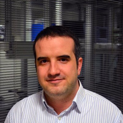 Jean-Paul Guillet合作论文数IMS Laboratory, University of Bordeaux20
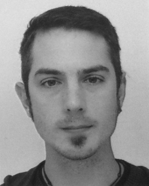 Hugo Balacey合作论文数17
 Pascal Desbarats合作论文数LaBRI, Université Bordeaux 117
 Jeanbaptiste Perraud合作论文数14
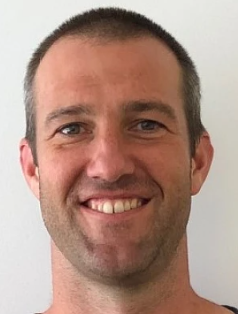 Andrew Kingston合作论文数X-Ray Tomography and Applications Group, Department of Materials Physics, Research School of Physics, Australian National University;College of Science, Australian National University12
 J Bou Sleiman合作论文数LOMA, Bordeaux Univ.;c;LOMA, Bordeaux Univ.11
 Jean-Philippe Domenger合作论文数Bordeaux University;Laboratoire Bordelais de Recherche en Informatique11
 Adrian Sheppard合作论文数Department of Materials Physics, Research School of Physics, College of Science, The Australian National University;Physics Education Centre, Research School of Physics, College of Science, The Australian National University10Glenn R. Myers合作论文数10
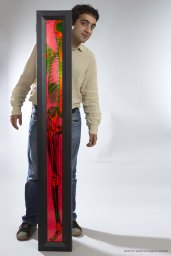 Henri Der Sarkissian合作论文数8Shane Latham合作论文数5I. Manek-Hoenninger合作论文数4Salort Simon合作论文数4N. Normand合作论文数4
 Emmanuel Abraham合作论文数283, Université Bordeaux I4
 Bruno Bousquet合作论文数Univ Bordeaux4M. Beurton-Aimar合作论文数LABRI UMR 5800, – Université Bordeaux 1 3514
 A. Younus合作论文数CPMOH, Université Bordeaux 1 and CNRS4Idris Dulau合作论文数3
 Heyang Li合作论文数Sch Math & Stat, Univ Canterbury3Benharbone W合作论文数3Frederique, L.合作论文数3Léna Bassel合作论文数3Chassagne Bruno合作论文数3
 Frederic Darracq合作论文数CNRS UMR 5818, Université Bordeaux 13
 Lionel Canioni合作论文数Department of Science and Technology, University of Bordeaux;Centre D’Optique, Photonique Et Laser, Université Laval;Argolight SA3
 Jean-Pierre Guédon合作论文数École polytechnique, l’Université de Nantes3Catherine Helmer合作论文数3
 Jean-Christophe Michel Delagnes合作论文数Centre de Physique;Moleculaire Optique et Hertzienne;Centre de Physique|Moleculaire Optique et Hertzienne3
 Cecile Delcourt合作论文数3
 Inka Manek-Hönninger合作论文数Université de Bordeaux3Jean-Pascal Caumes合作论文数3Jp Guedon合作论文数2Y. Amouriq合作论文数2
 Frank Ospald合作论文数2
 Yehuda Sternberg合作论文数2matthew geleta合作论文数2Michael Turner合作论文数2
 Yeukuang Hwu合作论文数2
 Venegas Pablo合作论文数2Vleugels, W.合作论文数2Durand, R.合作论文数2
 Joachim Jonuscheit合作论文数2Ziéglé Anne合作论文数2
 Imants Svalbe合作论文数School of Physics & Astronomy, Monash University2
 Myriam Servieres合作论文数École Centrale Nantes, LUNAM Université2
 Marijke Vandewal合作论文数2
 Cyril Petibois合作论文数Université de Bordeaux/ITTI-Consulting SAS2




