The efficacy of electrical stimulation facilitating peripheral nerve regeneration is evidenced extensively, while the associated secondary damage resulting from repeated electrode invasion and indiscriminate stimulation is inevitable. Here, we present an optogenetics strategy that utilizes upconversion nanoparticles (UCNPs) to convert deeply penetrating near-infrared excitation into blue emission, which activates an adeno-associated virus-encoding ChR2 photoresponsive ion channel on cell membranes. The induced Ca2+ flux, similar to the ion flux in the electrical stimulation approach, efficiently regulates viability and proliferation, secretion of nerve growth factor, and neural function of RSC96 cells. Furthermore, deep near-infrared excitation is harnessed to stimulate autologous Schwann cells in situ via a UCNP-composited scaffold, which enhances nerve sprouting and myelination, consequently promoting functional recovery, electrophysiological restoration, and reinnervation of damaged nerves. This developed postoperatively noninvasive optogenetics strategy presents a novel, minimally traumatic, and enduring therapeutic stimulus to effectively promote peripheral nerve repair.
Peripheral nerve injuries (PNI) are one of the most common nerve injuries, and graphene oxide (GO) has demonstrated significant potential in the treatment of PNI. GO could enhance the proliferation, adhesion, migration, and differentiation of neuronal cells by upregulating the expression of relevant proteins, and regulate the angiogenesis process and immune response. Therefore, GO is a suitable additional component for fabricating artificial nerve scaffolds (ANS), in which the slight addition of GO could improve the physicochemical performance of the matrix materials, through hydrogen bonds and electrostatic attraction. GO-composited ANS can increase the expression of nerve regeneration-associated genes and factors, promoting angiogenesis by activating the RAS/MAPK and AKT-eNOS-VEGF signaling pathway, respectively. Moreover, GO could be metabolized and excreted from the body through the pathway of peroxidase degradation in vivo. Consequently, the application of GO in PNI regeneration exhibits significant potential for transitioning from laboratory research to clinical use.
As an important transcription factor, c-Jun could upregulate growth factors expression in Schwann cells (SCs). Arginine-Glycine-Aspartate (RGD)-functionalized chitosan-graft-polyethyleneimine (RCP) gene vectors were prepared through the maleic anhydride & the carbodiimide methods, and electrostatically bound with c-Jun plasmids (pJUN), finally loaded on poly-L-lactic acid/silk fibroin parallel fiber films to fabricate nerve scaffold (RCP/pJUN-PSPF@PGA), which could locally deliver c-Jun plasmids into SCs via the mediation of RGD peptides, and upregulate the expression of nerve growth factor (NGF) and brain-derived neurotrophic factor (BDNF) in SCs. After the scaffold was bridged in sciatic nerve defect, the delivery of c-Jun plasmids from RCP/pJUN-PSPF@PGA facilitated SCs to sustain the expressions of NGF, BDNF and vascular endothelial growth factor in the injury field, promoting myelination, axonal growth and microvascular generation and nerve regeneration, muscle reinnervation and functional recovery. These results suggested that RCP/pDNA-PSPF@PGA, as an effective gene delivery platform, could provide a local gene therapy to improve nerve regeneration.
The study illustrates that graphene oxide nanosheets can endow materials with continuous electrical conductivity for up to 4 weeks. Conductive nerve scaffolds can bridge a sciatic nerve injury and guide the growth of neurons; however, whether the scaffolds can be used for the repair of spinal cord nerve injuries remains to be explored. In this study, a conductive graphene oxide composited chitosan scaffold was fabricated by genipin crosslinking and lyophilization. The prepared chitosan-graphene oxide scaffold presented a porous structure with an inner diameter of 18–87 μm, and a conductivity that reached 2.83 mS/cm because of good distribution of the graphene oxide nanosheets, which could be degraded by peroxidase. The chitosan-graphene oxide scaffold was transplanted into a T9 total resected rat spinal cord. The results show that the chitosan-graphene oxide scaffold induces nerve cells to grow into the pores between chitosan molecular chains, inducing angiogenesis in regenerated tissue, and promote neuron migration and neural tissue regeneration in the pores of the scaffold, thereby promoting the repair of damaged nerve tissue. The behavioral and electrophysiological results suggest that the chitosan-graphene oxide scaffold could significantly restore the neurological function of rats. Moreover, the functional recovery of rats treated with chitosangraphene oxide scaffold was better than that treated with chitosan scaffold. The results show that graphene oxide could have a positive role in the recovery of neurological function after spinal cord injury by promoting the degradation of the scaffold, adhesion, and migration of nerve cells to the scaffold. This study was approved by the Ethics Committee of Animal Research at the First Affiliated Hospital of Third Military Medical University(Army Medical University)(approval No. AMUWEC20191327) on August 30, 2019.
目的 本实验将混有卡铂的PECE水凝胶应用于动物腹腔粘连模型以了解卡铂-PECE是否具有预防腹腔粘连的效果且并未增加卡铂毒副作用.方法 制备卡铂-PECE水凝胶并检测其理化性质;建立大鼠腹壁缺损-盲肠摩擦模型,并将模型大鼠随机分为卡铂溶液组及卡铂-PECE水凝胶组.关腹前两组大鼠分别给予卡铂溶液、卡铂-PECE水凝胶处理创面,于术后15d处死两组大鼠并观察腹腔粘连情况.为探讨卡铂-PECE水凝胶防粘机制,用卡铂-PECE水凝胶处理腹膜创面后于术后1、3、5、7、9、12 d处死动物以观察水凝胶的降解、吸收及腹膜创面的修复过程并做组织病理学分析.以了解卡铂PECE水凝胶有无明显的毒副作用.结果 本实验所制备的卡铂PECE水凝胶10℃呈透明的溶胶态,37℃时呈不流动的凝胶态.将卡铂PECE水凝胶低压冻干后行扫描电镜观察.可见其水凝胶内有大量的微孔样骨架,镜下观察到此结构提示着卡铂-PECE水凝胶具有良好的药物缓释特性.应用卡铂-PECE水凝胶对模型大鼠治疗过程中,大鼠没有出现腹腔粘连,而卡铂溶液组全部出现右侧腹壁和盲肠的致密粘连(P<0.001).随着术后时间的延长,腹膜创面上的水凝胶逐渐降解,其中壁层消失时间约为5d,脏层约为7d;伴随凝胶降解腹腔中出现粘稠液体,术后第5天达到高峰,并于术后12d内完全吸收.伴随着凝胶的降解,手术创面逐渐愈合并于12 d完成了间皮化过程.卡铂-PECE水凝胶对大鼠各重要脏器无明显毒副作用.结论 卡铂-PECE水凝胶具有良好的防粘效果.同时伴随凝胶降解所产生的粘稠液体对于粘连预防也起到了一定作用.装载了卡铂的PECE水凝胶并未增加了卡铂毒副作用.
Postsurgical peritoneal adhesion is a major concern in clinical practice which causes significant morbidity and mortality. In this study, we investigated the efficacy of biodegradable and injectable thermosensitive poly(ethylene glycol)-poly(epsilon-caprolactone)-poly(ethylene glycol) (PEG-PCL-PEG) micelles in preventing postsurgical cauterization-induced peritoneal adhesion. The biodegradable PEG-PCL-PEG copolymer could form nano-sized micelles in water, which instantly turned into a non-flowing gel at body temperature due to micellar aggregation. Moreover, a novel sidewall and cecum cauterization rat model was developed and the micelles were assigned for adhesion prevention tests. The PEG-PCL-PEG micelles could be administered by an ordinary syringe and provided unrestricted coverage of the cauterized peritoneum. The micelles instantly formed a gel in situ at body temperature and the formed gel could adhere to the cauterized sites as a durable barrier during critical time of adhesion formation. All rats from the control group (n = 10) developed score 5 adhesion, whereas, eight out of ten rats in the micelle-treated group showed no adhesion at all. Besides, cauterization-induced adhesion formation, adhesiveness and degradation of micelles, remesothelization of peritoneum, and restoration of cauterized tissue were investigated in detail. Our results thus indicated that, it was feasible to use biodegradable and injectable thermosensitive PEG-PCL-PEG micelles for prevention of peritoneal adhesions after surgery.
Background Poly (ethylene glycol)-poly (ɛ-caprolactone)-poly (ethylene glycol) (PEG-PCL-PEG, PECE) hydrogel has been demonstrated to be biocompatible and thermosensitive. In this study, its potential efficacy and mechanisms of preventing postsurgical abdominal adhesions were investigated. Results PECE hydrogel was transformed into gel state from sol state in less than 20 seconds at 37°C. None of the animals treated with the hydrogel (n = 15) developed adhesions. In contrast, all untreated animals (n = 15) had adhesions that could only be separated by sharp dissection (P < 0.001). The hydrogel adhered to the peritoneal wounds, gradually disappeared from the wounds within 7 days, and transformed into viscous fluid, being completely absorbed within 12 days. The parietal and visceral peritoneum were remesothelialized in about 5 and 9 days, respectively. The hydrogel prevented the formation of fibrinous adhesion and the invasion of fibroblasts. Also, along with the hydrogel degradation, a temporary inflammatory cell barrier was formed which could effectively delay the invasion of fibroblasts during the critical period of mesothelial regeneration. Conclusion The results suggested that PECE hydrogel could effectively prevent postsurgical intra-abdominal adhesions, which possibly result from the prevention of the fibrinous adhesion formation and the fibroblast invasion, the promotion of the remesothelialization, and the hydroflotation effect.
The two major concerns after cytoreductive surgery of abdominal and pelvic malignancies are residual tumors and peritoneal adhesions, which are inevitable and have great impact on prognosis. Therefore, to improve the intraperitoneal chemotherapeutic effect and prevent postsurgical adhesions simultaneously after surgery, we developed a novel strategy that combines the controlled drug delivery system (CDDS) with an antiadhesion barrier. Biodegradable poly(ethylene glycol)-poly(ɛ-caprolactone)-poly(ethylene glycol) (PECE) copolymer formed micelles in water, which turned instantly into a nonflowing gel at body temperature as a result of micellar aggregation. Effectiveness of doxorubicin-loaded PECE micelles (Dox-M) in improving intraperitoneal chemotherapeutic effect and preventing adhesions was investigated. Subsequently, we established a novel mouse model for postsurgical residual tumors and peritoneal adhesions, in which Dox-M could improve intraperitoneal chemotherapeutic effect and prevent postsurgical peritoneal adhesions simultaneously. Thus, it is a promising strategy to combine the CDDS and barrier method to improve the intraperitoneal chemotherapeutic effect and prevent peritoneal adhesions simultaneously after surgery.
In this work, we investigated the efficacy of PECE-hydrogel in preventing postsurgical peritoneal adhesions in the rat uterine horn model. Standardized surgical traumas were applied to the peritoneum of the abdominal wall and the uterine horns. PECE hydrogel was applied to the two wound surfaces. Animals were euthanized at different times after surgery, and the gross necropsy findings were documented. Adhesion formation was assessed according to adhesion extent and adhesion severity, and the hydrogel degradation and healing of peritoneal wounds were observed. The remesothelialization was dynamically observed by scanning electron microscope. The results showed that none of the animals in the hydrogel-treated group (n = 12) developed adhesion. In contrast, all untreated animals (n = 12) had adhesions that could only be separated by sharp dissection (p < 0.001). The hydrogel could adhere to the peritoneal wounds and gradually disappear from the wounds within 7-9 days and transformed into viscous fluid being completely absorbed within 12 days. The injured parietal and visceral peritoneum was remesothelialized in about 7 and 9 days, respectively. This work confirmed that PECE hydrogel holds promise as a novel absorbable biomaterial for the reduction of postoperative adhesions after laparotomy.
In this paper, a series of porous chitosan scaffolds were successfully prepared by freeze-drying of chitosan hydrogel (pre-gelled with dibasic sodium phosphate at 37°C). Micro-structure, porosity, water adsorption and compressive strength were greatly affected by chitosan concentration. With the decrease of chitosan concentration, water adsorption and porosity of scaffold increased accordingly, while compressive strength of scaffold decreased. In vitro degradation test revealed that the chitosan scaffold was almost degraded by the lysozyme solution (1.5μg/ml) after 28day’s incubation. In vitro cytotoxicity test showed the prepared chitosan scaffolds were non-cytotoxicity against NIH3T3 cell. The cell viability as in function with time with acridine orange (AO) staining also demonstrated that NIH3T3 cell were metabolically active and well distributed throughout the scaffold after 5day’s incubation. Scanning electron microscopy (SEM) also showed that NIH3T3 cell appeared to adhere well and exhibited a normal morphology on the surface of scaffold after 24h cell culture.
Background Post-operative peritoneal adhesions are common and serious complications for modern medicine. We aim to prevent post-surgical adhesions using biodegradable and thermosensitive poly(ethylene glycol)-poly(ε-caprolactone)-poly(ethylene glycol) (PEG-PCL-PEG, PECE) hydrogel. In this work, we investigated the effect of PECE hydrogel on preventing post-surgical abdominal adhesions in mouse and rat models. Results The PECE hydrogel in sol state could be transformed into gel in less than 20 s at 37°C. In addition, the PECE hydrogel could be easily adhered to the damaged peritoneal surfaces, and be gradually degraded and absorbed by the body within 14 days along with the healing of peritoneal wounds. A notable efficacy of the PECE hydrogel in preventing peritoneal adhesions was demonstrated in the animal models. In contrast, all untreated animals developed adhesions requiring sharp dissection. Furthermore, no significant histopathological changes were observed in main organs of the hydrogel-treated animals. Conclusion Our results suggested that the thermosensitive PECE hydrogel was an effective, safe, and convenient agent on preventing post-surgical intro-abdominal adhesions.
In this work, biodegradable PCL–PEG–PCL (PCEC) triblock copolymers were successfully synthesized at one-step. Aqueous solution of PCEC copolymer displayed thermosensitive sol–gel–sol transition behavior, which is flowing sol at low temperature and turns into non-flowing gel at body temperature. The cytotoxicity of PCEC copolymer was evaluated by cell viability assay using HEK293 and WISH cells. In vivo gel-formation, degradation test, acute toxicity tests, and histopathological study of PCEC hydrogels were performed in BALB/c mice by subcutaneous administration. In acute toxicity test, the mice were observed continuously for 21 days. For histopathologic study, samples including heart, liver, spleen, lung, kidneys, and tissue of injection site were histochemical prepared and stained with hematoxylin–eosin. No mortality or significant signs of acute toxicity was observed during the whole observation period and there is no significant lesion to be shown in histopathologic study of major organs and tissue of injection site. The maximum tolerance dose (MTD) of PCEC hydrogel (20 wt%) by subcutaneous administration was calculated to be higher than 25 g/kg b.w. The results indicated that the obtained PCEC hydrogel was non-toxic after subcutaneous administration, and could be a safe candidate for in situ gel-forming controlled drug delivery system. © 2009 Wiley-Liss, Inc. and the American Pharmacists Association J Pharm Sci 98:4684–4694, 2009
 Xia Zhao (赵霞)合作论文数West China Second University Hospital, Sichuan University7
 Zhiyong Qian (钱志勇)合作论文数State Key Laboratory of Biotherapy, West China Hospital, Sichuan University7
 Yuquan Wei (魏雨泉)合作论文数College of Resources and Environmental Sciences, China Agricultural University6
 Changyang Gong (巩长旸)合作论文数State Key Laboratory of Biotherapy, Sichuan University6
 Feng Luo (罗锋)合作论文数West China Hospital, Sichuan University6
 Xiaorong Qi (綦小蓉)合作论文数West China Second University Hospital, Sichuan University5
 Guangfu Yin (尹光福)合作论文数College of Biomedical Engineering, Sichuan University5
 Zhongbing Huang (黄忠兵)合作论文数College of Biomedical Engineering, Sichuan University5
 Yulin Wang合作论文数4Gang Guo (郭刚)合作论文数State Key Laboratory of Biotherapy, Sichuan University4Zhaopu Yang合作论文数3Ning Mu合作论文数3
 Shengtao Zhou (周圣涛)合作论文数West China Second University Hospital, Sichuan University;State Key Laboratory of Biotherapy, Sichuan University3
 Tunan Chen (陈图南)合作论文数3
 Zhengyu Li (李征宇)合作论文数West China Second University Hospital, Sichuan University3Qian Zhong合作论文数3Min Deng合作论文数2
 Yu Nie (聂宇)合作论文数College of Biomedical Engineering, Sichuan University2
 Juan Wang (王娟)合作论文数College of Biomedical Engineering, Sichuan University2
 Qiang Ao (敖强)合作论文数College of Biomedical Engineering, Sichuan University;Institute of Regulatory Science for Medical Devices, Sichuan University2Qinjie Wu合作论文数2
 Xiaoming Liao (廖晓明)合作论文数College of Biomedical Engineering, Sichuan University2
 Shuai Shi (师帅)合作论文数School of Ophthalmology & Optometry, Wenzhou Medical University2YuJun Wang合作论文数2Pangbo Wang合作论文数1
 Hua Feng (冯华)合作论文数The Southwest Hospital of Army Medical University1
 Jirui Wen合作论文数1Ya Zhang (张亚)合作论文数1Chuanyan Yang合作论文数1
 Kang Ma合作论文数1Shi Wang合作论文数1Ying Lai合作论文数1
 Meijuan Huang (黄媚娟)合作论文数West China Hospital of Sichuan University1Liao JinFeng合作论文数National Clinical Research Center for Oral DiseasesWest China Hospital of Stomatology, Sichuan University1Yingchun Gu (顾迎春)合作论文数College of Biomass Science and Engineering, Sichuan University1
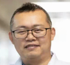 Xingyi Li (李星熠)合作论文数Xingyi Li ‘s Lab, Eye Hospital, Wenzhou Medical University1
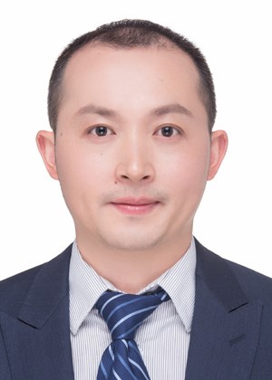 Kai Xiao (肖凯)合作论文数West China Hospital, Sichuan University1
 Jiang Wu (吴江)合作论文数West China School of Basic Medical Sciences & Forensic Medicine, Sichuan University;West China Hospital, Sichuan University1Ximing Pu (蒲曦鸣)合作论文数College of Biomedical Engineering, Sichuan University1Xiangye Kong合作论文数1Bing Kan合作论文数1Pengwei Dong合作论文数1




