In order to address the role of CD4 and CD8 Ag in the process of positive selection in the thymus, antibodies against these molecules, which do not result in the elimination of mature lymph node T cells, were injected in vivo. The results indicate that even long-term injection of nondepleting anti-CD4 and anti-CD8 antibodies does not cause the loss of CD4 or CD8 positive lymph node cells, but it completely blocks the development of the corresponding subpopulation of mature thymocytes. Thus, it appears that the interaction of the CD4 and CD8 accessory molecules on developing thymocytes with a ligand in the thymic environment (probably MHC Ag) is necessary for the positive selection of thymocytes into the appropriate T cell lineage.
Antigen-specific, major histocompatibility complex-restricted recognition by classical T cells is mediated by a T cell receptor (TCR) consisting of a disulfide-linked alpha beta heterodimer. During the search for the genes encoding the alpha and beta proteins, a third immunoglobulin-like gene, termed gamma, was uncovered. Like the TCR alpha and beta genes, the TCR gamma gene consists of variable and constant segments that rearrange during T cell development in the thymus. Although the physiological role of TCR gamma remains an enigma, much has been learned with the recent identification of the protein products of this gene family in both mice and humans. The gamma chain is associated with a partner chain, termed delta. The gamma delta heterodimer is associated with an invariant T3 complex, very similar to that associated with the alpha beta heterodimer, and appears predominantly, if not exclusively, on cells with a CD4-, CD8- phenotype both in the thymus and in the periphery. TCR gamma delta is the first T3-associated receptor to appear during thymocyte development and defines a separate T cell lineage distinct from alpha beta-bearing cells. Although TCR alpha beta-bearing cells and TCR gamma delta-bearing cells follow parallel developmental pathways, the diversity of expressed gamma delta receptors is extremely limited relative to that of alpha beta receptors.
A new lymphocyte cell surface alloantigen, provisionally designated Ly 9, is detected by flow microfluorometry(FMF), as an extra specificity in sera from anti-Lyt immunizations. Ly 9.2, one of the allelic specificities, commonly is detected as a unique immunofluorescence profile of thymocytes with anti-Lyt 3.1 sera. The alternative antigenic specificity, Ly 9.1, can be detected routinely in antisera prepared against Lyt 2.1 and Lyt 3.2. This lymphocyte alloantigen has both a unique strain and cell/tissue distribution. Quantitative measurement of Ly 9 immunofluorescence by FMF on heterozygote cells shows that Ly 9 antigen expression is co-dominant and reduced in levels relative to parental homozygote cells. Ly 9 is expressed on all thymocytes, peripheral lymphocytes, and on a subpopulation of cells in the bone marrow. This antigen is not expressed to any significant degree on erythrocytes, epidermal cells, sperm, or in suspensions of testis, brain, kidney, liver, or lung. FMF analysis and absorption typing reveals a quantitatively lower level of antigen expression on thymocytes compared with spleen or lymph node cells. Cytotoxic elimination experiments confirm that Ly 9 is expressed on at least 2 different T cell functional subsets and B cells.
 Bj Mathieson合作论文数6MJ WAXDAL合作论文数3SO SHARROW合作论文数2
 Drew Mark Pardoll合作论文数Department of Oncology, School of Medicine, Johns Hopkins University;Bloomberg~Kimmel Institute for Cancer Immunotherapy, School of Medicine, Johns Hopkins University;Sidney Kimmel Comprehensive Cancer Center, School of Medicine, Johns Hopkins University2Karen Laky合作论文数2WM LEISERSON合作论文数1K KOMSCHLIES合作论文数1D El-Khoury合作论文数1M WAXDAL合作论文数1F RAMSDELL合作论文数1Susan O. Sharrow合作论文数1Je Coligan合作论文数1Rh Schwartz合作论文数1DJ Kappes合作论文数1Am Kruisbeek合作论文数1
 Sandra M Hayes合作论文数1
 Randy Cron合作论文数Department of Pediatrics, Heersink School of Medicine, University of Alabama at Birmingham1
 Lawrence E. Samelson合作论文数Laboratory of Cellular and Molecular Biology, Center for Cancer Research, National Cancer Institute1
 Tiago Gregorio合作论文数1
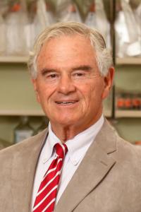 David H Sachs合作论文数Vagelos College of Physicians and Surgeons, Columbia University Irving Medical Center1
 Jeffrey Bluestone合作论文数School of Medicine, University of California, San Francisco;Helen Diller Family Comprehensive Cancer Center, University of California, San Francisco1Isabella Screpanti合作论文数Department of Experimental Medicine, University of Rome ‘La Sapienza’1
 Paul E. Love合作论文数National Institute of Child Health and Human Development, National Institutes of Health1Linette J. Edison合作论文数1




