Regulation of smooth muscle cell adhesion, proliferation, and motility on biomaterials is critical to the performance of blood-contacting implants and vascular tissue engineering scaffolds. The goal of this study was to examine the underlying substrate-smooth muscle cell response relations, using a selection of polymers representative of an expansive library of multifunctional, tyrosine-derived polycarbonates. Three chemical components within the polymer structure were selectively varied through copolymerization: (1) the content of iodinated tyrosine to achieve X-ray visibility; (2) the content of poly(ethylene glycol) (PEG) to decrease protein adsorption and cell adhesivity; and (3) the content of desaminotyrosyl-tyrosine (DT), which regulates the rate of polymer degradation. Using quartz crystal microbalance with dissipation, we quantified differential serum protein adsorption behavior because of the chemical components DT, iodinated tyrosine, and PEG: increased PEG content within the polymer structure progressively decreased protein adsorption but the simultaneous presence of both DT and iodinated tyrosine reversed the effects of PEG. The complex interplay of these components was next tested on the adhesion, proliferation, and motility behavior cultured human aortic smooth muscle cells. The incorporation of PEG into the polymer reduced cell attachment, which was reversed in the presence of iodinated tyrosine. Further, we found that as little as 10% DT content was sufficient to negate the PEG effect in polymers containing iodinated tyrosine, whereas in non-iodinated polymers, the PEG effect on cell attachment was reversed. Cross-functional analysis of motility and proliferation revealed divergent substrate chemistry related cell response regimes. For instance, within the series of polymers containing both iodinated tyrosine and 10% of DT, increasing PEG levels lowered smooth muscle cell motility without a change in the rate of cell proliferation. In contrast, for non-iodinated tyrosine and 10% of DT, increasing PEG levels increased cell proliferation significantly while reducing cell motility. Clearly, the polycarbonate polymer library offers a sensitive platform to modulate cell adhesion, proliferation, and motility responses, which, in turn, may have implications for controlling vascular remodeling in vivo. Additionally, our data suggests unique biorelevant properties following the incorporation of iodinated subunits in a polymeric biomaterial as a potential platform for X-ray visible devices.
Intro: We have developed the novel concept of using ePTFE vascular graft material as an underlying form for development of a scaffold. This scaffold is created by coating ePTFE with bioresorbable tyrosine-derived polycarbonates while maintaining porosity, but limiting exposure of the ePTFE to blood. We have developed a poly(ethylene glycol) (PEG)-containing, tyrosine-derived polycarbonate by copolymerizing DTE with 2.5% PEG (Mw= 2000) to obtain poly(DTE-co-2.5%PEG2k carbonate). The inhibitory effect of PEG on protein adsorption and cell adhesion has been established. This study was done to develop a porous scaffold for use in vascular applications and test the ability of this polymer to decrease thrombogenicity in a rat model. Methods: ePTFE was coated in a solution containing a specific concentration (0, 2, 4, 7, 10 and 15%) of polymer. The completeness of the coating was determined by SEM, and confocal microscopy of samples coated with fluorescent dye doped polymer. XPS spectra were acquired to observe changes in coating completeness as a function of polymer concentration. The ratio of % atomic concentrations of Fl to C were obtained, since the DTE contains no fluorine. Evaluation of thrombosis using an in vivo rat model was performed. Elliptical shaped patches (5 mm x 3 mm) of ePTFE were coated in 7% solutions of DTE, DTE-PEG, or uncoated ePTFE. An arteriotomy was made in the abdominal aorta of heparinized Sprague Dawley rats and a patch was sutured to the aorta. The rats were observed under anesthesia after restoration of blood flow and patency confirmed by Doppler. After 30 min, the segment of aorta containing the patch was removed. The aorta was opened longitudinally on the opposite side from the patch for imaging. A thrombosis score (0-5) was used to determine amount of clot present on the patch. Statistical differences among groups were determined by ANOVA with post-hoc comparison. Results: Increasing concentration of polymer used to coat the ePTFE resulted in visible changes by SEM with the nodes and fibrils appearing thicker with porosity maintained.
 Joachim Kohn合作论文数Department of Chemistry and Chemical Biology, School of Arts and Sciences, Rutgers University2
 Patrick A. Johnson合作论文数1Marybeth Concannon合作论文数1john kanzius合作论文数1Mary Helen Blackall合作论文数1Krista Csontos合作论文数1Michael Krein合作论文数1
 Steve A. Keller合作论文数1Paul Holmes合作论文数1Page Bouchard合作论文数1Kevin N Boykin合作论文数1Larry Carey合作论文数1Francine Breckler合作论文数1Derrick Chen合作论文数1Natasha D. Bir合作论文数1Shirong Chang合作论文数1
 Cliff P Connery合作论文数1cecilia crisanti合作论文数1Arnold Luk合作论文数1Tawnya L Bowles合作论文数1Bryan A. Cotton合作论文数1Tiffany Blake合作论文数1
 Charles W Acher合作论文数Division of Vascular Surgery, Department of Surgery, University of Wisconsin-Madison1Herbert Chen合作论文数1Joel T. Adler合作论文数1
 w chapman合作论文数Natl Ctr Atmospher Res1Theresa Boley合作论文数1Agnes Burris合作论文数1Mattia Branca合作论文数CTU Bern, Univ Bern1Brandon Cain合作论文数1Simon Gordonov合作论文数1Denise Carneiro-Pla合作论文数1Ciarán Bradley合作论文数1William G Kraybill合作论文数1
 Jeff Kiefer合作论文数Syst Oncol, Scottsdale, AZ 85255 USA1Anthony Kam合作论文数Dept Neurosurg, Loyola Univ Med Ctr1Trevor S Brown合作论文数NeuroCare Grp, Melbourne, Vic, Australia1
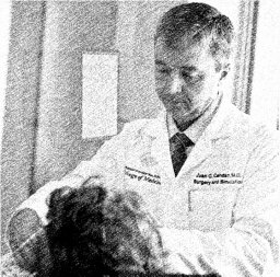 Juan Cendan合作论文数Department of Medical Education, University of Central Florida College of Medicine1
 Melina Kibbe合作论文数Office of the President, The University of Texas Health Science Center at Houston1
 Massimo Cristofanilli合作论文数Department of Medicine, Weill Cornell Medical College, Cornell University;Englander Institute of Precision Medicine, Weill Cornell Medicine, Cornell University;Meyer Cancer Center, Weill Cornell Medicine, Cornell University1
 Errol L. Bush合作论文数Department of Otolaryngology-Head and Neck Surgery, School of Medicine, Johns Hopkins University1
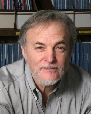 Stephen B. Baylin合作论文数Johns Hopkins School of Medicine;Cancer Genetics and Epigenetics, Johns Hopkins University1
 Joseph Kim合作论文数Medical Oncology, Internal Medicine, Yale School of Medicine, Yale University1
 John C. Kucharczuk合作论文数Surgery Faculty, Perelman School of Medicine, University of Pennsylvania1
 Nikunj Chokshi合作论文数Oakland Medical Center, The Permanente Medical Group, Inc.1Cassuto James R合作论文数1Kelz Rachel R合作论文数Dept Surg, Univ Penn1Kulp Gabriela A合作论文数1
 A. Benedict Cosimi合作论文数1




