Reliable control of rehabilitation and assistive devices using High-Density surface Electromyography (HD-sEMG) remains limited by poor robustness to electrode shifts, changes in skin condition, and variability across users. This study evaluates the performance of the Recursive Prosthetic Control Network (RPC-Net)/High-Density Electrode Array (HDE-Array) system, defined in previous studies, under conditions that reflect real-life usage, including electrode repositioning and cross-subject generalization. The first test evaluated whether the RPC-Net/HDE-Array system maintained stable performance when trained without electrode repositioning and evaluated on data from a different session with altered electrode placement. The study further examined whether explicitly incorporating electrode repositioning during training mitigates the performance degradation typically observed when testing is performed in a separate session. Finally, the effects of inter-subject training were assessed. Experimental results demonstrate that the RPC-Net/HDE-Array system is highly sensitive to electrode repositioning and skin condition variability when trained under static conditions. However, robustness improves significantly when such variability is included during training. The results indicate that performance improves with an increasing number of subjects in the training pool, provided the training set includes only data from subjects other than the one tested, suggesting a strong dependency on subject-specific patterns These findings demonstrate that the RPC-Net/HDE-Array system can achieve robust performance across sessions and users when trained under realistic conditions. This work represents a key step toward practical deployment of muscle-computer interfaces.
This paper investigated the suitability of the integrated Recursive Rehabilitation Control Network (RRC-Net)/ High-Density Electrode Array (HDE-Array) system for performing two multi-Degree of Freedom (DoF) control tasks, developed as proxies for Functional Electrical Stimulation control: (1) a cursor-based task and (2) a 3-DoF hand kinematic model control task. The goal of this study is enhancing rehabilitation independence for individuals with spinal cord injuries. The system was validated on both healthy and tetraplegic subjects. The hypotheses that users could successfully perform these tasks using the system and that there would be no significant performance differences between healthy and tetraplegic participants were assessed. The experiment involved 10 tetraplegic and 8 healthy subjects who completed a training phase followed by two testing phases. High-Density surface Electromyography (HD-sEMG) signals recorded from the neck during the training phase were used to train RRC-Net, a neural network designed to estimate multi-DoF movements. Subjects then performed the two control tasks in the testing phase, and performance metrics were analysed and compared between groups. Healthy and tetraplegic subjects achieved high performance in both control tasks. Hand position control performance between the two groups presented no statistically significant differences in Mean Global Distance (MGD) (p = 0.93) or Mean Angular Distance (MAD) (p = 0.77). Similarly, cursor control task performance showed no significant differences in Task Completion Score (TCS) (p = 0.68) or Normalised Distance (ND) (p = 0.63). The system’s simplicity, comfort, and effectiveness highlight its potential for rehabilitation, providing a non-invasive method for controlling assistive devices.
Spatial selectivity remains a major limitation of non-invasive functional electrical stimulation (FES), particularly in anatomically dense regions of the upper limb. This study investigates the design, modelling, and validation of concentric surface electrodes to improve spatial selectivity compared to conventional square geometries. A six-stage workflow was implemented, including concentric electrode design, finite element method (FEM) modelling of electric field (EF) distributions in a forearm model, axonal activation simulations using the McIntyre-Richardson-Grill (MRG) model, additive manufacturing of prototypes, impedance characterisation, andin-vivoevaluation of motor response and subjective discomfort. FEM analysis demonstrated that concentric electrodes enhanced EF focality and reduced lateral current spread relative to square configurations. Coupled MRG simulations showed improved neural selectivity, with a broader high-selectivity range (Δ = 2.7 mA). Prototypes were fabricated using low-cost, accessible materials (∼$1.64 per unit).In-vivotesting in 11 participants revealed significantly higher selectivity indices for concentric electrodes (0.723 ± 0.175) compared to square electrodes (0.546 ± 0.098,p< 0.05), without increased discomfort (p> 0.05). These findings indicate that concentric surface electrodes provide a practical and scalable strategy to enhance spatial selectivity in surface FES, and that the proposed modelling-to-prototyping workflow supports rapid optimisation of personalised neuroprosthetic interfaces.
BACKGROUND:Interferential current stimulation (ICS) has long been employed in neuromodulation and physical therapy, yet its mechanisms and potential applications in functional electrical stimulation (FES) remain under-explored. We present new data, including neural activation reported by muscle force measurement, to evaluate the potential advantages of ICS, such as selective targeting and reduced discomfort. METHODS:Experiments were conducted on human participants, focusing on ulnar and median nerve activation with stainless steel and commercial conductive hydrogel electrodes. Key parameters, including electrode configuration, frequency, current amplitude, and skin preparation, were investigated to test their effects on activation thresholds, force generation, and user comfort. RESULTS:Our results revealed that ICS can achieve proportional muscle force control, although its efficiency was lower than rectangular biphasic pulse stimulation. The application of moisturizing cream and gel significantly improved comfort and reduced activation thresholds, underscoring their importance in optimizing ICS protocols. However, ICS required higher electrical power and caused discomfort during burst initiation with all electrodes, presenting challenges for its practical use in FES. Furthermore, our findings indicated that ICS did not exclusively activate neural regions at the difference or "beat" frequency but also by the carrier frequency, challenging some prior assumptions in the literature. CONCLUSION:These results highlight the need for further research and practical measurements of neural recruitment and muscle fatigue and into the mechanisms of nerve activation and neuromuscular junction transmission with stimulation via the skin surface. Innovations in electrode design, stimulation waveforms, and protocols are also needed to enhance the efficacy and comfort of ICS.
This paper presents methods for FEM modelling the peripheral and central nervous systems with considerations for meshing and computational constraints. FEM models in this context are convenient for testing hypothesises about the effects of different stimulation parameters and exploring different electrode designs before moving to in vitro and in vivo experiments. The methods presented in this paper are motivated by assessing differentiation errors from different mesh sizes and the transitions between different materials in the model. We aim to support the development of transparent and reproducible modelling experiments. Accurate and reproducible models are essential, given the importance of the applications in which these models are used. However, a dearth of literature is devoted to promoting best practices in finite element modelling for biophysical models. We evaluate the impact of differentiation errors on calculating the Activating Function and predicting action potentials in a Hodgkin-Huxley (H-H) axon model. We found that poor spatial discretisation facilitates the generation of double-derivative noise. However, it does not generate false predictions of action potentials on the H-H model. Activation thresholds were higher (57.5 mA) for coarser meshes than Fine and Extremely Fine (55 mA). Implementing Multiscale meshes with the finest refined sizes reduced material transition discontinuities reflected in the activating function calculation. Our findings support using the finest spatial discretisations possible within computational constraints, which may rely on adaptive meshing techniques. We advocate coupling the extracellular field to H-H-based axons to further limit potential error sources.
PURPOSE:Functional electrical stimulation (FES) has been documented to provide invaluable, therapeutic effect. The success of FES applications relies on the positioning of stimulation electrodes closely to the muscle motor points. During joint movement, however, motor points may shift as the muscle length changes. Using an objective procedure to define motor points, we assessed how much and how relevantly knee extensors' motor point location changes with joint angle. METHODS:Current pulses were applied over 121 (11 × 11) equally spaced stimulation sites, determined according to the size and boundaries of the three superficial knee extensors of 17 healthy subjects. Five consecutive monophasic pulses (100 µs, 1pps) were delivered at each site for two knee joint angles (40° and 115°) and two intensities (120 % of motor threshold and maximum tolerable). The average peak torque greater than 60 % of the maximum torque was used to identify motor points across sites. The centroid of motor point clusters-motor zones-was considered to assess motor point displacement and peak torque for different knee joint angles. RESULTS:Significant centroid shifts were observed distally (2.5 % displacement; p < 0.031) and medially (4.4 % shift; p < 0.021), representing ∼ 1 cm shifts in both directions, when going from 40° to 115° of knee flexion. Mean differences in peak torque between joint angles were negligible, however, amounting to less than 2 % of maximal voluntary torque. CONCLUSION:The displacement of quadriceps motor points with knee position changes is of negligible practical relevance for justifying a variable position of stimulation electrodes in FES protocols.
OBJECTIVE:The purpose of this study was to develop and evaluate the performance of RPC-Net (Recursive Prosthetic Control Network), a novel method using simple neural network architectures to translate electromyographic activity into hand position with high accuracy and computational efficiency. METHODS:RPC-Net uses a regression-based approach to convert forearm electromyographic signals into hand kinematics. We tested the adaptability of the algorithm to different conditions and compared its performance with that of solutions from the academic literature. RESULTS:RPC-Net demonstrated a high degree of accuracy in predicting hand position from electromyographic activity, outperforming other solutions with the same computational cost. Including previous position data consistently improved results across subjects and conditions. RPC-Net showed robustness against a reduction in the number of electromyography electrodes used and shorter input signals, indicating potential for further reduction in computational cost. CONCLUSION:The results demonstrate that RPC-Net is capable of accurately translating forearm electromyographic activity into hand position, offering a practical and adaptable tool that may be accessible in clinical settings. SIGNIFICANCE:The development of RPC-Net represents a significant advancement. In clinical settings, its application could enable prosthetic devices to be controlled in a way that feels more natural, improving the quality of life for individuals with limb loss.
Presently manufacturing processes for 3D-printed electrodes require advanced fabrication techniques and time. In this study, we present a novel approach to manufacturing flexible and substrate-reusable electrodes through a streamlined 3D printing process. Here we demonstrate the technique for prototyping new shapes for FES research applications. This approach involves three key steps: Design, FEM modeling using COMSOL Multiphysics, and the actual manufacturing process. During the design phase, the electrode’s geometry is established. Subsequently, FEM modeling is performed to simulate and analyze the stationary electric field produced by the electrode within the body. The manufacturing step employs 3D CAD software, flexible resin and conductive paint for medical purposes. This method simplifies the manufacturing process for prototyping and allows the creation of custom shaped electrodes, enabling the exploration of novel FES techniques that are based on electrode geometry.
This paper aims to introduce HDE-Array (High-Density Electrode Array), a novel dry electrode array for acquiring High-Density surface electromyography (HD-sEMG) for hand position estimation through RPC-Net (Recursive Prosthetic Control Network), a neural network defined in a previous study. We aim to demonstrate the hypothesis that the position estimates returned by RPC-Net using HD-sEMG signals acquired with HDE-Array are as accurate as those obtained from signals acquired with gel electrodes. We compared the results, in terms of precision of hand position estimation by RPC-Net, using signals acquired by traditional gel electrodes and by HDE-Array. As additional validation, we performed a variance analysis to confirm that the presence of only two rows of electrodes does not result in an excessive loss of information, and we characterized the electrode-skin impedance to assess the effects of the voltage divider effect and power line interference. Performance tests indicated that RPC-Net, used with HDE-Array, achieved comparable or superior results to those observed when used with the gel electrode setup. The dry electrodes demonstrated effective performance even with a simplified setup, highlighting potential cost and usability benefits. These results suggest improvements in the accessibility and user-friendliness of upper-limb rehabilitation devices and underscore the potential of HDE-Array and RPC-Net to revolutionize control for medical and non-medical applications.
BACKGROUND:Ventilator-induced diaphragm dysfunction occurs rapidly following the onset of mechanical ventilation and has significant clinical consequences. Phrenic nerve stimulation has shown promise in maintaining diaphragm function by inducing diaphragm contractions. Non-invasive stimulation is an attractive option as it minimizes the procedural risks associated with invasive approaches. However, this method is limited by sensitivity to electrode position and inter-individual variability in stimulation thresholds. This makes clinical application challenging due to potentially time-consuming calibration processes to achieve reliable stimulation.METHODS:We applied non-invasive electrical stimulation to the phrenic nerve in the neck in healthy volunteers. A closed-loop system recorded the respiratory flow produced by stimulation and automatically adjusted the electrode position and stimulation amplitude based on the respiratory response. By iterating over electrodes, the optimal electrode was selected. A binary search method over stimulation amplitudes was then employed to determine an individualized stimulation threshold. Pulse trains above this threshold were delivered to produce diaphragm contraction.RESULTS:Nine healthy volunteers were recruited. Mean threshold stimulation amplitude was 36.17 ± 14.34 mA (range 19.38-59.06 mA). The threshold amplitude for reliable nerve capture was moderately correlated with BMI (Pearson's r = 0.66, p = 0.049). Repeating threshold measurements within subjects demonstrated low intra-subject variability of 2.15 ± 1.61 mA between maximum and minimum thresholds on repeated trials. Bilateral stimulation with individually optimized parameters generated reliable diaphragm contraction, resulting in significant inhaled volumes following stimulation.CONCLUSION:We demonstrate the feasibility of a system for automatic optimization of electrode position and stimulation parameters using a closed-loop system. This opens the possibility of easily deployable individualized stimulation in the intensive care setting to reduce ventilator-induced diaphragm dysfunction.
PurposeThe purpose of this paper is to investigate the design of skin surface electrodes for functional electrical stimulation using an isotropic single layered model of the skin and underlying tissue. A concentric ring electrode geometry was analysed and compared with a conventional configuration, specifically to localise and maximise the activation at depth and minimise the peak current density at the skin surface. Design/methodology/approachThe mathematical formulation determines the spatial electric potential distribution in the tissue, using the solution to the Laplace equation in the lower half space subject to boundary conditions given by the complete electrode model and appropriate asymptotic decay. Hence, it is shown that the electric potential satisfies a weakly singular Fredholm integral equation of the second kind which is then solved numerically in MATLAB for a novel concentric ring electrode configuration and the conventional two disk side-by-side electrode configuration. FindingsIn both models, the electrode geometry can be optimised to obtain a higher activation and lower maximum current density. The concentric ring electrode configuration, however, provides improved performance over the traditional two disk side-by-side electrode configuration. Research limitations/implicationsIn this study, only a single layer of medium was investigated. A comparison with multilayer tissue models and in vivo validation of numerical simulations are required. Originality/valueThe developed mathematical approaches and simulations revealed the parameters that influence nerve activation and facilitated the theoretical comparison of the two electrode configurations. The concentric ring configuration potentially may have significant clinical advantages.
We present an analytical approach to determining the nerve activation induced in multiple, layered, isotropic tissues by an unconventional planar concentric ring electrode geometry and assess the applicability of such electrodes for skin surface Functional Electrical Stimulation (FES). We model this problem using Laplace’s equation in a semi-infinite domain of piecewise constant conductivity subject to appropriate interface and surface boundary conditions given by the complete electrode model. This system of equations is solved by means of Hankel Transforms to determine the spatial potential distribution. In this paper, for simplicity, we present the detailed mathematical formulation for a three-layer medium, and we include the results of the numerical simulations obtained for a four-layered realistic tissue model consisting of skin, fat, muscle and bone. The approach can be easily extended to more layers. Different sets of tissue layer thicknesses were considered, corresponding to three standard categories of patient physical build. The results show that for each of these sets, the electrode design can be optimized to maximize the activating function at different depths and reduce the peaks of high current density that occur at the electrode edges. The impact of the thickness of the fat layer on the activating function is also highlighted.
BACKGROUND:Diaphragm muscle atrophy during mechanical ventilation begins within 24 h and progresses rapidly with significant clinical consequences. Electrical stimulation of the phrenic nerves using invasive electrodes has shown promise in maintaining diaphragm condition by inducing intermittent diaphragm muscle contraction. However, the widespread application of these methods may be limited by their risks as well as the technical and environmental requirements of placement and care. Non-invasive stimulation would offer a valuable alternative method to maintain diaphragm health while overcoming these limitations.METHODS:We applied non-invasive electrical stimulation to the phrenic nerve in the neck in healthy volunteers. Respiratory pressure and flow, diaphragm electromyography and mechanomyography, and ultrasound visualization were used to assess the diaphragmatic response to stimulation. The electrode positions and stimulation parameters were systematically varied in order to investigate the influence of these parameters on the ability to induce diaphragm contraction with non-invasive stimulation.RESULTS:We demonstrate that non-invasive capture of the phrenic nerve is feasible using surface electrodes without the application of pressure, and characterize the stimulation parameters required to achieve therapeutic diaphragm contractions in healthy volunteers. We show that an optimal electrode position for phrenic nerve capture can be identified and that this position does not vary as head orientation is changed. The stimulation parameters required to produce a diaphragm response at this site are characterized and we show that burst stimulation above the activation threshold reliably produces diaphragm contractions sufficient to drive an inspired volume of over 600 ml, indicating the ability to produce significant diaphragmatic work using non-invasive stimulation.CONCLUSION:This opens the possibility of non-invasive systems, requiring minimal specialist skills to set up, for maintaining diaphragm function in the intensive care setting.
Head and neck cancers represent the sixth most common cancer worldwide with approximately 630,000 new patients diagnosed annually1. Over that decade there has been a 20% increase in the incidence of HNC in the UK and worldwide. Up to 80% of patients will receive radiotherapy as part of their care2,3,4. Dry mouth (xerostomia) remains one of the most significant long-term effects on quality of life following radiotherapy5,6,7. The aim of the project is to develop a bespoke solution combing targeted optimal stimulation of the salivary glands, with minimal discomfort. The proposed system will incorporate a novel stimulating electrode, developed using COMSOL simulation, featuring focused activation with increased depth penetration and minimal discomfort.
Many COVID-19 patients need prolonged artificial ventilation. Skeletal muscle wastes rapidly when deprived of neural activation, and in ventilated patients the diaphragm muscle begins to atrophy within 24 hours (ventilator induced diaphragmatic dysfunction, VIDD). This profoundly weakens the diaphragm, complicating the weaning of the patient off the ventilator, and increasing the risk of complications such as bacterial pneumonia. 40% of the total duration of mechanical ventilation in ITU patients is accounted for by the weaning period, after the initial illness has resolved.
In functional electric stimulation, variables such as electrode size, shape, and inter-electrodes distance can produce different neural and functional responses. In this work, a computational model combining FEM and MRG axon models is implemented to replicate two experimental studies that compare the effect of changing inter-electrode distance when applying FES to induce knee flexion. One work affirms that the stronger torque happens for greater distances, while the other obtain its maximum at lower distances. Using a simplified computational model gave another study perspective to understand why these two stimulation methodologies obtain different results. According to our results, an anodic stimulation occurs with greater current intensities and inter-electrode distances. This anodic effect can activate other nerve or motor points in the vicinity of the anode, explaining that more muscle fibers are recruited and generate an increased torque. Clinical Relevance - This work gives another view to understanding how the distance between electrodes affects neural activation, which has implications for optimizing clinical and exercise protocols using electrical stimulation techniques.
Study design A training intervention study using standing dynamic load-shifting Functional Electrical Stimulation (FES) in a group of individuals with complete spinal cord injury (SCI) T2 to T10. Objectives Investigate the effect of FES-assisted dynamic load-shifting exercises on bone mineral density (BMD). Setting University Lab within the Biomedical Engineering Methods Twelve participants with ASIA A SCI were recruited for this study. Three participants completed side-to-side load-shifting FES-assisted exercises for 29 ± 5 weeks, 2× per week for 1 h, and FES knee extension exercises on alternate days 3× per week for 1 h. Volumetric Bone Mineral density ( v BMD) at the distal femur and tibia were assessed using peripheral quantitative computed tomography (pQCT) before and after the intervention study. Results Participants with acute and subacute SCI showed an absolute increase of f trabecular v BMD ( v BMD TRAB ) in the proximal (mean of 26.9%) and distal tibia (mean of 22.35%). Loss of v BMD TRAB in the distal femur was observed. Conclusion Improvements in v BMD TRAB in the distal tibia were found in acute and subacute SCI participants, and in the proximal tibia of acute participants, when subjected to anti-gravity FES-assisted load-bearing exercises for 29 ± 5 weeks. No v BMD improvement in distal femur or tibial shaft were observed in any of the participants as was expected. However, improvements of v BMD in the proximal and distal tibia were observed in two participants. This study provides evidence of an improvement of v BMD TRAB , when combining high-intensity exercises with lower intensity exercises 5× per week for 1 h.
Computational methods of determining the response of neural tissue to electrical stimulation have demonstrated value for the development of novel devices and the programming of neuromodulation therapies. Detailed biophysical models are excessively computationally intensive for many applications; simple metrics to approximate activation can speed up progress in this area. The activating function provides such a useful metric. However, this measure, defined for a specific axon orientation, is not immediately applicable to computed electric fields to assess their effects. We demonstrate a method for computation of the activating function generalized to a field in order to allow rapid computation of the effects of stimulation on neural tissue while preserving information on axon orientation. Clinical Relevance- This demonstrates a useful method of approximating the effect of electrical stimulation on nervous tissue for the development of devices and the optimization of parameters for electrical neuromodulation.
The Finetech-Brindley Sacral Anterior Root Stimulator (SARS) is a low cost and reliable system. The architecture has been used for various bioelectric treatments, including several thousand implanted systems for restoring bladder function following spinal cord injury (SCI). Extending the operational frequency range would expand the capability of the system; enabling, for example, the exploration of eliminating the rhizotomy through an electrical nerve block. The distributed architecture of the SARS system enables stimulation parameters to be adjusted without modifying the implant design or manufacturing. To explore the design degrees-of-freedom, a circuit simulation was created and validated using a modified SARS system that supported stimulation frequencies up to 600 Hz. The simulation was also used to explore high frequency (up to 30kHz) behaviour, and to determine the constraints on charge delivered at the higher rates. A key constraint found was the DC blocking capacitors, designed originally for low frequency operation, not fully discharging within a shortened stimulation period. Within these current implant constraints, we demonstrate the potential capability for higher frequency operation that is consistent with presynaptic stimulation block, and also define targeted circuit improvements for future extension of stimulation capability.
The transcutaneous stimulation of lower limb muscles during indoor rowing (FES Rowing) has led to a new sport and recreation and significantly increased health benefits in paraplegia. Stimulation is often delivered to quadriceps and hamstrings; this muscle selection seems based on intuition and not biomechanics and is likely suboptimal. Here, we sample surface EMGs from 20 elite rowers to assess which, when, and how muscles are activated during indoor rowing. From EMG amplitude we specifically quantified the onset of activation and silencing, the duration of activity and how similarly soleus, gastrocnemius medialis, tibialis anterior, rectus femoris, vastus lateralis and medialis, semitendinosus, and biceps femoris muscles were activated between limbs. Current results revealed that the eight muscles tested were recruited during rowing, at different instants and for different durations. Rectus and biceps femoris were respectively active for the longest and briefest periods. Tibialis anterior was the only muscle recruited within the recovery phase. No side differences in the timing of muscle activity were observed. Regression analysis further revealed similar, bilateral modulation of activity. The relevance of these results in determining which muscles to target during FES Rowing is discussed. Here, we suggest a new strategy based on the stimulation of vasti and soleus during drive and of tibialis anterior during recovery.
 Malcolm Granat合作论文数School of Health and Society, University of Salford16
 Rahman Davoodi合作论文数16
 Robin Gibbons合作论文数11
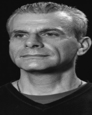 William Holderbaum合作论文数Department of Electronic Engineering, University of Reading11
 Adrian S. Poulton合作论文数11
 C. A. Kirkwood合作论文数10
 James J. Fitzgerald合作论文数Nuffield Department of Surgical Sciences, University of Oxford10RH BAXENDALE合作论文数9
 Michele Vanoncini合作论文数9
 Francisco Saavedra Rodriguez合作论文数8G.F. Phillips合作论文数8J P Paul合作论文数8
 Thierry Houdayer合作论文数7Ross Davis合作论文数7R Williamson合作论文数6
 Pablo Aqueveque合作论文数6
 Ben W. Heller合作论文数Bioengineering Unit, University of Strathclyde6
 James Shippen合作论文数Marlbrook6Dries Hettinga合作论文数5
 Rodrigo Osorio合作论文数5
 Mark Delargy合作论文数5
 Graham Creasey合作论文数Neurosurgery Department, Stanford University5
 Gary S. Beaupré合作论文数Department of Mechanical Engineering, Stanford University5
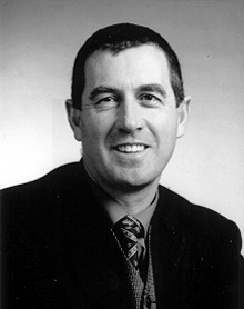 Garry D Wheeler合作论文数5Tadej Bajd合作论文数5
 Adam Thrasher合作论文数Institute of Biomaterials;Biomedical Engineering;University;Institute of Biomaterials, University5r s gibbons合作论文数4D. J. Nicol合作论文数4Andrew Barriskill合作论文数4
 Conor Keogh合作论文数Nuffield Dept Surg Sci, Univ Oxford4
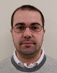 Giacinto Luigi Cerone合作论文数PoliToBIOMed Lab, Politecn Torino4
 Irene Tracey合作论文数Nuffield Department of Clinical Neurosciences, Medical Sciences Division, University of Oxford4
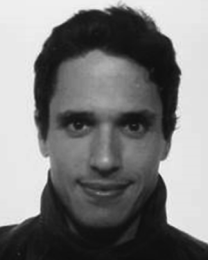 Vieira Taian M合作论文数Escola de Educação Física e Desportos, Universidade Federal do Rio de Janeiro;Laboratorio di Ingegneria del Sistema Neuromuscolare, Politecnico di Torino;Laboratorio di Ingegneria del Sistema Neuromuscolare, Universidade Federal do Rio de Janeiro4P.A. Freeman合作论文数4
 Amy B. Zavatsky合作论文数4Suzanne E. Halliday合作论文数4Woon Tsang合作论文数4Garry D. Wheeler合作论文数4
 Paulina Ortega合作论文数3Monica Armengol合作论文数3Daniela Pinto合作论文数Elect Engn Dept, Univ Concepcion3
 Enrique Germany合作论文数Dept Elect Engn, Univ Concepcion3
 Sebastian Dubo合作论文数3R. B. Stein合作论文数3
 Britam Gómez合作论文数Elect Engn Dept, Univ Concepcion3Ferguson A C合作论文数3Kischka Udo合作论文数3Patrick, J.合作论文数3




