Figure S4: Loss of stromal IL-33 alters the ST2+ immune cell secretome, resulting in a shift in CAF differentiation.
Supplementary Figure 1 provides additional details of orthotopic tumors treated with TCDD including histology, gross pictures and dose escalation. Data on additional cell lines including in vitro growth kinetics is provided.
Supplementary Figure 5 shows additional single cell sequencing data from orthotopic PDAC tumors in mice treated with TCDD or untreated.
Pancreatic ductal adenocarcinoma has a unique tumor microbiome, and the depletion of gut bacteria or fungi using antibiotic/antifungal cocktails has been shown to decrease pancreatic tumor burden in mice. However, functional studies evaluating the role of tumor-associated microbes are few due to the limited availability of clinically relevant microbiota. Here, we describe in detail an effective workflow for the isolation of bacteria and fungi from the duodenum and tumor of pancreatic cancer patients, specifically optimized for cryopreserved, low biomass samples. Using this workflow we also isolated microbiota from normal pancreatic tissue and duodenum from organ donors, and we confirmed the presence of bacteria and fungi isolated from tissue samples with 16S and ITS sequencing analysis. Isolation and sequencing results show distinct similarities between the pancreatic and duodenal microbiomes and highlight unique bacterial strains that survive in the tumor microenvironment. As a proof of concept, we characterized a select Klebsiella oxytoca strain (UMKO1) isolated from a pancreatic tumor, using whole genome sequencing, metabolomics, and ex- vivo tumor cultures to determine its potential impact on the pancreatic tumor microenvironment. In summary, this optimized workflow allows for the isolation of a variety of bacteria and fungi from low biomass, cryopreserved pancreatic and duodenal tissues, which can then be used for functional studies characterizing clinically relevant tumor-associated microbiota.
Figure S7: Tumor cell-initiated autocrine signaling drives IL-33 upregulation in pancreatic fibroblasts.
Supplementary Figure 2 shows RNA and protein expression of interferon gamma and IL22. An image of the standard ELISA curve is provided.
Figure S6: Expression of fibroblast IL-33 is extrinsically induced by epithelial KrasG12D and requires JAK1/2-STAT3 activation throughout tumorigenesis.
Supplementary Figure 3 shows gating strategies used for flow analysis of CD4 cells from orthotopic tumors as well as serum and tissue IL22 measured by ELISA. IHC and multiplex IHC analysis of TCDD treated tumors in the absence of T regulatory cells is shown.
The tumor microenvironment (TME) is composed of tumor cells and surrounding stroma, including immune, mesenchymal, and vascular cells, as well as soluble factors. Traditional techniques to interrogate cellular interactions and infiltration are limited by the loss of tissue architecture or by labeling only a small number of antigens. Multiplex fluorescent immunohistochemistry (mfIHC) is a tissue staining technique that involves tyramide-based signal amplification to covalently bind a fluorophore to an antigen of interest, followed by removal of the primary and secondary antibodies, thereby permitting co-staining and localization of multiple antigens on a single sample. mfIHC retains spatial information, allowing for in-depth analysis of cellular phenotypes and their interactions within the TME. Described here is an overview of the mfIHC experimental design, staining, and image analysis in murine tissues utilizing a manual technique and the Akoya OPAL system.
Supplementary Figure 4 shows histologic features of orthotic tumors placed in AhR knockout mice or those treated with an AhR inhibitor prior to TCDD administration.
Pancreatic intraepithelial neoplasia (PanIN) precedes pancreatic cancer, a deadly disease characterized by an extensive tumor microenvironment. How the microenvironment evolves during cancer progression is largely unknown, as PanINs are microscopic and non-diseased pancreas samples are exceedingly rare, while adjacent normal samples are disrupted by the presence of malignancy. Leveraging donor organs and spatial technologies we mapped the evolution of PanIN to cancer. The PanIN epithelial component falls on a continuum with cancer while the PanIN microenvironment is drastically distinct. Progression to cancer is accompanied by profound geographical reorganization of myeloid cells and lymphocytes and the formation of a cancer-specific fibroblast population characterized by high levels of Smooth Muscle Actin, LRRC15 and the WNT signaling component LEF1. Together, our data show asynchronous evolution of epithelial and stromal components during pancreatic carcinogenesis. Lack of stromal reprogramming might explain why most PanINs do not progress to cancer. Compiled data available at https://pascadimagliano-lab.github.io/PancAtlas.
Although smoking is a risk factor for pancreatic adenocarcinoma (PDAC), the underlying mechanisms promoting tumorigenesis and progression are unknown. In this study, we show that aryl hydrocarbon receptor (AHR) ligands found in cigarette smoke, like the carcinogen 2,3,7,8-tetrachlorodibenzo-p-dioxin, promote pancreatic dysplasia and PDAC progression in a mouse model of this disease. This effect is mediated by AHR activation in CD4+ T cells, leading to their polarization to IL22-producing TH22 cells and regulatory T cell accumulation, ultimately driving a blunted CD8+ T-cell effector response. Analysis of human pancreata from organ donors revealed that smokers have increased AHR activation relative to nonsmokers. Similarly, PDAC tumors from patients with a history of cigarette smoking presented with increased regulatory T-cell accumulation compared with nonsmokers. These findings support a model whereby AHR ligands (AHRL) in cigarette smoke promote tumorigenesis and progression of PDAC through dysregulation of immune responses. SIGNIFICANCE:Our study investigates the mechanistic link between AHRL and pancreatic cancer. We determined that AHRLs polarize naïve T cells, resulting in increased production of IL22 and immunosuppression. Our findings identify a novel signaling axis linking environmental chemicals to pancreatic tumorigenesis via the immune system. See related commentary by Zhao and Hill, p. 13.
Supplementary Figure 6 shows the dysplasia and AhR expression in a spontaneous murine model of PDAC. Additionally expression in human tissues is shown.
 Timothy L Frankel合作论文数30
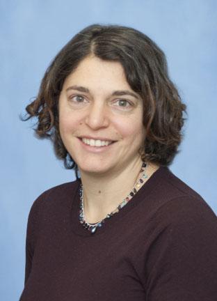 Marina Pasca Di Magliano合作论文数Department of Surgery, University of Michigan23Padma Kadiyala合作论文数Univ Michigan, Sch Med, Dept Neurosurg, Ann Arbor, MI USA21
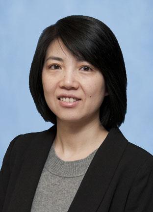 Yaqing Zhang合作论文数Department of Surgery, Medical School, University of Michigan;Department of Surgery Research, Medical School, University of Michigan20Eileen S. Carpenter合作论文数18
 Filip Bednar合作论文数General Surgery, Department of Surgery, Medical School, University of Michigan-Ann Arbor17
 Jiaqi Shi合作论文数Department of Pathology, Medical School, University of Michigan-Ann Arbor17
 Arvind Rao合作论文数Department of Biomedical Engineering, College of Engineering, University of Michigan-Ann Arbor17Carlos E Espinoza合作论文数15
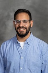 Ahmed Elhossiny合作论文数University of Michigan15Katelyn L Donahue合作论文数14
 Wenting Du (杜文婷)合作论文数14Lei Sun合作论文数13Jake McGue合作论文数13Hannah R Watkoske合作论文数13Kristee Brown合作论文数11Nicholas M. Nedzesky合作论文数10Rachael Baliira合作论文数10Nur M Renollet合作论文数10Paola I Medina-Cabrera合作论文数10Jacqueline Morales合作论文数10
 Ashley Velez合作论文数10Emily L. Lasse Opsahl合作论文数10Rosa E Menjivar合作论文数10Michael K Scales合作论文数10Benjamin L. Allen合作论文数10Jacob Edwards合作论文数8
 Fatima Lima合作论文数5Nicole Peterson合作论文数5Elizabeth Novak合作论文数UPMC Childrens Hosp Pittsburgh, Univ Pittsburgh5Jay Li合作论文数5
 Vaibhav Sahai合作论文数Division of Hematology and Oncology, Department of Internal Medicine, Medical School, University of Michigan-Ann Arbor5
 Kevin Mollen合作论文数5Vei Shaun Siow合作论文数4Valerie Gunchick合作论文数4Sarah Nelson合作论文数4Stephanie The合作论文数4
 Meredith Barrett合作论文数Propeller Hlth, San Francisco, CA USA4
 Christopher J. Sonnenday合作论文数Department of Health Management and Policy, School of Public Health, University of Michigan-Ann Arbor4Sun Lei合作论文数4
 Garret Vincent合作论文数4Chin-Tung Chen合作论文数4Michael Mattea合作论文数3Nicholas Olden合作论文数3Heather L Mentrup合作论文数3Kellie E Cunningham合作论文数3Allison Bischoff合作论文数3
 Hari Nathan合作论文数General Surgery, Department of Surgery, Medical School, University of Michigan-Ann Arbor3Santhoshi Krishnan合作论文数2




