A 56-year-old man with a history of hereditary multiple exostoses and secondary low-grade chondrosarcomas was diagnosed with a pigmented mass in his left eye involving the inferonasal ciliary body and peripheral choroid following a superotemporal visual field defect. The intraocular mass continued to grow following brachytherapy, prompting enucleation that revealed a low-grade chondrosarcoma (CHS), similar in appearance to his prior skeletal neoplasms. There are 3 reports of CHS metastatic to the eye, and all 3 individuals had metastases to the lungs and other body sites before or at the time of ocular metastasis. Our patient had no pulmonary or other metastases identified radiographically at the time his intraocular tumor was diagnosed, presenting a conundrum: was the intraocular tumor a metastasis or did it arise de novo in a patient with hereditary multiple exostoses? The intraocular CHS in our patient resembles that reported rarely in other mammals, suggesting that it was a primary neoplasm arising from induced chondrogenic differentiation of uveal mesenchymal stromal cells in a patient with hereditary multiple exostoses and a predilection for developing low-grade chondrosarcomas.
Background: Oncologic resections and reconstructions of the pelvis are among the most challenging procedures in orthopedic surgery due to complex anatomy, tumor variability, and the need for precise margins. Patientspecific instrumentation (PSI) created with 3D printing has emerged as a promising solution, but accurate intraoperative referencing remains difficult. This study evaluates the use of novel acetabular-referencing PSI cutting guides for pelvic tumor resections and reconstructions. Methods: Five patients (mean age 57.2 years) underwent pelvic resections with acetabular-referencing PSI between 2019 and 2023. Each guide incorporated a hemispherical feature seating within the acetabulum after femoral head removal. Postoperative CT reconstructions were compared with preoperative plans to assess resection accuracy, implant alignment, operative time, and margin status. Results: All patients achieved negative gross margins; two had microscopically positive margins (one planned). Mean operative time was 8 h 52 min, including two complex procedures. Mean deviations were 8.32 mm for center of rotation (CoR) and 7.37 mm for center of gravity (CoG). Directional CoR deviations averaged 2.62 mm anteroposteriorly, 6.30 mm mediolaterally, and 3.92 mm superoinferiorly. Angular deviations averaged 1.88 degrees in anteversion and -2.54 degrees in inclination. All implants were positioned within or near accepted thresholds for pelvic reconstruction accuracy. Conclusions: Acetabular-referencing PSI enabled reproducible guide placement and accurate osteotomies in complex pelvic resections without increasing operative time. This technique offers a reliable alternative to surface-contoured guides and may enhance precision in oncologic reconstruction by mitigating challenges of intraoperative interfacing. Larger studies with long-term follow-up are needed to confirm functional outcomes and implant survivorship.
Background Oncologic resections and reconstructions of the pelvis are among the most challenging procedures in orthopedic surgery due to complex anatomy, tumor variability, and the need for precise margins. Patient-specific instrumentation (PSI) created with 3D printing has emerged as a promising solution, but accurate intraoperative referencing remains difficult. This study evaluates the use of novel acetabular-referencing PSI cutting guides for pelvic tumor resections and reconstructions. Methods Five patients (mean age 57.2 years) underwent pelvic resections with acetabular-referencing PSI between 2019 and 2023. Each guide incorporated a hemispherical feature seating within the acetabulum after femoral head removal. Postoperative CT reconstructions were compared with preoperative plans to assess resection accuracy, implant alignment, operative time, and margin status. Results All patients achieved negative gross margins; two had microscopically positive margins (one planned). Mean operative time was 8hours 52minutes, including two complex procedures. Mean deviations were 8.32mm for center of rotation (CoR) and 7.37mm for center of gravity (CoG). Directional CoR deviations averaged 2.62mm anteroposteriorly, 6.30mm mediolaterally, and 3.92mm superoinferiorly. Angular deviations averaged 1.88° in anteversion and –2.54° in inclination. All implants were positioned within or near accepted thresholds for pelvic reconstruction accuracy. Conclusions Acetabular-referencing PSI enabled reproducible guide placement and accurate osteotomies in complex pelvic resections without increasing operative time. This technique offers a reliable alternative to surface-contoured guides and may enhance precision in oncologic reconstruction by mitigating challenges of intraoperative interfacing. Larger studies with long-term follow-up are needed to confirm functional outcomes and implant survivorship.
Ewing sarcoma and osteosarcoma constitute 36% of all primary bone cancers. However, these 2 subtypes represent the most commonly diagnosed bone cancer types in the pediatric and adolescent population. Although still largely unknown, certain genetic mutations, rearrangements, and/or predisposition syndromes likely play a role in the pathogenesis of bone cancer. Osteosarcoma may also develop as a direct result of the long-term side effects of radiation therapy. With the implementation of a multimodality approach to treatment, including multiagent neoadjuvant and adjuvant chemotherapy regimens, targeted therapy options, surgery, and radiation, individuals with Ewing sarcoma and osteosarcoma are showing higher cure rates and improved overall survival. The NCCN Guidelines for Bone Cancer provide a consensus and evidence-based framework for the workup, management, and surveillance of local and recurrent/metastatic disease.
There is discrepancy in the histologic concordance of pre-operative biopsy grading and post-surgical resection grading of periacetabular chondrosarcoma. This discrepancy poses a question as to whether the chosen treatment should depend upon on the initial assessment of the tumor grade. This is a retrospective review of patients with periacetabular cartilaginous tumors who were treated between the years 2000 and 2022 at a single institution. All patients included in the analysis underwent surgical resection. The pre-operative biopsy and post-surgical resection histological grades were compared. Concordance and treatment implications were evaluated. Twenty-two pre-operative Image Guided Core Needle Biopsies (IGCNB) were performed. There was concordance between pre-operative and surgical resection histological grade in only 9 cases, giving a (9/22) 40.9% accuracy rate. In total there were 13 cases of discrepancy between pre-operative biopsy and surgical resection regarding histological grade. In 12 cases of discrepancy, surgical resection histopathology revealed a higher grade than that of pre-operative biopsy histopathology. In one case of discrepancy, pre-operative IGCNB biopsy histology revealed a high-grade chondrosarcoma while the surgical resection histology revealed an intermediate-grade. There were no situations in which the plan made based on a preoperative biopsy resulted in an inappropriate treatment decision, even when there was not concordance between preoperative diagnosis and diagnosis after definitive resection. Significant discrepancy in grading was observed with pre-operative IGCNB of periacetabular cartilaginous lesions. However, while 12 out of the 13 discordant cases (92.3%) underestimated the true grade of the tumor, there was no impact on appropriateness of the selected treatment, suggesting that accuracy of grade is not an important factor to consider in this location.
Objective:To manufacture and characterize a modified niclosamide stearate (NS) prodrug therapeutic (mNSPT) for use in a small clinical trial in a metastatic canine osteosarcoma (OS) model. Animals:10 dogs that presented for treatment of nondetectable metastatic OS underwent resection of primary tumors prior to systemic therapy. Four cycles of IV carboplatin (300 mg/m2, IV, q 3 wk) followed by 4 cycles of the experimental mNSPT (10 mg/kg, IV, weekly). Posttreatment surveillance included physical examination and thoracic radiographs every 3 months for 2 years. Samples for pharmacokinetic analysis were taken at the end of the 0.5- to 1-hour IV infusion of the mNSPTs and followed for 2 hours. Clinical Presentation:The NS average concentration at the end of infusion was 134.88 ± 13.32 μg/mL; the average area under the curve was 211.62 ± 27.89 h·µg/mL. The niclosamide concentration at the end of infusion was 23.11 μg/mL ± 3.77 μg/mL, and the area under the curve was 27.52 ± 5.92 h·µg/mL, well above the NSPT 0.75 μg/mL (1.27 μM) cell-effective concentrations for OS cells in culture. The average terminal half-life was 4.57 hours for NS and 3.23 hours for niclosamide. Three dogs developed metastatic disease on carboplatin and did not receive mNSPT. The median time to tumor progression and OS of the 7 treated dogs was 510 and 632 days, respectively, with 3 dogs living > 4 years. Results:The results support further translational investigation of this novel therapeutic approach to OS treatment in a randomized, prospective phase III study in dogs and, if successful, ultimately in OS patients. Clinical Relevance:These results suggest that niclosamide-when transformed into the highly bioavailable NSPT-may have potential as a novel therapeutic agent for treating OS.
The addition of pembrolizumab to preoperative radiotherapy (RT) improved disease-free survival (DFS) for patients with stage III undifferentiated pleomorphic sarcoma (UPS) and dedifferentiated/pleomorphic liposarcoma (LPS) in the randomized SU2C-SARC032 trial. To precisely identify patients who benefit from pembrolizumab and RT, we performed comprehensive multi-omics profiling of pre- and post-treatment tumor and blood samples, including bulk RNA-seq, flow cytometry, and cytometry by time of flight. Additionally, we built a single-cell RNA-seq atlas spanning 65,786 cells from UPS and LPS to recover single-cell states in bulk tumor samples using digital cytometry. Two opposing tumor microenvironments (TMEs), immune-cold sarcoma ecotype 1 (SE1) and immune-hot sarcoma immune class E (SIC E), benefited from pembrolizumab. Pembrolizumab combined with RT depleted PD-1+ exhausted T cells in SIC E sarcomas and increased effector memory CD4+ T cells in SE1 sarcomas with an overall increase in CD8+ early activated T cells, CD4+ follicular helper T cells, and T cell receptor diversity. Matrix-remodeling stromal and epithelial-like sarcoma cell programs were associated with worse outcomes and diminished with pembrolizumab and RT. Our findings identify different mechanisms of response to pembrolizumab in localized, high-risk UPS/LPS and suggest that sarcoma TME signatures may identify patients most likely to benefit from adding pembrolizumab to preoperative RT.
BACKGROUND:While megaprosthetic reconstructions have been instrumental in restoring patients' functional abilities, surgeons remain cautious of revision risk. This is the first study to assess the impacts of preoperative bone mineral density (BMD) on outcomes following lower extremity megaprosthetic reconstructions. METHODS:This was a retrospective cohort study of patients who underwent lower extremity oncologic megaprosthetic reconstruction between March 2005 and January 2022. Hounsfield units of the first lumbar vertebrae within a year prior to surgery were blindly collected for assessment of BMD. Patient demographics, megaprostheses characteristics and postoperative outcomes were recorded. Primary outcomes were complications according to the Henderson classification and implant survival rate. RESULTS:Eighty-four patients underwent a lower extremity megaprosthetic reconstruction. Mean age was 46.6 ± 22.5 years and average follow-up was 53.5 ± 52.9 months. Indication for surgery included primary bone tumor (81.3 %) and secondary bone tumor (18.8 %). Twenty-three patients (28.8 %) had low BMD. The rate of revision was 27.5 % in the total cohort and not significantly different between groups. The most common indication for revision was infection. While no patients with low BMD developed aseptic loosing or structural failure, they were more likely to receive femoral cement fixation (OR = 5.7; 95 %CI = 1.2,55.6). Five-year implant survivorship was 66.6 % (95 %CI [54.9 %- 80.8 %]). CONCLUSION:There was no significant difference in revision rate following oncologic lower extremity megaprosthetic reconstruction in patients with or without low BMD. No patients with low BMD in this cohort experienced aseptic loosening or structural failure; however, there was a bias toward cement fixation in patients with low BMD. This may reflect calculated decisions by the surgeon when evaluating the patient's bone quality and their specific risk profile. Our findings suggest that patients with low BMD have similar outcomes to patients without low BMD undergoing oncologic megaprosthetic reconstructions by experienced surgeons. The specific effect of cemented vs uncemented fixation in patients with low BMD in the oncologic population deserves further investigation.
Background/Objectives: Locally recurrent osteosarcoma is associated with high patient morbidity and mortality, yet the risk factors for local recurrence remain incompletely understood. Therefore, the objective of this study was to comprehensively assess the impact of tumor histology, patient demographics, surgical resection, and chemotherapy delivery on the risk of local recurrence, development of metastases, and overall patient survival in osteosarcoma. Methods: This single-center retrospective review included 102 patients with skeletal osteosarcoma who underwent primary surgical resection between August 2001 and August 2021. Primary outcomes included the development and timing of local recurrence following primary resection. Data was abstracted from the electronic medical record, and statistical analyses were performed to identify demographic, tumor, or management (surgical and chemotherapy) related factors associated with an increased risk of local recurrence. Results: Locally recurrent osteosarcoma developed in 13.7% (n = 14) of patients after primary resection. Patients with local recurrence were more likely to have the chondroblastic subtype (57.1% versus 19.3% in those without local recurrence, p = 0.005). The chondroblastic subtype was associated with shorter local recurrence-free survival and overall survival than other histologic subtypes (p < 0.001). Resection approach, surgical margin status, histologically assessed response to neoadjuvant chemotherapy, and type and dose intensity of chemotherapy delivered were not statistically associated with risk of local recurrence. Conclusions: Chondroblastic histology is a risk factor for local recurrence in osteosarcoma and is associated with poor overall survival in our patient cohort.
Orthopedic procedures often require removing bone or pathological tissue, with traditional methods involving instruments like curettes and rongeurs. However, these methods can be time-consuming and lead to increased blood loss. To mitigate these side effects, vacuum-assisted tools have been developed to aid in tissue removal. These devices enable surgeons to suction tissue without discarding it, potentially improving outcomes in conditions such as osteomyelitis or tumor removal while enabling collection of the material for downstream applications. Despite limited research, vacuum-assisted devices show promise beyond bone marrow harvesting. This study assesses infection and clearance rates, estimated blood loss, and total procedure time associated with the use of vacuum-assisted tissue removal, with a goal to understand if these devices can be used for tissue removal across a variety of pathologic conditions. A retrospective cohort study was conducted on patients undergoing orthopedic procedures with the Avitus® Bone Harvester repurposed from its original design from December 1, 2021, to July 1, 2023. Procedures were categorized into oncology, and debridement for infection cases. Infection cases were further categorized into those secondary to trauma and those involving primary infections (osteomyelitis and periprosthetic joint infection). Clinical variables, including demographics, intraoperative details, complications, and follow-up, were reviewed. Statistical analysis included descriptive statistics computed with R Studio. The study included 44 patients, with debridement for infection cases being the most common (primary infection: 45.5
Benign, locally aggressive tumors of the distal femur are typically treated with intralesional curettage and polymethylmethacrylate (PMMA) cementation. However, it is not known whether plate fixation should be added to biomechanically augment these PMMA-filled defects. The purpose of this study was to evaluate the performance of two competing techniques for reconstruction of a distal femoral defect. For this biomechanical study, we used 12 composite femurs with properties comparable to bone. In nine femurs, identical contained medial distal femoral defects were created using a robotic arm. Group A contained three intact femurs, Group B three femurs with an unfilled defect, Group C three femurs reconstructed with PMMA alone, and Group D three femurs reconstructed with PMMA plus a medial locking plate. Locations of greatest stress concentration were determined by PhotoStress analysis, then three strain gauges were applied to each specimen at these high-stress locations. Specimens were loaded within a physiologic range followed by loading to failure. Outcome measures included construct stiffness, strain along the distal femur, and load at failure. Results showed that stiffness and strain were not significantly different between reconstructive techniques; however, both techniques reduced tensile strain along the popliteal surface by approximately 40% compared to non-reconstructed specimens. All specimens failed at the femoral neck before failing at the distal femur. These findings suggest that plate augmentation of PMMA-filled distal femoral defects like the one in this study offers insignificant biomechanical benefit within physiologic loads and therefore may be unnecessary.
Background In the United States, socioeconomic status (SES), race, and ethnicity are factors used to predict adverse outcomes in various carcinomas as well as in soft tissue sarcoma. However, existing evidence is unclear if race is an independent predictor of overall survival in common primary malignant bone tumors, including Ewing sarcoma, osteosarcoma, and chondrosarcoma. Methods This is a retrospective analysis of the National Cancer Database (NCDB) of patients diagnosed with primary Ewing sarcoma, osteosarcoma, and chondrosarcoma between 1998 and 2016. Race/ethnicity was divided into White, Black, Asian, and “Hispanic”. Baseline and treatment characteristics between groups were compared using Pearson’s chi-square test for categorical variables and analysis of variance (ANOVA) for continuous variables. Long-term survival for each group was evaluated with the Kaplan-Meier (KM) method with comparisons between races based on the log-rank test. A multivariable Cox proportional hazard model was used to predict variables associated with lower overall survival. Results Black Ewing sarcoma patients had significantly poorer overall survival than did White, Asian, or Hispanic patients (p < 0.001) with a KM 5-year survival of 48.2% vs 61.0% vs 68.3% vs 63.0%, respectively, and a 123.5% (HR: 2.235, p = 0.009) higher independent likelihood of mortality in a multivariate analysis. Independent predictors of overall survival for Ewing sarcoma, osteosarcoma, and chondrosarcoma included older age (HR: 1.029, p < 0.001; HR: 1.020, p < 0.001; HR: 1.034, p < 0.001), more advanced cancer stage (HR: 3.315, p < 0.001; HR: 6.167, p < 0.001; HR: 10.201, p < 0.001), and positive surgical margins (HR: 1.679, p = 0.001; HR: 1.540, p < 0.001; HR: 1.992, p < 0.001), respectively. A significant racial disparity in survival for Black patients with Ewing sarcoma was identified. Conclusions Our study identifies a significant racial disparity in survival for Black patients with Ewing sarcoma. Our study reinforces previously established literature about predictors of overall mortality for Ewing sarcoma, osteosarcoma, and chondrosarcoma populations, while also providing new insight into the role of race, cancer-specific factors and treatment, and SES on long-term survival.
Purpose. Recently, the association between ATRX and a more aggressive sarcoma phenotype has been shown. We performed a retrospective study of sarcomas from an individual institution to evaluate ATRX as a prognosticator in soft tissue sarcoma. Experimental Design. 128 sarcomas were collected from a single institution and stained for ATRX. The prognostic significance of these markers was evaluated in a smaller cohort of primary soft tissue sarcomas (n = 68). Kaplan–Meier curves were created for univariate analysis, and Cox regression was utilized for multivariate analysis. Results. High expression of ATRX was found to be a positive prognostic indicator for overall survival and metastasis-free survival in our group of soft tissue sarcomas both in univariate analysis and multivariate analysis (HR: 0.38 (0.17–0.85), P=0.02 and HR: 0.49 (0.24–0.99), P=0.05, respectively). Conclusions. High expression of ATRX is a positive prognostic indicator of overall survival and metastasis-free survival in patients with STS. This is consistent with studies in osteosarcoma, which indicate possible mechanisms through which loss of ATRX leads to more aggressive phenotypes. Future prospective clinical studies are required to validate the prognostic significance of these findings.
CIC-rearranged sarcomas comprise a group of exceptionally aggressive round-cell sarcomas. These tumors most commonly demonstrate CIC::DUX4 fusion and show similar histopathology to Ewing sarcomas, though lesions mimicking vascular neoplasms have recently been described. Here, we describe a case of a patient with CIC::DUX4 fusion sarcoma identified using RNA-based molecular testing who was initially diagnosed with an endothelial neoplasm. The tumor showed extensive vasoformative growth, complete WT1 negativity, and global positive staining for ERG, CD31, and DUX4 by immunohistochemistry. Methylation testing of the tumor clustered more closely with angiosarcomas than with CIC-rearranged sarcomas. Our findings suggest that CIC::DUX4 fused neoplasms may demonstrate a more diverse phenotypic range than previously appreciated and offer evidence that both molecular and immunohistochemical studies are needed for accurate diagnosis.
11504 Background: Surgery & radiation therapy (RT) yield high local control rates for soft tissue sarcoma (STS) of the extremity and limb girdle. However, patients (pts) with high-grade stage III STS are at significant risk for developing metastasis. Median survival for pts with metastatic STS is < 2 years. SARC028 (NCT02301039) evaluated the efficacy of pembrolizumab (pembro) for metastatic STS, showing 20% and 8.7% response rates in undifferentiated pleomorphic sarcoma (UPS) and pleomorphic/dedifferentiated liposarcoma (LPS), respectively. We hypothesized that neoadjuvant pembro with concurrent RT followed by surgery and adjuvant pembro for stage III UPS, including myxofibrosarcoma, or LPS would stimulate an anti-tumor immune response to eliminate micrometastatic disease & improve disease-free survival (DFS). SU2C-SARC032 (NCT03092323) is a multi-institutional, international, randomized phase 2 trial evaluating the safety and efficacy of adding pembro to standard of care (SOC) RT & surgery for pts with stage III UPS or LPS. Methods: Pts aged > 12 yo with stage III (FNCLCC grade 2 or 3) UPS or LPS of the extremity and limb girdle were enrolled. Pts were randomized (1:1, stratified by grade) to neoadjuvant RT (50 Gy/25 fx) then surgery (SOC arm) or neoadjuvant pembro and RT then surgery & adjuvant pembro (EXP arm). Pembro was given 200 mg IV Q3 wk for 3 doses (before, during & after RT) & up to 14 adjuvant cycles. The primary endpoint was 2-yr DFS. Secondary endpoints included local recurrence-free survival (LRFS), distant disease-free survival (DDFS), & overall survival (OS). Target enrollment of 126 evaluable patients (max 144 total) provided 80% power (1-sided α = 0.05) to distinguish between a null hypothesis of 50% 2-yr DFS rate & alternative hypothesis of 75% 2-yr DFS rate by log-rank test, with initial analysis at 45 DFS events. Cox models were stratified by grade; primary analysis was a one-sided stratified log-rank test. Results: Between July 2017-November 2023, 143 patients were enrolled, predominantly with UPS (85%) & grade 3 (64%) histology. Median follow-up for alive patients is 24.1 mo. DFS in the EXP arm is significantly higher than the SOC arm (p = 0.023; HR 0.57, 90% CI: 0.35, 0.91). Estimated 2-yr DFS is 53% (90% CI: 43, 66%) for SOC vs 70% (90% CI: 61, 81%) for EXP arm. Currently, there is no statistically significant difference in LRFS (HR 0.55, 95% CI: 0.21, 1.42), DDFS (HR 0.57, 95% CI: 0.32, 1.01), or OS (HR 0.39, 95% CI: 0.14, 1.12). Pts with grade 3 sarcomas had improved DFS with pembro (HR 0.47, 95% CI: 0.25, 0.89), but no difference in DFS was observed in grade 2 tumors (HR 1.21, 95% CI: 0.35, 4.18). The proportion of patients with grade 3+ adverse events was significantly higher in EXP (52%) vs SOC arm (26%) (p = 0.002). Conclusions: Theaddition of neoadjuvant & adjuvant pembro to RT and surgery significantly improves DFS for pts with stage III UPS and LPS of the extremity and limb girdle. Clinical trial information: NCT03092323 .
Background:Percutaneous tripod fixation of periacetabular lesions is performed at our institution for patients with metastatic bone disease and a need for quick return to systemic therapy. We have begun to use the IlluminOss Photodynamic Bone Stabilization System instead of the metal implants previously described in the literature because of the success of the IlluminOss implant in fixing fragility fractures about the pelvis. Description:At our institution, the procedure is performed in the interventional radiology suite in order to allow for the use of 3D radiographic imaging and vector guidance systems. The patient is positioned prone for the transcolumnar PSIS-to-AIIS implant and posterior column/ischial tuberosity implant or supine for the anterior column/superior pubic ramus implant. Following a small incision, a Jamshidi needle with a trocar is utilized to enter the bone at the chosen start point. A hand drill is utilized to advance the Jamshidi needle according to the planned vector; alternatively, a curved or straight awl can be utilized. The 1.2-mm guidewire is placed and reamed. We place both the transcolumnar and posterior column wires at the same time to ensure that there is no interference. The balloon catheter for the IlluminOss is assembled on the back table and inserted according to the implant technique guide. The balloon is inflated and observed on radiographs in order to ensure that the cavity is filled. Monomer is then cured, and the patient is flipped for the subsequent implant. Following placement of the 3 IlluminOss devices, adjunct treatments such as cement acetabuloplasty or cryoablation can be performed. Alternatives:Alternative treatments include traditional open fixation of impending or nondisplaced acetabular fractures in the operating room, or percutaneous implant placement in the operating room. Implant placement may be performed with the patient in the supine, lateral, or prone position, depending on surgeon preference. Alternative implants include standard metal implants such as plates and screws, or cement augmentation either alone or with percutaneous screws. Finally, ablation alone may be an alternative option, depending on tumor histology. Rationale:Open treatment of acetabular fractures is a more morbid procedure, given the larger incision, increased blood loss, longer time under anesthesia, and increased length of recovery. Percutaneous fixation may be performed in either the operating room or interventional radiology suite, depending on the specific equipment setup at an individual institution. At our institution, we prefer utilizing the interventional radiology suite as it allows for more precise implant placement through the use of an image-based vector guidance system and 3D fluoroscopy to accurately identify safe corridors. The use of percutaneous fixation allows for faster recovery and earlier return to systemic therapy. Because the IlluminOss implant is radiolucent, it allows for better evaluation of disease progression and can better accommodate nonlinear corridors or fill a lytic lesion to provide stability. Expected Outcomes:Postoperatively, we expect the patient to be weight-bearing as tolerated with use of an assistive device. We expect the small incisions to fully heal within 2 weeks. Patients should be able to return to systemic therapy as indicated earlier than with an open procedure. Important Tips:The use of a hand drill with the Jamshidi needle and trocar can help adjust a drilled pathway and allow for close adherence to a planned vector.Vector guidance systems can be useful to fully capture the area at risk for fracture and to provide maximal stability with the expandable implant, but they are not necessary to perform the procedure.Placing both posterior implants at the same time can be helpful to avoid interference. This is accomplished by drilling and placing the guidewire for both implants prior to reaming and placing the balloon implant. Acronyms and Abbreviations:CT = Computed tomographyPSIS = posterior superior iliac spineAIIS = anterior inferior iliac spine.
Loddo et al. (Br J Cancer 100:959–70, 2009) established the prognostic significance of cell cycle markers and "Cell-Cycle Phenotypes" in breast carcinoma. This study aims to 1) identify prognostic cell-cycle markers in sarcoma, and 2) assess the prognostic potential of specific cell-cycle phenotypes in sarcoma. Tissue samples from 128 soft tissue sarcomas were stained for four cell cycle-specific markers: Mcm2, Geminin, Plk1, and H3S10ph. Only primary soft tissue tumors (liposarcoma, leiomyosarcoma, synovial sarcoma, and undifferentiated pleomorphic sarcoma) were included in the analysis. Any tumor coming from a recurrent or metastatic lesion were excluded from the analysis. Three cell-cycle phenotypes (I, II, III) were derived from marker expression patterns. Prognostic significance was evaluated in a subset of primary soft tissue sarcomas using Cox regression for survival analysis. Compared to phenotype I, the phenotype III tumors had a decreased 5-year overall survival (HR 6.81 [2.36–19.61]; p = < 0.001), 5-year disease-free survival (HR 1.07 (1.02–1.18); p = 0.004), and 5-year metastasis-free survival (HR 4.34 [1.58–11.93]; p = 0.004). High expression of Plk1 was associated with decreased 5-year overall survival (HR: 4.04 CI [1.21–6.67; p = 0.02) and 5-year metastasis-free survival (HR: 2.91 CI [1.15–7.37]; p = 0.03). Geminin was also found to have a decreased 5-year overall survival (HR:2.84 CI [1.21–6.67]; p = 0.02). No statistical difference in prognostication were noted between phenotypes and the AJCC system. We identified three unique sarcoma cell cycle phenotypes that have prognostic significance. This performs similarly to the AJCC staging system.
Abstract BACKGROUND: Psoralen is a natural ultraviolet activated compound that has been used for over 70 years to treat skin cancer, psoriasis, and vitiligo. Recently, to expand its use beyond external treatment, a novel therapeutic called X-PACT (X-ray psoralen activated cancer therapy) was created to treat internal tumors. A combination of 8-methoxypsoralen and phosphors that convert low doses of x-ray radiation to ultraviolet (UV) light are injected into solid tumors and subsequently activated by x-ray photons with considerable antineoplastic effect. Notably, canine clinical trials have observed significant abscopal effects of X-PACT therapy on metastatic lesions throughout patients, but the immunomodulatory mechanisms of action are unclear (Nolan et al, in submission). Here, we present preliminary data investigating the immune response to X-PACT therapy in a murine soft tissue sarcoma (STS) model. METHODS: Six mice of the inducible KrasG12D/+; p53flox/flox (KP) murine STS model with palpable, gastrocnemius tumors were used in this initial study. Four mice underwent treatment with X-PACT. In this group, psoralen was mixed with energy converters and injected into the tumor. The mice then received low-level radiation to activate the drug. Treatment was performed five times over a 15-day period and the animals were sacrificed within 48 hours of final treatment. The other two mice with induced tumors received no treatment prior to sacrifice. For both groups, a cardiac blood draw was performed at the time of sacrifice to obtain plasma serum for analysis. Luminex xMAP technology was used for multiplexed quantification of 45 mouse cytokines, chemokines and growth factors. RESULTS: There were five markers that showed significantly different levels between the treated and control groups. Results indicated significantly higher (p = 0.006) plasma concentrations of macrophage derived chemokine (MDC) following five rounds of treatment with X-PACT therapy. Additionally, the treated mice displayed significantly lower concentrations of granulocyte colony-stimulating factor (G-CSF, p = 0.003), eotaxin (p = 0.016), tissue inhibitor of matrix metalloproteinase 1 (TIMP1, p = 0.024), and interleukin-1β (IL-1β, p = 0.033) compared to untreated mice with STS. CONCLUSION: In humans, MDC levels are associated with longer survival and lower cancer severity. G-CSF, eotaxin, TIMP1, and IL-1β are upregulated in cancer patients and correlate with disease severity. Together, the lower concentrations of G-CSF, Eotaxin, TIMP1, and IL-1β suggest a softening of cancer severity following X-PACT treatment. Further studies and larger sample sizes are necessary to confirm and expand upon these preliminary findings. Citation Format: Beatrice Schleupner, Alexandra Hunter Aitchison, Elle MacAlpine, Nicole Cantor, Jason Somarelli, Brian Brigman, Julia Visgauss, Michael Monument, William Eward. Exploring the x-ray psoralen activated cancer therapy mechanism of action in a genetically engineered mouse model of soft tissue sarcoma [abstract]. In: Proceedings of the American Association for Cancer Research Annual Meeting 2024; Part 1 (Regular Abstracts); 2024 Apr 5-10; San Diego, CA. Philadelphia (PA): AACR; Cancer Res 2024;84(6_Suppl):Abstract nr 6642.
 William C. Eward合作论文数Duke University School of Medicine77
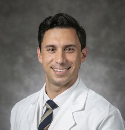 Lazarides Alexander L合作论文数School of Medicine, Duke University31
 David Kirsch合作论文数Department of Radiation Oncology, University of Toronto29
 Julia Visgauss合作论文数Department of Orthopaedic Surgery, Duke University School of Medicine23
 Daniel German Blazer合作论文数Division of Behavioral Medicine & Neurosciences, Department of Psychiatry & Behavioral Sciences, Duke University School of Medicine16
 Diana Cardona合作论文数Department of Pathology, Duke University School of Medicine15
 Jeffrey K. Mito合作论文数15
 Richard Francis Riedel合作论文数Department of Medicine, School of Medicine, Duke University13
 Leslie G. Dodd合作论文数Department of Pathology and Laboratory Medicine, School of Medicine, The University of North Carolina at Chapel Hil11Jorge M. Ferrer合作论文数9
 Jason Andrew Somarelli合作论文数Department of Medicine, School of Medicine, Duke University School of Medicine9
 Kyle Cuneo合作论文数Department of Radiation Oncology, Medical School, University of Michigan;Rogel Cancer Center, University of Michigan Health9
 Moungi Bawendi (莫吉·巴旺迪)合作论文数Department of Chemistry, Massachusetts Institute of Technology9
 David L Kerr合作论文数8
 Nicole A. Larrier合作论文数School of Medicine, Duke University8
 Ivan Spasojevic合作论文数Department of Medicine, School of Medicine, Duke University School of Medicine8
 Linda G. Griffith合作论文数Department of Biological Engineering, School of Engineering, Massachusetts Institute of Technology8
 Harrison R Ferlauto合作论文数School of Medicine, Duke University7
 Detlev Erdmann合作论文数Department of Orthopaedic Surgery, Duke University School of Medicine7Rebecca D. Dodd合作论文数7Anthony A Catanzano合作论文数6Mark M Cullen合作论文数6
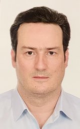 Vasileios Kontogeorgakos合作论文数School of Health Sciences, University Hospital of Thessalia6
 Melodi Whitley合作论文数6Salutario Martinez合作论文数6Brian L Dial合作论文数5Etienne Flamant合作论文数5
 J. Sybil Biermann合作论文数Medical School, University of Michigan5
 Sin-Ho Jung合作论文数Division of Integrative Genomics, Department of Biostatistics & Bioinformatic, Duke University School of Medicine;Duke Cancer Institute, Duke University5
 Sean V Mcgarry合作论文数5Carter Jessica E合作论文数5
 Matt Van De Rijn合作论文数Maternal & Child Health Research Institute, School of Medicine, Stanford University;Stanford Cancer Institute, Stanford University5
 Joel Mayerson合作论文数College of Medicine, The Ohio State University5
 Paul Joseph Mosca合作论文数Department of Surgery, School of Medicine, Duke University;Duke Quality Network, Duke University Health System5
 Robert L. Satcher合作论文数Department of Orthopaedic Oncology, Division of Surgery, The University of Texas MD Anderson Cancer Center;Ken Kennedy Institute of Information Technology, Rice University;Department of Biomedical Engineering, Northwestern University5
 David B Strasfeld合作论文数5
 Richard J O'Donnell合作论文数4
 Junheng Gao合作论文数Department of Biostatistics & Bioinformatics, Duke University School of Medicine4
 Chang Lung Lee合作论文数Department of Radiation Oncology, Duke University School of Medicine;Duke Cancer Institute, Duke University4
 Howard Levinson合作论文数Department of Dermatology, Duke University School of Medicine4
 Whitney O'Neill Lane合作论文数Division of Plastic, Maxillofacial, and Oral Surgery, School of Medicine, Duke University;Duke University Medical Center4
 Herrick Siegel合作论文数Orthopedic Specialties Building, University of Alabama at Birmingham4
 Robert S. Benjamin合作论文数Department of Sarcoma Medical Oncology, Division of Cancer Medicine, The University of Texas MD Anderson Cancer Center4
 Monga Varun合作论文数Division of Hematology/Oncology and Bone Marrow Transplantation, University of Iowa Hospitals and Clinics4
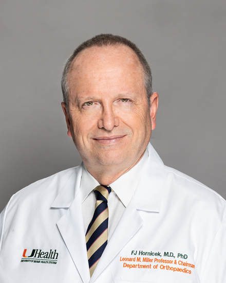 Francis John Hornicek合作论文数Sarcoma Research Laboratory, University of Miami4Daniel P. Nussbaum合作论文数Keck School of Medicine, University of Southern California4
 R. Lor Randall合作论文数Department of Orthopaedics, Huntsman Cancer Institute4
 Warren Chow合作论文数Department of Medical Oncology and Therapeutics Research, City of Hope National Medical Center4




