PURPOSE:Donation after circulatory death (DCD) as a donor for heart transplantation is a new practice. We sought to determine if center volume (high vs low) influenced perioperative outcomes and short-term survival. METHODS:The United Network for Organ Sharing registry was used to identify DCD heart recipients from 2019 to 2025. Recipients were stratified based on center volume: high-volume (>15 transplants over the observational period from 2019 to 2020) or low volume (≤15 transplants). Comparative statistics and Kaplan-Meier methods with the log-rank test were used to compare groups and determine survival. A Cox regression analysis using select donor and recipient criteria was created to determine the association of center volume and mortality. RESULTS:Low-volume group recipients had significantly longer waitlist times, temporary mechanical circulatory support use, and were more likely to be status 1 or 2 compared to the high-volume group. However, high-volume centers had significantly higher exceptions granted per center compared to low-volume centers. There were no significant differences in perioperative outcomes or short-term survival. Following adjustment, center volume was not independently associated with increased mortality, while increasing ischemic time was. CONCLUSIONS:Transplant center volume had no significant effect on perioperative outcomes or survival after DCD heart transplantation. The discrepancy in presumed medical urgency and increased waitlist time in the low-volume cohort is highlighted by significantly more exceptions granted to high-volume recipients. This should be a noted area for improvement in the new allocation system to make heart transplantation more equitable for all recipients.
BACKGROUND:Donor to recipient size matching is an essential part of lung transplantation, with significant mismatch leading to worse patient outcomes. The current practice is based on limited data along with broadly accepted themes that have not been articulated in the form of objective and definitive guidelines. The objective of the American Association for Thoracic Surgery Clinical Practice Standards Committee expert panel was to develop evidence- and expert-based recommendations for optimal donor to recipient lung allograft size matching based on review of the existing literature. METHODS:The American Association for Thoracic Surgery Clinical Practice Standards Committee assembled an expert panel of 18 lung transplant surgeons from 15 centers who developed a consensus document of recommendations. The panel was divided into subgroups covering size-matching in (1) bilateral-lung transplantation, (2) single-lung transplantation, (3) lobar transplantation, (4) unique situations, and (5) management of complications after severe size mismatch. After a focused literature review, each subgroup formulated recommendation statements for each subtopic, which were reviewed and further refined using a Delphi process until consensus was achieved on each final statement by the voting group. RESULTS:The expert panel achieved consensus on 20 recommendations for current best practices in donor and recipient size matching. These recommendations include the use of a ratio of donor-to-recipient predicted total lung capacity between 0.8 and 1.2, with special considerations based on recipient pathology, single-lung or lobar transplantation, and anatomic variations such as chest wall abnormalities or significant mediastinal shift. Furthermore, oversized allografts can be reduced in size via nonanatomic or anatomic resection in select cases when required. CONCLUSIONS:Consistent practice guidelines regarding donor to recipient size matching will be helpful and important to achieve optimal outcomes in lung transplantation. The recommendations described here provide guidance for professionals involved in the care of patients with end-stage lung disease considered for transplantation.
Background Atrial amyloid is frequently encountered incidentally on surgical specimens and often attributed to isolated atrial amyloidosis. However, atrial amyloid may also represent early transthyretin amyloid (ATTR) cardiac amyloidosis (CA), a treatable condition. Case Summary A 70-year-old man presented with worsening dyspnea, lower-extremity edema, and atrial fibrillation. He underwent coronary artery bypass grafting with MAZE procedure and left atrial appendage ligation. Pathology of the atrial appendage revealed Congo red-positive amyloid deposits. Mass spectrometry confirmed transthyretin-type amyloid. Transthyretin gene sequencing was negative, and technetium-99m pyrophosphate scintigraphy demonstrated significant cardiac uptake, confirming wild-type ATTR CA. He was treated with tafamidis and spironolactone, maintained sinus rhythm after cardioversion, and remained clinically stable at 1-year follow-up. Discussion This case highlights the diagnostic value of atrial tissue histopathology in detecting occult ATTR amyloidosis. Further, it emphasizes the importance of comprehensive evaluation and early initiation of disease-modifying therapy. Proper evaluation of atrial amyloid can alter management and improve patient outcomes. Take-Home Messages Atrial amyloid identified on surgical pathology should prompt comprehensive evaluation for CA. Atrial amyloid should not be assumed to represent isolated atrial amyloidosis, as it may provide an opportunity for early diagnosis and initiation of disease-modifying therapy.
The evolving landscape of heart transplantation requires cardiac anesthesiologists to adapt to new paradigms aimed at expanding donor and recipient pools such as utilizing more donor hearts from older donors, those with higher BMI, left ventricular hypertrophy (LVH), or coronary artery disease, as well as grafts obtained through long-distance procurement and from donors with infectious conditions such as hepatitis C, COVID-19, and HIV. The promising use of devices such as Organ Care System (OCS), Paragonix SherpaPak (SCTS), and Hypothermic Oxygenated Machine Perfusion (HOPE) for preservation of donor hearts, as well as recent increases in donation after circulatory death (DCD) transplants, exemplifies these advancements. In particular, the introduction of beating heart DCD transplants offers the opportunity to minimize ischemic times. However, perioperative complications such as catecholamine-sensitive and resistant vasoplegia, acute right-sided heart dysfunction, primary graft dysfunction (PGD), surgical bleeding, and coagulopathy complicate the picture. The expansion of donor and recipient pools increases the risk of ischemia and reperfusion injury. Despite these challenges, cardiac anesthesiologists must be vigilant in recognizing and managing these complications to ensure the best short and long-term outcomes. For example, novel prediction tools for PGD such as RADIAL and PREDICTA may assist in facilitating earlier intervention in high-risk patients. Finally, the interplay between surgical bleeding, vasoplegia, acute RV dysfunction/failure, and PGD immediately post-transplant provides a significant challenge to cardiac anesthesiologists. By addressing these challenges, they can improve outcomes and play a pivotal role in advancing heart transplantation. Their efforts will make the expansion worthwhile, increasing the availability of donor hearts and enhancing patient care.
INTRODUCTION:Socioeconomic status can affect outcomes following cardiac surgery. The Distressed Communities Index (DCI) is a comprehensive comparative measure for economic well-being. This study aimed to explore the association of DCI scores and outcomes following surgical aortic valve replacement (SAVR). METHODS:Seven humdred forty-six consecutive patients who underwent SAVR at our Institution from 2012 to 2020 were retrospectively reviewed. These patients were categorized into 2 groups based on their DCI scores: distressed (DCI>80 [n = 144]) and nondistressed (DCI ≤ 80 [n = 602]). Key variables were used for a 1:1 propensity-match, followed by Kaplan-Meier and conditional survival analyses. Multivariable Cox regression models were also constructed in both unmatched and matched cohorts to evaluate the independent association between DCI and survival. RESULTS:Before matching, the distressed group had a significantly lower median age (61.0) as well as a higher median Society of Thoracic Surgeons risk score (2.13%) compared to the nondistressed group (65.0, 1.74%, P = 0.0253, P = 0.0031). Kaplan-Meier estimated mortality was also significantly higher for the distressed group (P = 0.0004). After matching, 144 patients were identified in each group, with all standardized mean differences less than 0.10. Kaplan-Meier estimated mortality remained significantly higher for the distressed group in this matched cohort (P = 0.03). Multivariable Cox regression also confirmed DCI>80 was independently associated with higher mortality in both cohorts. CONCLUSIONS:These findings reveal that distressed DCI scores correlate with worse clinical outcomes post-SAVR. These survival disparities highlight a need for improved patient care and suggest that DCI could be a useful tool for better risk stratification and postdischarge planning to improve outcomes for high-risk populations.
Right ventricular failure (RVF) is a common complication following left ventricular assist device (LVAD) implantation and increases patient morbidity and mortality. Due to the complex and limited understanding of RVF pathophysiology, efforts to prognosticate RVF after LVAD have been challenging. To fill the gaps, current efforts have been focused on identifying molecular drivers and physiological mechanisms of right ventricular dysfunction in this population. Recent work suggests that pro-inflammatory and oxidative stress pathways contribute to the development and progression of RVF post-LVAD, and elevation of inflammatory indices have been correlated with poor prognosis. Current prediction models are limited and performed only modestly in validation studies and do not include immunologic and molecular parameters, which could enhance pre-operative risk stratification towards reducing post-operative burden of RVF post-LVAD. In this review, we identified and summarized clinically relevant molecular and inflammatory markers of RVF and RVF following LVAD placement. Correlating these markers with current haemodynamic and echocardiographic parameters provides an avenue to reduce RVF after LVAD.
BACKGROUND Traditional limitations of cold static storage (CSS) on ice at 4 °C during lung transplantation have necessitated limiting cold ischemic time (CIT) to 4-6 hours. Ex vivo lung perfusion (EVLP) can extend this preservation time through the suspension of CIT and normothermic perfusion. As we continue to further expand the donor pool in all aspects of lung transplantation, teams are frequently traveling further distances to procure organs. AIM To determine the effect of CSS or EVLP on donors with extended travel distance [> 750 nautical miles (NM)] to recipient. METHODS Lung transplants, whose donor traveled greater than 750 NM, were identified from the United Network for Organ Sharing Database. Recipients were stratified into either: CSS or EVLP, based on preservation method. Groups were assessed with comparative statistics and survival was assessed by Kaplan-Meier methods. A 3:1 propensity match was then created, and same analysis was repeated. RESULTS Prior to matching, those in the EVLP group had significantly increased post-operative morbidity to include dialysis, ventilator use, acute rejection, and treated rejection in the first year (P < 0.05 for all). However, there were no significant differences in midterm survival (P = 0.18). Following matching, those in the EVLP group again had significantly increased post-operative morbidity to include dialysis, extracorporeal membrane oxygenation use, ventilator use, and treated rejection in the first year (P < 0.05 for all). As before, there were no significant differences in midterm survival following matching (P = 0.08). CONCLUSION While there was no significant difference in survival, EVLP patients had increased peri-operative morbidity. With the advent of changes in CSS with 10 °C storage further analysis is necessary to evaluate the best methods for utilizing organs from increased distances.
Background:Ex vivo lung perfusion (EVLP) of donor lungs not otherwise acceptable for transplantation can provide outcomes similar to standard-criteria lung transplantation and has been reported to increase transplant volume by approximately 20% in some transplant centers. Evidence to support decisions about use of EVLP is limited, so expert opinion can be a useful decision aid. This study developed expert consensus recommendations for EVLP with acellular perfusate using a modified Delphi method. Methods:A panel of 18 physicians with expertise in lung transplantation and EVLP who practice in North America completed three surveys on EVLP: Survey 1 used open-ended questions; Survey 2 used primarily Likert-scale questions; and Survey 3 repeated Survey 2 while providing panelists with the Survey 2 results. A follow-up meeting after Survey 3 probed open questions. Results:The primary goal for EVLP is expanding the number of donor lungs available for transplant. Lungs that are acceptable after EVLP are equivalent to lungs that met standard criteria initially. Lungs with unclear or marginal quality should be placed on EVLP for evaluation, including lungs received from third party organizations with incomplete or concerning information. Decisions on whether to put lungs on EVLP require nuanced clinical judgement and should consider compliance and deflation, the ratio of PaO2 to fraction of inspired oxygen (P/F ratio), peak inspiratory pressure (PIP), edema on imaging, and bronchoscopy, with additional parameters considered as appropriate if lung quality is unclear. EVLP lungs are appropriate for transplant if all relevant parameters are acceptable and may be appropriate if some parameters are borderline depending on clinical judgment. Decisions about transplanting EVLP lungs should consider radiography, delta PO2, overall movement, STEEN Solution™ loss, bronchoscopy, peak airway pressure, and palpation, along with other parameters as appropriate. Key open areas for research include evidence-based criteria for lung selection and assessment, the role of biomarkers, and enhanced techniques and perfusion solutions. In addition, the role of EVLP is unclear in lungs with pulmonary emboli and lungs procured with normothermic regional perfusion (NRP), as is the maximal duration of cold ischemia time (CIT). Conclusions:Decisions about EVLP require nuanced consideration of numerous parameters. Expert opinion from this study may help optimize use of EVLP.
Microvascular dysfunction is a key contributor to the development of acute inflammatory diseases, characterized by heightened vascular hyperpermeability and leukocyte infiltration into interstitial tissues. Despite substantial research efforts, the precise mechanisms remain partially elucidated. Here, it is identified that USP30 is a critical regulator of lung microvascular inflammation and endothelial cell (EC) barrier integrity. Lipopolysaccharide (LPS) induces deubiquitinase activity of USP30. It is demonstrated that USP30 activation exacerbates EC dysfunction. Inhibiting USP30 leads to a 50% attenuation of inflammatory responses in ECs. In vivo, EC-specific USP30-deficient mice exhibit reduced microvascular dysfunction in models of endotoxin-induced and ischemia-reperfusion lung injury. Inhibition of USP30 preserves EC function via a mitophagy-independent mechanism involving the S-adenosylmethionine (SAM) cycle, DNA methylation, and miR-30a-5p expression. Mechanistically, USP30 depletion destabilizes and reduces methionine adenosyltransferase 2A (MAT2A) by deubiquitination, which in turn lowers SAM levels by ≈40%, and decreases global DNA methylation by roughly 35%, thereby resulting in a fourfold upregulation of miR-30a-5p. Elevated miR-30a-5p suppresses MDM2 and NFAT5 expression, contributing to the maintenance of EC function. These findings highlight that targeting USP30 may represent a potential therapeutic strategy warranting further preclinical and clinical exploration in acute lung injury.
INTRODUCTION:Extracorporeal membrane oxygenation (ECMO) is a life support system composed of a pump, an oxygenator, hemocompatible component coating, and integrated monitoring. ECMO systems have evolved greatly since the initial intraoperative cardiopulmonary bypass circuits. AREAS COVERED:This device profile describes the VitalFlow ECMO system, which has been cleared for ECMO use in the United States. This integrated system is designed for the care of the ECMO patient in the intensive care unit. This profile reviews design improvements to the centrifugal pump, the blood flow path and monitoring capabilities of the oxygenator, the hemocompatible surface coating, as well as the user-friendly console and the mobility-focused caddy. Capabilities and advantages over older designs are discussed. EXPERT OPINION:All the components in modern ECMO machines (ie, centrifugal pumps, membrane oxygenators, coated blood circuits, integrated hemodynamic monitoring, and control devices) are individually important. The VitaFlow system integrates these components while still maintaining a degree of modularity, allowing for a small, highly human-compatible, highly physiologically supported system that causes minimal blood trauma and facilitates in-hospital transport and early mobilization.
 Nahush A. Mokadam合作论文数School of Medicine, Washington University85Ganapathi Asvin M合作论文数Dept Surg, Duke Univ78
 Sylvester M. Black合作论文数Department of Surgery, University of Minnesota55
 Ahmet Kilic合作论文数Department of Chemistry, University of Harran52
 Don Hayes合作论文数Cincinnati Children's Hospital Medical Center, University of Cincinnati College of Medicine51
 Matthew C. Henn合作论文数44Paul Ml Janssen合作论文数Ohio State Univ, Dept Physiol & Cell Biol, Columbus, OH 43210 USA42Michael A. Maddaus合作论文数38Kirkby Stephen合作论文数36Lampert Brent C合作论文数32
 Doug A Gouchoe合作论文数27
 Peter Mohler合作论文数Departments of Physiology & Cell Biology & Internal Medicine, College of Medicine, The Ohio State University27
 Vadim V Fedorov合作论文数G. V. Karpenko Physicomechanical Institute, Academy of Sciences of the Ukrainian SSR25Rosenheck Justin P合作论文数24
 Jonathan D'Cunha合作论文数Division of Cardiovascular and Thoracic Surgery, University of Minnesota Department of Surgery24
 Ning Li合作论文数23
 Shawn S. Groth合作论文数University of Minnesota23
 Michael Essandoh合作论文数Ohio State University22
 Chittoor B Sai-Sudhakar合作论文数20Emani Sitaramesh合作论文数20
 Joseph Tobias合作论文数Department of Life Sciences, Faculty of Natural Sciences, Imperial College London20
 Smith Sakima合作论文数19Lee Yong Gyu合作论文数Sch Biosci & Biotechnol, Kangwon Natl Univ18Reader Brenda F合作论文数17
 Brian Hansen合作论文数Yale School of Medicine17Rafael S. Andrade合作论文数17
 Yazhini Ravi合作论文数Ohio State University16
 Eliza Beal合作论文数Karmanos Cancer Center;Michael & Marian Ilitch Department of Surgery, School of Medicine, Wayne State University16Anuradha Kalyanasundaram合作论文数Department of Cell Biology and Physiology, The Ohio State University College of Medicine and Public Health15
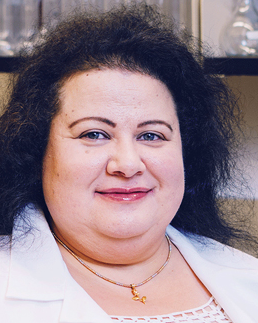 Heidi M. Mansour合作论文数Center for Translational Science, Florida International University;Department of Environmental Health Sciences, Robert Stempel College of Public Health and Social Work, Florida International University;Department of Cell and Molecular Medicine, Herbert Wertheim College of Medicine, Florida International University15
 Brian C. Keller合作论文数14
 Robert Higgins合作论文数14Jung-Lye Kim合作论文数Department of Food and Nutrition and Korean Institute of Nutrition, Hallym University14
 John Hummel合作论文数From the Department of Internal Medicine, The University of Michigan Medical Center14
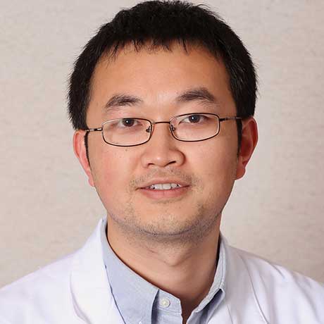 Hua Zhu合作论文数Division of Cardiac Surgery, College of Medicine, The Ohio State University13
 Sara Shumway合作论文数13
 Jichao Zhao (赵继超)合作论文数Auckland Bioengineering Institute, Faculty of Engineering, The University of Auckland12
 Jianjie Ma合作论文数School of Medicine, University of Virginia;Department of Surgery, The Ohio State University12
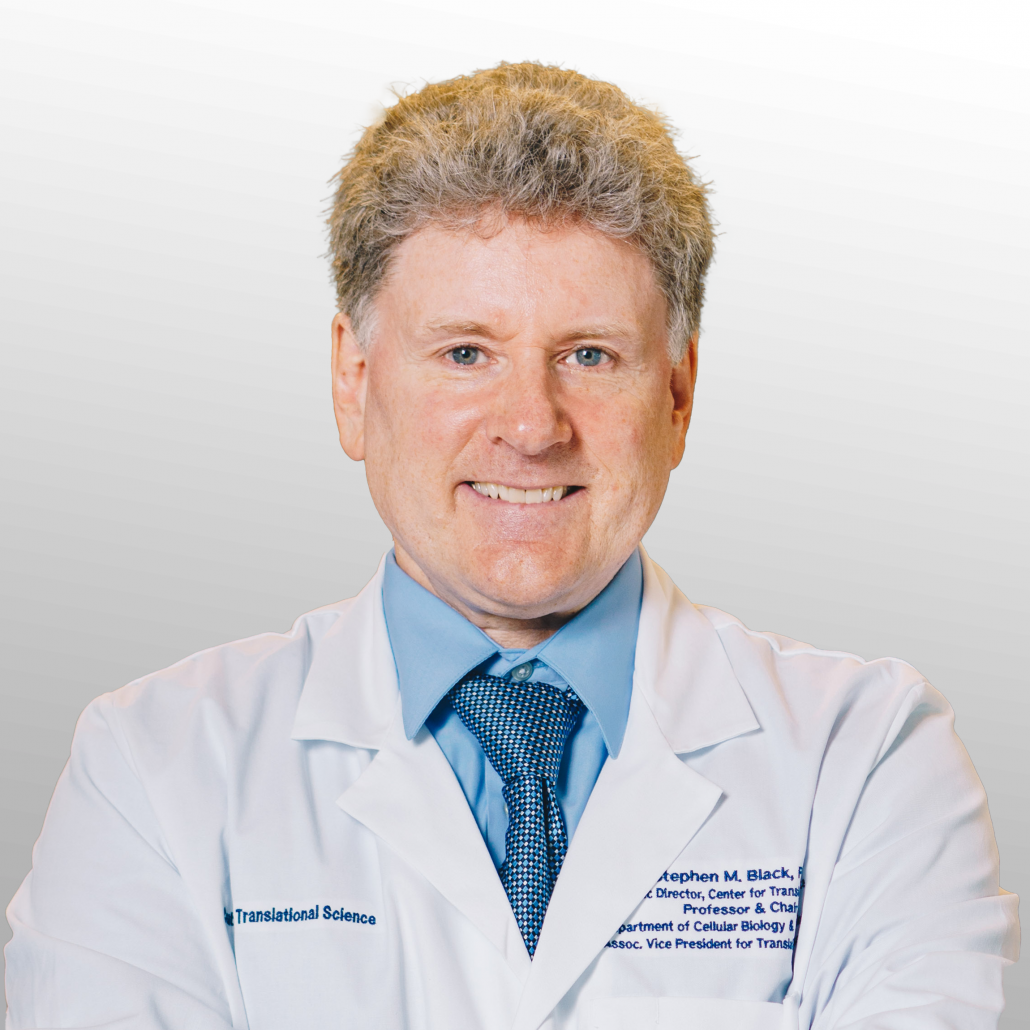 Stephen M. Black合作论文数Department of Environmental Health Sciences, Robert Stempel College of Public Health and Social Work, Florida International University;Center for Translational Science, Florida International University;Department of Cellular Biology & Pharmacology, Herbert Wertheim College of Medicine, Florida International University12Divyaam Satija合作论文数11Ervin Y. Cui合作论文数11
 David Ray Nunley合作论文数11
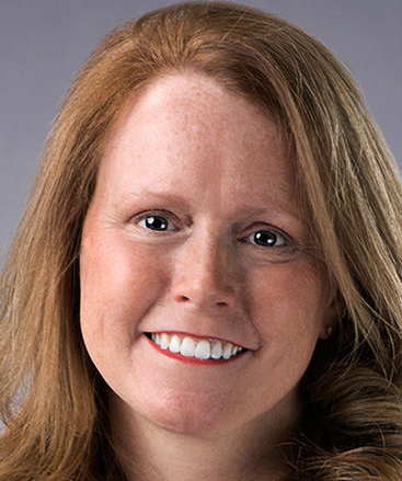 Rosemary Kelly合作论文数Department of Surgery, Medical School, University of Minnesota11
 Robert Kratzke合作论文数Division of Hematology, Oncology, and Transplantation, Department of Medicine, Medical School, University of Minnesota Twin Cities11David R Nunley合作论文数11Pope-Harman Amy合作论文数9Esthela J. Artiga合作论文数9




