Background: EUS plays an important role in local staging of rectal cancer and may help in the evaluation of perineal Crohn's disease. The role of EUS-FNA in the evaluation of pelvic masses however has not been well defined. Aim: To evaluate the diagnostic yield, safety and accuracy of EUS-FNA in patients (pts) with pelvic lesions of unknown etiology. Methods: A retrospective review of our EUS database identified 24 pts (14 women; mean age 58 years, range 28-83) referred for EUS-FNA to assess pelvic masses extrinsic to the rectum or distal colon over a 10-year period (1994-2004). Pts underwent radial followed by linear array EUS; FNA utilized a 22G needle. An on-site cytopathologist reviewed all samples and classified specimens as benign (inflammatory/infectious) or malignant. EUS-FNA diagnostic accuracy was confirmed by surgical pathology or clinical follow-up. Results: FNA was trans-rectal (n = 17), trans-sigmoid (n = 6) or through the descending colon (n = 1); samples were considered adequate for diagnosis in 21/24 (87.5%) pts. There were 12/21 (57%) EUS-FNA diagnoses of malignancy (n = 11) or atypical cells (n = 1) and 9/21 (43%) of benign lesions. Mean number of needle passes was 3.9 for benign vs. 3.2 for malignant lesions (range 1-7). In the 21 pts with adequate specimens, EUS-FNA accurately predicted the presence of malignant disease (sensitivity 92%, specificity 100%, PPV 100%, NPV 88%, accuracy 95%), with only one false negative diagnosis. One infectious complication occurred that was successfully managed with medical therapy. Conclusions: EUS-FNA is an accurate and safe method to diagnose pelvic masses. Prophylactic antibiotics should be used to reduce infectious complications.
Background: The finding of “dilated duct(s) of unknown cause” in asymptomatic individuals usually prompts further evaluation. EUS has been advocated as a useful tool to rule out an obstructive lesion, but there is insufficient data to fully evaluate the utility of EUS in this clinical scenario. Aim: To determine the diagnostic yield and accuracy of EUS in patients with asymptomatic biliopancreatic ductal dilation and normal liver enzymes. Methods: A retrospective review of our EUS database identified over a 10-year period (1994-2004) 30 asymptomatic patients with normal liver biochemistry referred for EUS to evaluate bile duct (CBD) and/or pancreatic duct (PD) dilation detected by US, CT or MRI. Mean age: 69 ± 9 (range 50-84); sex: 26F/4M. 18/30 (60%) had isolated CBD dilation, 4/30 (13%) isolated PD dilation, and 8/30 (27%) CBD and PD dilation. 10/30 (33%) patients had a previous cholecystectomy. EUS diagnoses were confirmed with pathology (FNA or surgery) or extended clinical/imaging follow-up. Results: Mean duct diameters at EUS: CBD = 12.2 mm (7-22), PD = 4.4 mm (2-9). Follow-up: Patients with significant EUS findings (n = 4); pancreatic masses, died of cancer (1), on chemoradiotherapy (1); IPMN (1) and focal narrowing of PD (1) alive, respectively, after 42 and 11 months of follow-up; EUS without significant findings (n = 26); died of unrelated causes (3), lost for follow-up (3), and asymptomatic (20) after a mean follow-up of 22 ± 24 months (2-102). Conclusion: The incidental finding of isolated bile duct dilatation in asymptomatic individuals with normal liver enzymes may not warrant further investigation, but EUS accurately rules out significant pathology. The finding of unexplained pancreatic duct dilation should not be ignored and EUS accurately defines the pathology.
Background: Complete pharyngoesophageal disconnect is a rare complication of radiation therapy for head and neck malignancies. Surgery has been the mainstay of treatment for this condition. Lew et al (Head and Neck, Feb 2004) described a combined endoscopic/laryngoscopic approach to establishing luminal patency using a stiff guidewire. However, repeated dilation may be required to achieve maximum diameter, and the restenosis rate for radiation-induced strictures has been high. Mitomycin-C has been used with success in the prevention of recurrent laryngeal and tracheal stenosis and is thought to have an antiproliferative effect. We describe a novel one-step method of reconnection, dilation, and treatment for pharyngoesophageal disconnect. Methods: Retrograde endoscopy through an existing gastrostomy tube tract is performed to the level of the disconnected proximal esophagus. Direct laryngoscopy is performed at the same time to visualize the light source below. An EUS needle (Echotip, Wilson Cook Corp) is advanced by the laryngoscopist with endoscopic guidance. A .035 mm ERCP guidewire (Jagwire, Microvasive corp) is passed through the EUS needle into the esophagus and grasped with a forcep. The wire is pulled back thru the gastrostomy and fixed in place. Savary style dilators are then passed per os, serially from the smallest to a minimum of 51 Fr. Before removing the guidewire, mitomycin-C pledgets are applied via laryngoscopy. Results: Four patients with pharyngoesophageal disconnect after radiation therapy for head and neck malignancies underwent the combined procedure. Luminal patency was achieved in all patients, and all were able to progress to teaching for self dilation. One patient suffered a minimal localized esophageal perforation for which conservative management was used. Luminal patency was maintained in all patients with follow-up to 7 months after the initial procedure. Conclusions: This combined one-step approach offers a less invasive and technically superior option to surgery. Dilation in one setting to 51 Fr can be performed with minimal complications. The EUS needle allows for a controlled puncture and passage of a guidewire. Despite their prior disconnect, these patients can be taught to perform self-dilation to prevent or delay recurrence without forming false tracts. Mitomycin-C topical treatments may provide an antiproliferative effect for radiation-induced strictures, however further long term studies are warranted.
 Brenda J. Hoffman合作论文数3
 Sandra Faias合作论文数2Sarto C. Paquin合作论文数2Andre K. Chong合作论文数2Joseph Romagnuolo合作论文数2
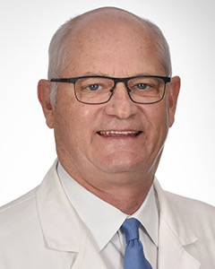 Robert H. Hawes合作论文数Orlando Health Digestive Health Institute Center for Advanced Endoscopy, Research and Education;University of Central Florida College of Medicine1D.O. Castell合作论文数1Rh Hawes合作论文数1Brenda J Hoffman合作论文数1Margaret Leveen合作论文数1




