OBJECTIVE:To assess 25-year implant survival, success rates, peri-implant conditions, and variables influencing peri-implant mucositis (PM), peri-implantitis (PI), and loss of osseointegration. METHODS:Partially edentulous patients rehabilitated with tissue-level implant-supported prostheses were evaluated. Calibrated examiners performed clinical and radiographic assessments, and potential variables for PM and PI were analyzed at the final follow-up. RESULTS:At the 10- and 25-year follow-ups, 303 and 159 patients with 511 and 252 implants, respectively, were included. Implant failure (IF) was 1.2% at 10 years and 6.0% at 25 years, with implant survival rates of 98.8% and 94.0%, respectively (p < 0.001). Implant success rates ranged from 97.0% at 10 years to 90.8% at 25 years (p < 0.001). IF due to loss of osseointegration increased from 0.6% to 3.6% at the implant level and from 1.0% to 2.5% at the patient level, and were located only in premolar or molar sites. At implant level, peri-implant health (PH) was 14.4% at 10 and 25 years (p = 1.00), while PM decreased from 82.1% to 76.5% (p = 0.08), and PI increased from 3.5% to 9.1% (p = 0.001). At patient level, PH remained stable between 10 and 25 years, ranging from 10.0% to 10.3% (p = 0.94), while PM decreased from 80.0% to 71.6% (p = 0.06), and PI increased from 10.0% to 18.1% (p = 0.01). In the multivariate analysis, history of periodontitis (OR = 3.87) and facial mucosal thickness (OR = 0.42) were associated with PM, whereas facial mucosal thickness was associated with PI (OR = 0.02). CONCLUSIONS:After 25 years, implant survival and success exceeded 90%. Loss of osseointegration accounted for approximately 60% of IF. While PI and IF increased over time, PM decreased but remained prevalent.
OBJECTIVES:The 1st Global Consensus for Clinical Guidelines (GCCG) in Implant Dentistry introduced an innovative, evidence-based approach to developing patient-centered and practical recommendations for the rehabilitation of the edentulous maxilla. Within this framework, Group 2 aimed to formulate clinical recommendations on the use of short, standard-length, and zygomatic implants in atrophic maxillae. MATERIALS AND METHODS:Group 2 followed the S2k-level guideline framework of the Association of the Scientific Medical Societies in Germany (AWMF), applying a structured nominal group technique. The evidence base included two systematic reviews synthesizing patient-reported outcomes (PROs), clinician-reported outcomes (ClinROs), and their respective measures, as well as single-round international surveys involving expert clinicians, patients, and cross-disciplinary experts. Draft recommendations were discussed during the in-person consensus meeting in Boston (June 16-18, 2025) and finalized through anonymous plenary voting. Consensus thresholds were predefined at ≥ 75% and ≤ 95% agreement for consensus and > 95% agreement for strong consensus. RESULTS:The Group 2 participants formulated 6 clinical recommendations addressing the domains patient selection, surgical treatment options (short, standard-length, and zygomatic implants), and treatment planning. All 6 recommendations reached consensus. CONCLUSIONS:The Group 2 consensus provides practical guidance for the use of short, standard-length, and zygomatic implants in atrophic maxillae, balancing surgical complexity, prosthetic feasibility, and patient-centered care. Remaining evidence gaps-especially regarding standardized outcome sets, loading protocols, prosthetic strategies for zygomatic implants, the use of short implants for full-arch restorations and maintenance frameworks-should be prioritized in future research.
Objective Long-term stability of peri-implant hard and soft tissues is a primary goal of implant treatment. Studies have shown that peri-implant tissue stability is influenced by factors such as the implant characteristics, implant-abutment interface, abutment type/length, and prosthetic design. To enhance tissue stability while offering clinical versatility, this case report utilizes a dual-hybrid approach. This combines (1) a hybrid implant surface design, combining a micro-rough and machined surface, and (2) a hybrid connection where one implant connection allows for two different implant-abutment interfaces (matching platform/non-matching with platform switching).Clinical Considerations A patient presented with two posterior teeth deemed "hopeless" due to periodontal and endodontic pathologies. Following extractions, immediate implant placements were performed. Tissue-level implants were placed subcrestally and restored with non-matching abutments. The 3-year follow-up examination showed stable marginal bone levels maintained at the implant shoulder.Conclusions This dual-hybrid approach represents an alternative strategy for supporting peri-implant tissue stability. It underscores the importance of versatile implant-abutment connections that preserve tissues while allowing future restorative modifications without compromising the implant. This method retains the option to transition to a matching abutment if clinically indicated in the future, for example if any pathologies lead to changes in the peri-implant tissue levels.Clinical Significance The presented dual-hybrid approach represents a treatment strategy designed to support peri-implant tissue stability while maintaining the flexibility to transition to a matching abutment when clinically indicated. Given the prevalence of peri-implantitis reported in the literature, treatment concepts that not only aim to reduce the risk of disease onset but also facilitate effective reintervention without compromising the implant are of critical importance.
AIM:The high prevalence of peri-implantitis is concerning, with a growing consensus that the majority of cases are complications initiated by clinician-related errors rather than classic pathology. A primary predisposing factor for peri-implantitis is exposure of the micro-rough implant surfaces to the peri-implant sulcus after treatment. OBJECTIVES:To identify surgical/prosthetic factors causing micro-rough surface exposure and advocate for prevention and evidence-based protocols. MATERIALS AND METHODS:This paper reviews evidence linking surgical and prosthetic errors to micro-rough surface exposure to the sulcus and subsequent peri-implantitis development. RESULTS:Surgical factors for surface exposure include malposition, avascular necrosis, and incomplete bone regeneration of peri-implant defects. Prosthetic factors include cement remnants and wide prosthetic emergence angles. Clinician-influenceable co-factors-including patient compliance, history of periodontitis, uncontrolled systemic factors/habits, lack of keratinized mucosa, prosthetic misfit, overcontoured/uncleansable prostheses, failure to detect early bone loss or mucosal changes, and inadequate maintenance-contribute to the initiation and progression of peri-implantitis once micro-rough surfaces are exposed. CONCLUSIONS:Most peri-implantitis cases are preventable complications. Professional education and research must prioritize identifying clinician-related errors and adherence to foundational treatment principles. This requires a paradigm shift toward a complication-based model of peri-implantitis. CLINICAL RELEVANCE:To reduce the risk of peri-implantitis, clinicians should avoid clinical errors that lead to exposure of the micro-rough implant surface. Hybrid surface designs and subcrestal micro-rough surface placement should be considered as safety buffers. Success depends on meticulous diagnostics and planning, proper surgical/prosthetic execution, cleansable prosthesis design, and proactive maintenance to detect early bone loss and soft tissue changes.
AIM:To assess buccal vertical bone resorption following implant placement in healed sites with varying buccal bone wall thicknesses. MATERIALS AND METHODS:In 11 miniature pigs, three tapered hybrid titanium implants were placed per hemi-maxilla in healed bone. Sites were randomised into three groups based on buccal bone wall thickness: G1 (< 1.0 mm), G2 (1.0-1.5 mm) and G3 (> 1.5 mm). Animals were euthanised at 24 h and 2, 4 or 8 weeks. Histological and histometric analyses were performed. The primary outcome was the vertical distance from the transition point (TP) between the machined and moderately rough implant surfaces to the first bone-to-implant contact (fBIC). RESULTS:Healing was uneventful. At 2 weeks, all groups showed buccal resorption, although G3 exhibited earlier bone apposition and fewer resorptive signs. By 8 weeks, all G1 implants displayed exposure of the moderately rough surface, while only one implant in G3 showed exposure. TP-fBIC values at 8 weeks were 0.92 ± 0.63 mm (G1), 0.27 ± 0.54 mm (G2, p = 0.041) and -0.16 ± 0.17 mm (G3, p = 0.0002). Bone-to-implant contact increased over time across all groups. CONCLUSION:Thin buccal bone walls (< 1 mm) were associated with greater vertical bone loss and implant surface exposure, whereas thick walls (> 1.5 mm) preserved the buccal bone better and protected the implant surface.
OBJECTIVE:A cross-sectional study was made to evaluate the role of local factors, including surgical, implant, and prosthesis-related parameters, in the presence of peri-implantitis. METHODS:Consecutive partially edentulous patients with ≥ 1 implant presenting peri-implantitis were included. Clinical and radiographic data were collected to characterize local factors. Overall, 28 site-specific variables were analyzed for peri-implantitis and non-peri-implantitis implants. Simple and multiple logistic regression analyses were performed to explore potential associations. The diagnostic accuracy of those variables that demonstrated statistical significance in the multiple logistic regression analysis was further tested. RESULTS:In total, 100 patients with 452 implants met the inclusion criteria. Of these, 227 implants (50.2%) were diagnosed with peri-implantitis, while 225 (49.8%) did not present peri-implantitis. The multiple logistic regression analysis demonstrated statistical significance for the following factors: (1) medial implant position within the prosthesis (OR = 3.9) was more prone to develop peri-implantitis; (2) internal Morse taper implant connection was the least likely to develop peri-implantitis (OR = 0.43); (3) mesio-distally centered implants were less prone to exhibit peri-implantitis (OR = 0.46); (4) bucco-lingual centered implants were less prone to exhibit peri-implantitis (OR = 0.29); (5) implants positioned inside (≥ 2 mm) the bony housing were less prone to exhibit peri-implantitis (OR = 0.13); and (6) implants tilted 10°-30° (OR = 5.84) and ≥ 30° (OR = 7.43) were more prone to exhibit peri-implantitis. Moreover, a prediction model based on the aforementioned factors yielded a high level of accuracy. CONCLUSIONS:Implant malpositioning is a critical factor associated with peri-implantitis.
AIM:Different approaches have been proposed for implant placement following tooth extraction. A Consensus conference was organised to provide expert-based recommendations for the treatment of the postextraction site in the aesthetic zone in conjunction with implant therapy. METHODS:A panel of eight experts with a documented longstanding clinical and research experience in the field of implant therapy in the aesthetic zone were invited to participate in a structured survey. Participants were asked to select their preferred treatment approach for different clinical scenarios of the postextraction site from a list of different treatment options. Results were summarised and discussed in person at a 2 day consensus conference. Based on the outcome, treatment recommendations were phrased and are reported here. RESULTS:The group agreed that in case of an intact alveolus, immediate implant placement with immediate prosthetics represents the reference choice if proper primary stability can be achieved and the buccal bone plate is present. A bone-to-implant gap more than 2 mm should be seeked and grafted. Alveolar ridge preservation and early placement with contour augmentation may represent an alternative. If the alveolus is compromised, a staged approach (early or delayed placement) with bone augmentation may be preferred. CONCLUSIONS:The characteristics of the site, in terms of the available bone volume, the integrity of the buccal bone plate and the periodontal phenotype are determining factors in the therapeutic choice. Therefore, case selection based on well-defined selection criteria is extremely important and is the adequate way to guide the clinician in choosing the most appropriate approach to postextraction site management and timing for implant placement.
OBJECTIVES:To evaluate the impact of the marginal fit of implant-supported prostheses (ISP) on peri-implant bone levels. Additionally, the study aimed to determine a clinically relevant threshold for the radiographic vertical misfit gap at the ISP, when present, and to identify potential risk factors associated with changes in bone levels. METHODS:This study involved subjects who received ISPs for tooth replacement therapy. Standardized intraoral periapical radiographs were taken 10 years after loading to assess the radiographic distance between the implant shoulder and the most coronal point of crestal bone (DIB). ISP marginal gaps were categorized as no gap or gap, with vertical dimensions categorized as 0 mm, > 0- < 0.1 mm, and ≥ 0.1 mm. A multivariable linear mixed-effect model was applied to control for potential confounders. RESULTS:A total of 301 patients and 505 implants with a 10.6 ± 0.7 years follow-up were analyzed. ISPs without gaps exhibited statistically significantly lower DIB values (3.22 ± 0.8 mm) than those with gaps (3.43 ± 0.6 mm; p = 0.001). Gaps ≥ 0.1 mm were associated with statistically significantly higher DIB values (3.45 ± 0.7 mm; p = 0.001) compared with gaps between > 0 and < 0.1 mm (3.36 ± 0.5 mm; p = 0.001), or no gaps (3.22 ± 0.7 mm; p = 0.001). Each increment of 0.1 mm in the vertical crestal gap corresponded to a significant increase in DIB values (0.08 mm; p = 0.03). Finally, smoking and a history of periodontitis were independent risk factors for increased DIB. CONCLUSIONS:Marginal misfit of ISP affects peri-implant bone stability, with gaps ≥ 0.1 mm linked to higher DIB. Smoking and periodontitis are independent risk factors for increased DIB.
Dental implants are commonly used to replace missing single teeth. However, esthetic rehabilitation of an adjacent tooth may also be required due to diastemas, crowding, or existing large direct restorations to improve the final esthetic outcome. With the advancements in ceramics and bonding techniques, minimally invasive esthetic approaches have become viable for compromised spacing issues. This case report describes a dental technique for the esthetic rehabilitation of compromised anterior spacing with a customized zirconia implant abutment at a maxillary central incisor site and a partial ceramic veneer bonded to the adjacent central incisor.
AIM:The aim of this randomized, prospective, and clinical multicenter study was to compare the overall clinical performance of two restorative options over a 5-year period: individualized CAD/CAM abutments veneered with a hand-layered ceramic, and prefabricated zirconium dioxide abutments veneered with press ceramic and inserted into a single edentulous gap in the anterior maxilla. MATERIALS AND METHODS:Forty subjects were recruited from two universities: 20 from the University of XX and 20 from the University of XY. Each subject received an implant to restore a single edentulous gap in the maxillary anterior region (14-24 FDI). 20 patients were randomized into each Group. Group A received a one-piece single crown produced from a prefabricated zirconia abutment with pressed ceramic and Group B received an individualized CAD/CAM zirconia abutment with a hand-layered technique. After 5 years, the aesthetic and radiographic parameters were assessed. RESULTS:Group A had four dropouts and one failure, resulting in a 95% survival rate and 95% success rate. Group B had two dropouts and two failures which resulted in a 90% survival rate and 90% success rate. No crestal bone level changes were observed, with a mean DIB of 0.06 mm in Group A and 0.09 mm in Group B. No statistically significant differences were present at baseline, 6 months, 1 year, 3 years, or 5 years for DIB values between time points and groups. Pink aesthetic score/white aesthetic score, Peri-Implant and Crown Index, and Implant Crown Aesthetic Index values were stable over time at all five points for both groups. CONCLUSION:Both implant-supported restorative options represent a valuable treatment option for the restoration of implant crowns in the anterior maxilla. CLINICAL SIGNIFICANCE:In general, the use of ceramic abutments in the anterior zone represents a valuable treatment procedure with both standardized and CAD/CAM individualized abutments and following the recommendations from the respective manufacturer(s).
To analyze the trends in the context of implant therapy in a 3-year patient population and compare it with data obtained over the last 20 years. All adult subjects who received treatment in the context of implant therapy between 2020 and 2022 were included in this retrospective study. Data regarding patient demographics, indications and location of implant therapy, implant characteristics, surgical techniques, complications, and early implant failures were recorded and compared to data obtained in the years 2002–2004, 2008–2010, and 2014–2016. Between 2020 and 2022, n = 1555 implants were placed in n = 1021 patients. The mean age at implant placement was 59.9 + 15.1 years, demonstrating an increase over time in the age group 61–80 years of 23.1
OBJECTIVES:The aims of Working Group 1 were to address the role (i) of the buccolingual bone dimensions after implant placement in healed alveolar ridge sites on the occurrence of biologic and aesthetic complications, and (ii) of soft tissue augmentation (STA) on the stability of clinical, radiographic, and patient-related outcomes of implant treatments.MATERIALS AND METHODS:Two systematic reviews were prepared in advance of the Consensus Conference and were discussed among the participants of Group 1. Consensus statements, clinical recommendations, recommendations for future research, and reflections on patient perspectives were based on structured group discussions until consensus was reached among the entire group of experts. The statements were then presented and accepted following further discussion and modifications as required by the plenary.RESULTS:Dimensional changes of the alveolar ridge occurred after implant placement in healed sites, and a reduction in buccal bone wall thickness (BBW) of 0.3 to 1.8 mm was observed. In healed sites with a BBW of <1.5 mm after implant placement, increased vertical bone loss, and less favorable clinical and radiographic outcomes were demonstrated. Implants with buccal dehiscence defects undergoing simultaneous guided bone regeneration, showed less vertical bone loss, and more favorable clinical and radiographic outcomes, compared to non-augmented dehiscence defects during initial healing. At healthy single implant sites, probing depths, bleeding and plaque scores, and interproximal bone levels evaluated at 1 year, remained stable for up to 5 years, with or without STA. When single implant sites were augmented with connective tissue grafts, either for soft tissue phenotype modification or buccal soft tissue dehiscence, stable levels of the soft tissue margin, and stable or even increased soft tissue thickness and/or width of keratinized mucosa could be observed from 1 to 5 years. In contrast, non-augmented sites were more prone to show apical migration of the soft tissue margin in the long-term. Favorable aesthetic and patient-reported outcomes after STA were documented to be stable from 1 to 5 years.CONCLUSIONS:It is concluded that dimensional changes of the alveolar ridge occur after implant placement in healed sites and that sites with a thin BBW after implant placement are prone to exhibit less favorable clinical and radiographic outcomes. In addition, it is concluded that STA can provide stable clinical, radiographic, aesthetic, and patient-reported outcomes in the medium and long-term.
BackgroundThe significance on the association between the peri-implant bucco-lingual dimension (BLD) at the stage of implant placement and the occurrence of biological and esthetic complications is yet unknown. Material and methodsSystematic screening of electronic sources was carried out to identify clinical and preclinical studies reporting on the baseline BLD and/or buccal bone thickness (BBT) values. A secondary objective was to assess the effect of simultaneous grafting at sites with deficient or no buccal bone wall (BBW) at baseline. The primary outcome variables were BBT, BLD, and buccal vertical bone loss (VBL) at re-evaluation. Moreover, radiographic, clinical, and patient-reported outcome measures (PROMs) were evaluated. ResultsOverall, 12 clinical and four preclinical studies met the inclusion criteria. Inconsistencies were found in defining the critical BBT across the clinical and preclinical data evaluated. The clinical evidence demonstrated that during healing, dimensional changes occur in the alveolar bone and in the BBW that may compromise the integrity of the peri-implant bone, leading to VBL and mucosal recession (MR), particularly in scenarios exhibiting a thin BBW. The preclinical evidence validated the fact that implants placed in the presence of a thin BBW, are more prone to exhibit major dimensional changes and VBL. Moreover, the clinical data supported that, in scenarios where dehiscence-type defects occur and are left for spontaneous healing, greater VBL and MR together with the occurrence of biologic complications are expected. Furthermore, the augmentation of dehiscence-type defects is associated with hard and soft tissue stability. PROMs were not reported. ConclusionsDimensional changes occur as result of implant placement in healed ridges that may lead to instability of the peri-implant hard and soft tissues. Sites presenting a thin BBW are more prone to exhibit major changes that may compromise the integrity of the buccal bone and may lead to biologic and esthetic complications.
This case report describes a novel approach combining orthodontic and implant digital treatment planning workflows to enable prosthetically driven implant placement before orthodontic treatment is performed or completed. The computer-assisted implant placement is performed using a static surgical guide based on the future positions of the teeth after completion of orthodontic treatment. This innovative approach allows for select partially edentulous patients to undergo fixed prosthetic rehabilitation before orthodontic treatment is completed.
Incorrect implant positioning can lead to functional and aesthetic compromise. Implant positioning errors can occur in three dimensions: mesiodistal, corono-apical, and orofacial. Treatment solutions to manage adverse outcomes through positioning errors require an understanding of the underlying conditions and of those factors that may have led to the error being committed in the first place. These types of complications usually occur because of human factors. If errors do occur with adverse aesthetic outcomes, they are difficult and sometimes impossible to correct. Connective tissue grafts to reverse recession defects are only feasible in defined situations. The option to remove and replace the implant may be the only recourse, provided the removal process does not further compromise the site. Error in judgment by the clinician.
Bone augmentation procedures are frequent today in implant patients, since an implant should be circumferentially anchored in bone at completion of bone healing to have a good long-term stability. The best documented surgical technique to achieve this goal is guided bone regeneration (GBR) utilizing barrier membranes in combination with bone fillers. This clinical review paper reflects 35 years of development and progress with GBR. In the 1990s, GBR was developed by defining the indications for GBR, examining various barrier membranes, bone grafts, and bone substitutes. Complications were identified and reduced by modifications of the surgical technique. Today, the selection criteria for various surgical approaches are much better understood, in particular, in post-extraction implant placement. In the majority of patients, biodegradable collagen membranes are used, mainly for horizontal bone augmentation, whereas bioinert PTFE membranes are preferred for vertical ridge augmentation. The leading surgeons are using a composite graft with autogenous bone chips to accelerate bone formation, in combination with a low-substitution bone filer to better maintain the augmented bone volume over time. In addition, major efforts have been made since the millenium change to reduce surgical trauma and patient morbidity as much as possible. At the end, some open questions related to GBR are discussed.
BACKGROUND:This study aimed to evaluate the reliability and reproducibility of different non-invasive methods for the assessment of peri-implant mucosal thickness. METHODS:Subjects with two adjacent dental implants in the central maxillary region were included in this study. Three different methods to assess facial mucosal thickness (FMT) were compared: digital file superimposition using Digital Imaging and Communication in Medicine (DICOM) and stereolithography (STL) files of the arch of interest (DICOM-STL), DICOM files alone, and non-ionizing ultrasound (US). Inter-rater reliability agreements between different assessment methods were analyzed using inter-class correlation coefficients (ICCs). RESULTS:A total of 50 subjects with 100 bone-level implants constituted the study population. Assessment of FMT using STL and DICOM files demonstrated excellent inter-rater reliability agreement. Mean ICC values of 0.97 and 0.95 were observed in the DICOM-STL and DICOM groups, respectively. Comparison between the DICOM-STL and US revealed good agreement, with an ICC of 0.82 (95% CI: 0.74 to 0.88) and a mean difference of -0.13 ± 0.50 mm (-1.13 to 0.86). Comparison between DICOM files alone versus US showed good agreement, with an ICC of 0.81 (95% CI: 0.73 to 0.89) and a mean difference of -0.23 ± 0.46 mm (-1.12 to 0.67). Comparison between DICOM-STL and DICOM files revealed excellent agreement, with an ICC of 0.94 (95% CI: 0.91 to 0.96) and a mean difference of 0.1 ± 0.29 mm (LOA -0.47 to 0.46). CONCLUSIONS:Quantification of peri-implant mucosal thickness via analysis of DICOM-STL files, DICOM files, or US assessment are comparably reliable and reproducible methods.
AIM:To histologically evaluate the influence of (1) loading and (2) grafting on osseointegration and peri-implant soft-tissue healing at immediately placed, self-cutting progressive tissue-level implants (TLX) in a minipig model.MATERIALS AND METHODS:TLX implants (n = 56) were immediately placed following the extraction of the mandibular first and second premolars, bilaterally, in a total of n = 14 minipigs. In each animal, the implant sites were allocated to the following four groups: (1) unloaded with simultaneous grafting using a bovine bone mineral; (2) unloaded without grafting; (3) loaded with simultaneous grafting; and (4) loaded without grafting. Histomorphometric assessments at 4 and 12 weeks (n = 7 animals each) included primary (i.e., bone-to-implant contact [BIC]) and secondary outcome measures (e.g., first BIC [fBIC], junctional epithelium length [JE], connective tissue contact length [CTC], biological width [BW = JE + CTC]).RESULTS:At 4 weeks, mean BIC values ranged from 74.5 ± 11.6% in Group 2 to 83.8 ± 13.3% in Group 1, and, at 12 weeks, from 75.5% ± 7.9% in Group 2 to 79.9 ± 8.6% in Group 1. Multivariate linear mixed regression did not reveal any associations between BIC and implant loading or grafting at 4 and 12 weeks. At 12 weeks, significantly higher fBIC values were noted in Group 2 when compared with Group 1. All groups showed comparable JE, CTC, and BW values.CONCLUSIONS:Implant loading and grafting had no major effects on osseointegration and peri-implant soft tissue healing at TLX implants.
AbstractObjectivesTo evaluate the long‐term effectiveness of 6 mm implants in various indications with a micro‐rough surface after 4.6–18.2 years in function and to assess key factors associated with implant survival, success, and biologic/technical complications.Materials and methodsFifty‐five patients with seventy‐four 6 mm implants placed from 2000 to 2013 attended the re‐examination assessing well‐established clinical and radiographic parameters, biologic and prosthetic complications, and patient‐reported outcome measures.ResultsFive implants were lost after a mean follow‐up period of 9.1 years resulting in a survival rate of 93.2%. All losses occurred in free‐end situations in the mandible. Smoking habit significantly reduced implant survival (hazard ratio 36.25). Two implants exhibited a history of peri‐implantitis, and one implant showed progressive marginal bone loss (MBL) resulting in a success rate of 89.2%. The mean MBL amounted to 0.029 mm. Increased MBL was found for implants placed in the maxilla (0.057 mm) and for implants with a diameter of 4.1 mm (0.043 mm). Soft tissue thickness (1.39 mm) and width of keratinized mucosa (1.91 mm) had no effect on MBL. Patient‐reported outcome measures showed high satisfaction (mean VAS scores 88%) and high quality of life (mean OHIP‐G14 score 2.2).ConclusionThe present study demonstrated survival and success rates of 93.2% and 89.2% for 6 mm implants used in various indications. A factor leading to higher implant failure was smoking, whereas modulating factors increasing annual MBL included implants placed in the maxilla and implants with a diameter of 4.1 mm compared to 4.8 mm.
Statement of problem. Vertical eruption of teeth adjacent to an implant has been reported clinically and might affect the esthetic outcome over time. The prevalence of the problem is unclear. Purpose. The purpose of this prospective clinical trial was to evaluate the vertical eruption of anterior maxillary teeth adjacent to single-implant crowns after a 3-year follow-up period.Material and methods. Thirty single dental implants were inserted in maxillary anterior sites including the first premolar. The mean age of the participants at implant-supported crown insertion was 48.4 years (range 23 to 79 years). Each implant was restored with a 1-piece screw-retained ceramic single crown. The vertical changes of 60 adjacent anterior maxillary teeth were evaluated from periapical radiographs and casts at baseline, 6, 12, and 36 months. Results. A global ANOVA test showed statistically significant differences for the outcomes of the distance-implant platform and cement-enamel junction (DPC) (P<.001), crown length distal (P=.021) and mesial (P=.035), implant crown length (P=.022), and incisal edge to edge (P<.001).Conclusions. Continuous vertical tooth eruption next to a single dental implant was observed in adult participants. (J Prosthet Dent 2022;128:605-10)
 Michael Marc Bornstein合作论文数Faculty of Dentistry, The University of Hong Kong;Department of Oral Health and Medicine, University Center of Dental Medicine Basel75
 Dieter Bosshardt合作论文数Department of Oral Surgery and Stomatology, University of Bern;Robert K. Schenk Laboratory of Oral Histology, School of Dental Medicine, University of Bern58
 Urs Belser合作论文数Klinik für Oralchirurgie und Stomatologie, Universität Bern;Department of Oral Surgery and Stomatology, School of Dental Medicine, University of Bern;Department of Fixed Prosthodontics, School of Dental Medicine, University of Bern46
 David L. Cochran合作论文数Department of Periodontics, School of Dentistry, UT Health San Antonio45
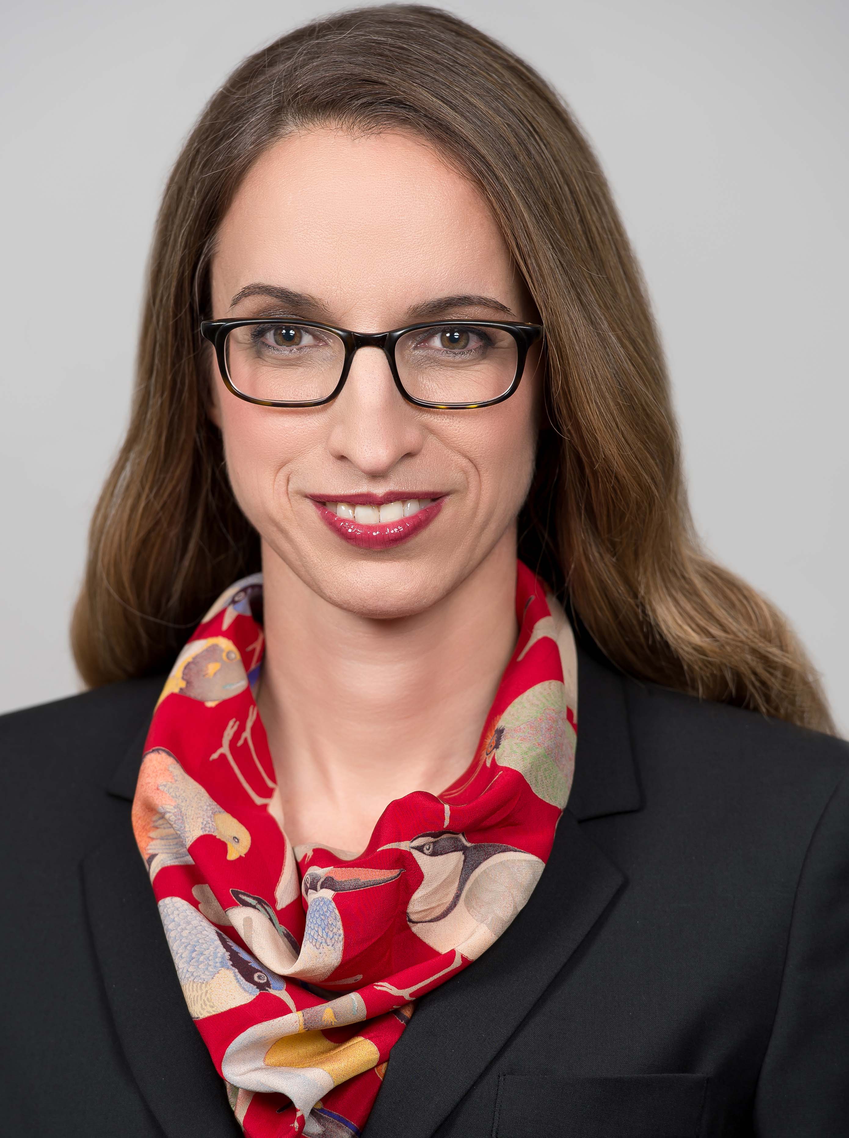 Vivianne Chappuis合作论文数Department of Oral Surgery and Stomatology, School of Dental Medicine, University of Bern44
 Reinhard Gruber合作论文数Universitätszahnklinik Wien, Medical University of Vienna33
 Anton Sculean合作论文数Department of Periodontology, School of Dental Medicine, Faculty of Medicine, University of Bern31
 Niklaus Lang合作论文数Centre for Oral Clinical Research, Institute of Dentistry, Faculty of Medicine and Dentistry, Queen Mary University of London;Universität Zürich;University College London;Universidad Complutense de Madrid29
 Richard J. Miron合作论文数Miron Lab;Department of Periodontology, Nova Southeastern University27
 Thomas Von Arx合作论文数Department of Oral Surgery and Stomatology, School of Dental Medicine, University of Bern24
 Jordi Caballe-Serrano合作论文数Dept Oral Surg & Stomatol, Univ Bern22
 R K Schenk合作论文数22Stephen T. Chen合作论文数21Dula K合作论文数19JS Hermann合作论文数18Simone F M Janner合作论文数17
 Simon Storgård Jensen合作论文数Department of Odontology, Faculty of Health and Medical Sciences, University of Copenhagen16
 Urs Bragger合作论文数16H Berthold合作论文数15
 Julia Wittneben合作论文数Department of Restorative Dentistry and Biomaterials Sciences, Harvard School of Dental Medicine (HSDM),15
 Lussi Adrian合作论文数University of Bern;University of Innsbruck14
 Monje Alberto合作论文数University of Michigan School of Dentistry13Hans-Peter Weber合作论文数12
 Martin Schimmel合作论文数Department for Reconstructive Dentistry and Gerodontology, School of Dental Medicine, University of Bern;Medizinische Fakultät, Universität Bern12
 Nina Broggini合作论文数12
 Valerie Geraldine Anne Suter合作论文数School of Dental Medicine, University of Bern12U Brägger合作论文数11
 Yufeng Zhang (张玉峰)合作论文数Hospital of Stomatology, Wuhan University11Giovanni E. Salvi合作论文数11Saulacic Nikola合作论文数10Robert K. Schenk合作论文数10Frank L. Higginbottom合作论文数10
 Archie Jones合作论文数10Benoit Schaller合作论文数Bern University Hospital, University of Bern10H P Hirt合作论文数10Braut Vedrana合作论文数School of Dental Medicine, University of Bern9
 Ernst B. Hunziker合作论文数Department of Orthopaedic Surgery and Traumatology, Universitatsspital Bern9Edouard Stauffer合作论文数9
 Andreas Filippi合作论文数University of Basle, University of Gießen9S. Chen合作论文数8
 Ulrike Kuchler合作论文数8Altermatt Hans Jörg合作论文数8Weber Hans-Peter合作论文数8
 German O. Gallucci合作论文数Division of Regenerative and Implant Sciences, Department of Restorative Dentistry and Biomaterials Sciences, Harvard School of Dental Medicine, Harvard University8John D. Schoolfield合作论文数8Joseph P. Fiorellini合作论文数8Jean-Pierre Bernard合作论文数8
 Kholy Karim El合作论文数Forsyth Institute;Harvard University;Forsyth Institute, Harvard University7Sawada Kosaku合作论文数7




