There has been increasing interest in the use of platelet-rich plasma and microneedling for the treatment of various clinical presentations in aesthetic medicine and surgery. PRP and Microneedling in Aesthetic Medicine attempts to review the current literature on these modalities and present individual author protocols when available. The authors correctly recognize the paucity of evidence in the literature. Therefore, this work represents a reference to key literature published to date. In addition, this work offers practical considerations when reviewing the variety of delivery methods and modalities. Frequently, when there are no evidence-based data available, this textbook presents experience-driven protocols to help the reader with available best practices with platelet-rich plasma and microneedling and relevant multimodal combination treatments.The authors provide an in-depth review on the basic science mechanisms behind autologous platelet-rich plasma with associated clinical indications. While reviewing the options for platelet-rich plasma preparation, the authors highlight the basic steps but steer away from specific protocols, likely because of their proprietary nature in commercialized systems. However, a valuable comparative discussion on the different commercialized platelet-rich plasma systems is included. This information is helpful to a reader who may be considering purchasing a system for his or her practice. The textbook is divided into three parts. The first section focuses on platelet-rich plasma. For the plastic surgeon, the authors provide their individual experience with platelet-rich plasma as a sealant for hemostasis and to aid in healing. They again reiterate the absence of evidence-based data, but they share their clinical experience with what is essentially a case series with facial rejuvenation surgical procedures. The conflicting literature on the role of platelet-rich plasma in autologous grafting is also reviewed. This work also includes a useful assessment of the variety of other clinical indications for platelet-rich plasma. For instance, the authors explore platelet-rich plasma’s role in collagenesis and nonsurgical facial rejuvenations. Again, they correctly underscore the lack of standardized preparation techniques and delivery methods. Applications considered include skin, rhytide, and periocular rejuvenation. The authors also explore platelet-rich plasma multimodal therapies involving microneedling, fat grafting, and/or hyaluronic acid fillers with important technical considerations. A thorough literature review is presented with respect to the role of platelet-rich plasma for alopecia and hair restoration. In the second section, the authors focus on microneedling. In a fashion similar to that used in the platelet-rich plasma section, the basic science behind microneedling is presented. However, the authors accurately highlight that a complete understanding of the mechanism of action of microneedling remains unknown. In addition, the different devices and related technical considerations are reviewed. A comprehensive review of clinical applications based on an experience-driven protocol is helpful for those considering microneedling in their practice. The authors should be credited for a thorough examination of microneedling with radiofrequency. Clinical applications for lower face and neck laxity; periorbital, nasal, and perioral rhytides; axillary hyperhidrosis; rosacea; and striae distensae are included. The authors also discuss the different microneedling with radiofrequency technologies. However, the newer InMode Morpheus8 (Inmode Ltd., Yokneam, Israel) microneedling with radiofrequency and subdermal adipose remodeling is not included, as this technology was likely not available at the time of print. In the last section, the authors discuss applications and safety in skin of color. For physicians looking to include platelet-rich plasma and/or microneedling with radiofrequency or multimodal treatments, this textbook has an excellent chapter on complications and adverse reactions, with management recommendations. Recognizing the paucity of evidence-based data, this textbook provides a comprehensive review of the literature to date on platelet-rich plasma and microneedling. It is an excellent primer for resident/fellows or any physicians and surgeons in aesthetics looking to add these modalities to their practice.
BACKGROUND:Human acellular dermal matrix (HADM; previously termed "acellular cadaveric dermis") may limit inflammatory changes believed to play a role in capsular contracture, a common complication of implant-based breast reconstruction.OBJECTIVES:Differences between HADM and native breast capsule specimens were evaluated by immunohistochemical analysis of key inflammatory markers involved in capsule formation.METHODS:Twenty consecutive patients underwent immediate, 2-stage, implant-based breast reconstruction with dual-plane HADM. During tissue expander-implant exchange, full-thickness biopsies of biointegrated HADM and native breast capsule (internal control) from the tissue-expander envelope were obtained. Immunohistochemical analysis was performed for endothelial cells (CD31), B cells (CD20), T cells (CD3), macrophages (CD68), collagen I and III, and myofibroblasts (α-smooth muscle actin). Observed levels of marker labeling were semiquantitatively scored from 0 (none) to 3 (severe) by a blinded histopathologist and were statistically analyzed with the Wilcoxon rank sum test.RESULTS:A bilateral sample was obtained from 1 patient; all other samples were unilateral. Compared with capsule samples from native breast tissue, HADM samples had significantly lower levels of all inflammatory markers (P < .001).CONCLUSIONS:These lower levels of inflammatory markers support previous evidence that HADM may inhibit inflammatory and profibrotic signaling characteristics of breast capsule development and decrease the risk of capsular contracture. Further investigation is needed to determine the mechanism by which HADM inhibits these inflammatory cells, whether HADM reduces the incidence of breast capsular contracture, and if so, the longevity of this effect.
Background: Professional and social changes have resulted in decreased involvement in organizations. Little is currently known about young plastic surgeons’ attitudes toward the Plastic Surgery Foundation and its sponsored activities. The authors gathered opinions of young plastic surgeons to determine factors related to participation. Methods: A 21-question online survey was e-mailed to all 2155 members of the Young Plastic Surgeons Forum. Questions were related to demographics, current involvement, and initiatives in education, research, funding, and health policy. Results: Of 2155 forum members, 397 responded (19 percent response rate). Most had not contributed to the Plastic Surgery Foundation. The primary reason cited was financial hardship, and respondents noted this would change with increased practice revenue. Involvement in American Society of Plastic Surgeons committees correlated with contribution to Plastic Surgery Foundation. The main educational initiatives favored by Young Plastic Surgeons included critical analysis of literature/evidence-based medicine, statistical analysis, and compensation issues. According to respondents, primary areas for organizational focus should be clinical research, increased representation of young surgeons, and leadership development. Respondents would be more willing to donate if they could earmark their contributions for specific purposes, including leadership training, clinical research, and medical missions. Conclusions: Methods to recruit and retain young surgeons into the American Society of Plastic Surgeons and in contributing to the Plastic Surgery Foundation should include opportunities to participate at a decreased cost, focus on compensation issues, clinical research, leadership development, and increased young surgeon representation. These data should be used to guide efforts to increase young member involvement.
BACKGROUND:Given the continued evolution of the American health system, the authors explored young plastic surgeons' attitudes on challenges affecting the specialty and the future role of organized plastic surgery and its advocacy mission.METHODS:A 21-question online survey was distributed to all members of the Young Plastic Surgeons Forum. Questions were related to demographics, attitudes toward policy issues, participation in the American Society of Plastic Surgeons, and its advocacy efforts.RESULTS:The survey was e-mailed to 2155 Forum members, of which 397 responded (19% response rate). Young plastic surgeons appear to be interested in American Society of Plastic Surgeons and PlastyPAC, as evidenced by a higher than normal response rate to this survey and rate of contribution. However, a lack of awareness about the details of the organizations and mechanisms for involvement remains. Scope-of-practice issues and encroachment on the specialty by noncore providers remain the top concern of young plastic surgeons. Other top concerns were financial barriers to participation in advocacy efforts and concerns with return on investment on PlastyPAC contributions. A majority received minimal or no public policy education on issues affecting plastic surgery during training. A minority currently participate in American Society of Plastic Surgeons advocacy efforts.CONCLUSIONS:These data represent the first such collection of opinions from Young Plastic Surgeons members regarding goals and directions of the American Society of Plastic Surgeons and PlastyPAC. These organizations are in a uniquely strong position to enlist participation from and provide for the future success of the profession's younger members.
INTRODUCTION: Use of acellular cadaveric dermis (ACD) in implant-based breast reconstruction provides an alternative to total submuscular placement. We previously reported that biointegrated ACD samples from implant-based breast reconstruction patients had statistically diminished levels of granulation tissue formation, vessel proliferation, chronic inflammatory changes, capsule fibrosis, fibroblast cellularity, and foreign body giant cell inflammatory reaction compared with native breast capsule samples. These findings suggested that certain properties intrinsic to ACD may diminish inflammatory changes that may promote peri-prosthetic capsule formation.1 We sought to further evaluate differences between ACD and native breast capsule specimens through immunohistochemical analysis of key inflammatory markers involved in capsule formation. METHODS: Twenty patients underwent implant-based breast reconstruction using the “dual-plane” ACD (AlloDerm® Regenerative Tissue Matrix, LifeCell Corporation, Branchburg, NJ) technique. During exchange of the tissue expander for the implant, intraoperative biopsies of biointegrated ACD and native subpectoral capsule from the tissue expander envelope were obtained. Immunohistochemical analysis was performed for inflammatory markers: macrophages (CD68), T-cells (CD3), B-cells (CD20), myofibroblasts (αSMA), endothelial cells (CD31), and collagen I and III. Masked biopsy specimens were semi-quantitatively scored by a histopathologist to reflect observed levels of marker staining. Scores were statistically analyzed using the Wilcoxon rank test. RESULTS: ACD samples had statistically diminished levels of all inflammatory markers assessed compared with corresponding native breast capsule samples (P<0.005) (Figure 1). Although all variables were significantly diminished in biointegrated ACD capsules compared with native breast capsules, fibroblast cellularity, granulation tissue, endothelial cells, vessel proliferation, collagen I, and capsule fibrosis had substantially lower P values, suggesting that for those variables, there was an even greater difference between ACD and the native capsule control. Interestingly, although myofibroblasts, collagen III, and B-cell lymphocytes were also decreased in ACD compared with capsular control, these differences were not as significant as the previous variables.Figure 1: Semiquantitative analysis of inflammatory markers with acellular cadaveric dermis (ACD; AlloDerm®) vs. native breast capsule samples (0=none, 3=severe levels of marker staining). *n=21 samples (a bilateral sample was obtained from 1 patient; all other samples were unilateral). P<0.005 for all comparisons.CONCLUSION: This study further supports the theory that ACD has certain intrinsic properties that may limit capsule formation. The decreased inflammatory changes in the ACD samples as evidenced by decreased fibroblast activity, collagen I deposition, and capsular fibrosis, suggest that use of ACD may result in decreased capsule formation. Further investigation is needed to determine the mechanism by which ACD inhibits these inflammatory cells, whether ACD reduces the incidence of breast capsular contracture, and the longevity of this effect.
Summary: Despite advances in breast implant surgery, capsular contracture remains a challenging sequela of reconstructive and cosmetic breast implant surgery. Although there are established modalities for treatment, most recently, acellular dermal matrix products have been suggested to have a role in preventing or diminishing the pathologic process of capsular contracture. In this article, the author presents a review of the literature to highlight the level of evidence on the role of acellular dermal matrices in the treatment of capsular contracture.
BACKGROUND:Acellular cadaveric dermis in implant-based breast reconstruction provides an alternative to total submuscular placement. To date, there has been no detailed in vivo human analysis of the histopathologic sequelae of acellular cadaveric dermis in implant-based breast reconstruction. Based on clinical observations, we hypothesize that acellular cadaveric dermis decreases the inflammatory response and foreign body reaction normally seen around breast implants.METHODS:Twenty patients underwent tissue expander reconstruction using the "dual-plane" acellular cadaveric dermis technique (AlloDerm). During implant exchange, intraoperative biopsy specimens were obtained of (1) biointegrated acellular cadaveric dermis and (2) native subpectoral capsule (internal control). Histopathologic analysis was performed. Masked biopsy specimens were scored semiquantitatively by an experienced histopathologist to reflect observed granulation tissue formation, vessel proliferation, chronic inflammatory changes, capsule fibrosis, fibroblast cellularity, and foreign body giant cell inflammatory reaction. Scores were analyzed statistically using the Wilcoxon signed rank test.RESULTS:Acellular cadaveric dermis (AlloDerm) had statistically diminished levels for all parameters compared with corresponding native breast capsules (p<0.001).CONCLUSIONS:This represents the first detailed histopathologic comparative analysis between biointegrated acellular cadaveric dermis and native capsules in implant-based breast reconstruction. These histopathologic findings suggest that certain properties intrinsic to acellular cadaveric dermis may limit capsule formation by diminishing inflammatory changes that initiate capsule formation. Further investigation is needed to determine whether acellular cadaveric dermis reduces the incidence of breast capsular contracture.
Summary: Despite the many benefits of ambulatory surgery, there remain inherent risks associated with any surgical care environment that have the potential to jeopardize patient safety. This practice advisory provides an overview of the preoperative steps that should be completed to ensure appropriate patient selection for ambulatory surgery settings. In conjunction, this advisory identifies several physiologic stresses commonly associated with surgical procedures, in addition to potential postoperative recovery problems, and provides recommendations for how best to minimize these complications.
A potentially devastating sequela of brachial artery injury in the setting of upper extremity trauma is the development of compartment syndrome (CS). We performed a retrospective review of 139 trauma patients with brachial artery injury from 1985-2001. Objective characteristics of each case were extracted and analyzed using multivariate logistic regression. Three variables were found to be significant in the final model: estimated intraoperative blood loss as a continuous variable, and presence of a multiple arterial injury and presence of an open fracture as categorical variables. Odds ratio were 1.12, 5.79, and 2.68, respectively. We used these variables to create a summative score for the development of CS with weights assigned proportional to the adjusted odds ratio. Odds of having CS for subjects in group 2 and group 3 are 5.3 and 15.1 times the odds for subjects in group 1, respectively. Applying multivariate regression analysis to the largest series of brachial artery injuries to date, we have developed a predictive scoring model of CS.
Maintaining patient safety in the operating room is a major concern of surgeons, hospitals and surgical facilities. Circumventing preventable complications is essential, and pressure to avoid these complications in cosmetic surgery is increasing. Traditionally, nursing and anesthesia staff have managed patient positioning and safety issues in the operating room. As the number of office-based procedures in the plastic surgeon's practice increases, understanding and implementing patient safety guidelines by the plastic surgeon is of increasing importance. A review of the Joint Commission's Universal Protocol highlights requirements set forth to prevent perioperative complications. In the present paper, the importance of implementing these guidelines into the cosmetic surgery practice is reviewed. Key aspects of patient safety in the operating room are outlined, including patient positioning, ocular protection and other issues essential for minimization of postoperative morbidity. Additionally, as the demand for body contouring surgery in the cosmetic practice continues to increase, special attention to safety considerations specific to the obese and massive weight loss patients is mandatory. After review of the present paper, the reader should be able to introduce the Joint Commission's Universal Protocol into their daily practice. The reader will understand key aspects of patient positioning, airway management and ocular protection in cosmetic surgery. Finally, the reader will have a better understanding of the perioperative care of unique populations including the morbidly obese, massive weight loss patients and the elderly. Attention to detail in these aspects of patient safety can help avoid unnecessary complication and significantly improve the patient's experience and surgical outcome.
BACKGROUND: Despite increasing female veteran numbers, literature regarding reconstruction after breast cancer is lacking. The purpose of this study was to examine breast reconstruction referral rates and reconstruction outcomes at a tertiary Veterans Affairs hospital.METHODS: Female breast cancer patients (1997-2008) were identified. Demographics, tumor stage, oncologic therapies, reconstructive timings and procedures, and complications were noted.RESULTS: Eighty-two women underwent mastectomy (46%) or breast conservation (43%). The referral rates to plastic surgery were 61% (mastectomy) and 32% (overall). Reconstruction rates were 42% (mastectomy) and 22% (overall). Sixty-nine percent were suitable candidates and chose immediate (67%) or delayed (33%) reconstruction, with implant-based (44%), autologous (39%), or autologous plus implants (17%). There were complications (28%) but no mortalities. Comorbidifies were not correlated with outcomes.CONCLUSIONS: Breast reconstruction can be effectively delivered within the Veterans Affairs system. It is essential that sufficient Veterans Affairs resources be deployed to address the increasing reconstructive needs of female veterans. Published by Elsevier Inc.
Through its oversight of residency education in the United States, the Accreditation Council for Graduate Medical Education has mandated new structural changes in resident education with its newly created core competencies and an emphasis on outcomes-based education. These core competencies represent the central areas in which the Accreditation Council for Graduate Medical Education believes a plastic surgery resident should receive adequate and appropriate education and training. In addition, as part of this outcomes-based education, residents are to be evaluated on their level of mastery in these core competencies. Increasingly, the Accreditation Council for Graduate Medical Education will assess the ability of residency programs to integrate the teaching and evaluating of the core competencies in their accreditation process of plastic surgery residency programs. This shift in residency evaluation initiated by the Outcomes Project by the Accreditation Council for Graduate Medical Education will have a significant impact in how plastic surgery residents are taught and, as importantly, evaluated in the coming years. The objectives of this work were as follows: (1) to outline the different methods available to foster a core competency-based plastic surgery training curriculum and (2) to serve as a primer to help both full-time academic and clinical faculty to further develop their curriculum to successfully teach and constructively evaluate their residents in the core competencies in accordance with the Accreditation Council for Graduate Medical Education guidelines. At the conclusion of this review, the reader should have a better understanding of what is necessary to formulate and help foster a plastic surgery core competency curriculum, particularly with an emphasis on the contemporary methods used for outcomes evaluations.
Surgical treatment of breast cancer can have a profound impact on patients both physically and psychologically. Postmastectomy breast reconstruction can significantly decrease the psychologic distress that a breast cancer patient experiences. Whereas breast mound reconstruction was initially thought to be sufficient, surgeons and patients have recognized the importance of nipple-areolar reconstruction. The following is a case report of a patient who developed Paget disease of a left nipple graft after left mastectomy and reconstruction with a TRAM flap and nipple sharing from the right breast. This case report provides a unique 14-year retrospective review of a clinical course and to our knowledge is the first reported case in the English literature of Paget disease developing in a nipple reconstructed from the contralateral nipple using the nipple-sharing technique.
Negative pressure therapy (NPT) has been accepted as a valuable adjunct for wound closure in adults; however, reports on its effectiveness in young children and infants, including neonates, are limited. A retrospective chart review was conducted on children treated with NPT at a single institution between January 2003 and December 2005. Wound volumetric measurements were calculated at the start and end of therapy. Sixty-eight patients with 82 wounds were identified. The mean age was 8.5 years (range 7 days-18 years). Twenty patients (29%) were 2 years of age or younger, including eight neonates. Wound types included: pressure ulcers (n=13), extremity wounds (n=18), dehisced surgical wounds (n=19), open sternal wounds (n=10), wounds with fistulas (n=3), and complex abdominal wall defects (n=6). Low suction pressures (<100 mmHg) were generally used in children younger than 4 years of age. Following NPT, 93% of wounds decreased in volume. The average wound volume decrease was 80% (p<0.01, n=56). NPT can be effectively used to manage a variety of wounds in children and neonates. No major complications were identified in our retrospective review. Prospective studies are required to better refine the use of this technology in children.
 Leong Mimi合作论文数5Lynn L.C. Jeffers合作论文数Department of Surgery, The University of Michigan Medical Center5Jon P Ver Halen合作论文数4
 M John Hicks合作论文数Baylor College of Medicine3Ronald E Iverson合作论文数2
 Larry Harold Hollier合作论文数Baylor College of Medicine;Department of Plastic Surgery, Texas Children's Hospital2
 Loren S. Schechter合作论文数2Andre N. Gay合作论文数1Cara Downey合作论文数1Pratik Rastogi合作论文数1
 Emily Van Kouwenberg合作论文数1Berry Fairchild合作论文数1M Shuja Shafqat合作论文数1Shannon Stone McCord合作论文数1Sami U. Khan合作论文数1Kathy McLane合作论文数1Steven Woodward合作论文数1Matthew J. Wall合作论文数1Michael F McGuire合作论文数1Jordan Kaplan合作论文数1Sharona Czerniak合作论文数1Clark F. Schierle合作论文数1
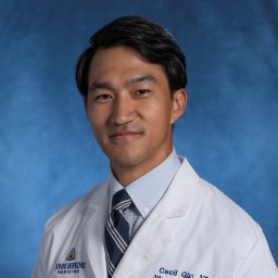 Cecil S Qiu合作论文数Department of Plastic and Reconstructive Surgery, Johns Hopkins Hospital1Robert Dorfman合作论文数1
 John Stranix合作论文数Department of Plastic Surgery, University of Virginia Health System1Richard M Elledge合作论文数1John Y S Kim合作论文数1
 W. Grant Stevens合作论文数1
 John Y Kim合作论文数Department of Dermatology, Feinberg School of Medicine, Northwestern University1
 Jamal Bullocks合作论文数1Slavin Sumner A合作论文数1
 Chuma J Chike-Obi合作论文数1
 Maurice Nahabedian合作论文数National Center for Plastic Surgery;Johns Hopkins University;Virginia Commonwealth University1
 Maia Munique合作论文数Department of Plastic Surgery, University of Texas Southwestern Medical Center1
 Epstein Michael合作论文数Auditory Modeling & Proc Lab, Northeastern Univ1Bancroft Gregory N合作论文数1Wahba Melissa合作论文数1Carruthers Katherine H合作论文数1Lee Edward I合作论文数1
 Antonio J. Forte合作论文数Mayo Clinic Florida1
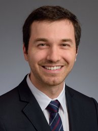 Winocour Julian合作论文数Vanderbilt University, University of Alabama at Birmingham1Patel Ashit合作论文数Division of Plastic and Reconstructive Surgery, Albany Medical Center1
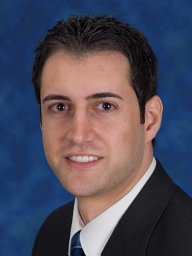 Hammoudeh Ziyad S合作论文数Department of Surgery, Wayne State University1Bucknor Alexandra合作论文数1Chandawarkar Akash合作论文数1
 Kent K Higdon合作论文数1
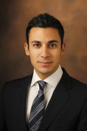 Christodoulos Kaoutzanis合作论文数Department of Surgery, Saint Joseph Mercy Health System1
 Megan Fracol合作论文数Perelman School of Medicine, University of Pennsylvania1
 Pankaj Tiwari合作论文数1




