Biochemical and clinical heterogeneity in long chain 3-hydroxyacyl-CoA dehydrogenase deficiency: identification of a new disorder of mitochondrial fi-oxidation. R. J. A. Wanders, L Ijlst, A. Munnich and J.-M. Saudubray Neonatal symptoms are common in medium chain acyl coenzyme A dehydrogenase deficiency. B. Wilcken, K. Carpenter and J. Hammond Choline deficiency in non-ketotic hyperglycinaemia. J. L K. Van Hove, F. Lazeyras, S. C. Garner, C. H. Charles, S. G. Kahler, R.-M. Boustany and S. H. Zeisel Dextromethorpban, a N-methyl-D-aspartate antagonist, in the treatment ofnon-ketotic hyperglycinaemia. B. Schmitt, B. Steinmann, R. Gitzelmann, L. Thun-Hohenstein and G. Dumermuth Complex I (NADH dehydrogenase) activity and biosynthesis in fibroblasts and lymphoblasts from patients with Leber's hereditary optic neuropathy. W. d. Rhead, E. Freneaux, A. Shires and E. Stone Porphyria due to 5-aminolevulinic acid dehydratase deficiency: a novel clinical and biochemical presentation. H. Mandel, M. Jaffe, N. Schoenfeld, A. Aizin and M. Berant Hereditary tyrosinaemia type I: self-induced correction of the genetic defect? E. A. Kvittingen, H. Rootwelt, P. Bradtzaeg, A. Bergan and R. Berger Galactosaemia, a carbohydrate-deficient glycoprotein syndrome. L d. M. Spaapen, T. Vulsma, P. M. V. M. Theunissen, S. B, van der Meer and J. daeken Alanine effect on phenylketonuric rats. M. Wajner, C. G. Pedron, F. C. Moreira, M. H. Xavier, B. d. dos Santos, A. T. S. Wyse and C. M. D. Wannmacher Possible high frequency of tetrahydrobiopterin deficiency in South Brazil. L B. Yardim, M. G. Burin, R. Giugliani and N. Blau Screening of PKU mutations in Spain and South America. B. Perez, L R. Desviat, M. Die, M. J. Garcia and M. Ugarte Evaluation of dental health in phenylketonuric children. M. Roupa, C. Demars and B. Francois A mutation tightly linked to haplotype 6 in the Turkish PKU alleles. M. Ozgiig, E. I~lmaz, H. Erdem, T. Coskun, S. Ayter and I. Ozalp fl-Endorphin system in chemically-induced phenylketonuria. C. M. D. Wannmaeher, A. R. Bolner, J. Engelmann and M. Wajner Is there still a place for the Guthrie test? A. H. Wilcox, J. Stern and J. M. Whitaker Magnetic resonance imaging, biochemical control and genetic analysis in patients with phenylketonuria. J. H. Walter, L A. Tyfield, J. B. Holton and C. Johnson Neuroradiological (NMR) examination in PKU patients: clinical and biochemical correlation. V. Leuzzi, F. Fabbrizi, I. Antonozzi, G. Gualdi, C. Di Biasi and G. Trasimeni Localized proton NMR spectroscopy in patients with phenylketonuria. K. Johannik, P. Van Hecke, B. Francois, M, Smet, J. Jaeken and A. Baert Central motor conduction time in phenylketonuria after cessation of dietary treatment. M. A. Cleary, J. H. Walter, J. E. Wraith and S. M. Alani Supplementation of phenylketonuria children with a bacterial selenosource. M. Calomme, J. Hu, B. Francois, M. Van Caillie-Bertrand and D. A. Van den Berghe Phenylalanine feto-material gradients by cordocentesis at 19 to 33 weeks gestation. W. E. Schoonheyt, W. B. Hanley, J. T. R. Clarke and J. Johnson Fumarylacetoacetate lyase-enzyme stability and its relevance to tyrosinaemia type I and glutathione synthetase deficiency. R. G. F. Gray, A. J. Lloyd and A. Green Cerebrospinal fluid amines and pterins in tyrosinaemia type II. S. J. R. Heales, A. Bowron, I. Smith, R. A. H. Surtees and P. T. Clayton Molecular basis of phenylketonuria in Central and Southern Italian population. C. Carducei, A. Pontecorvi, L. Ellul, M. Scuderi, F. Fabbrizi, V. Leuzzi and I. Antonozzi Maple syrup urine disease diagnosis and treatment in Bulgaria. B. Radeva and I. Sinigerska Intermittent form of maple syrup urine disease in brother and sister. M. Hrebicek, S. Stastna, U. Wendel, P. Verner, J. Zeman, V. Kozich, J. H yanek, Z. Rytir, D. Nemcova and J. Rytir Management and CT-findings in a case of neonatal (classical) maple syrup urine disease. J. G. M. Huijmans, J. B. C. de Klerk, S, G. F. Robben, W. J. Kleijer, R. Jankie, R. Slotema and R. Spritzer Maple syrup urine disease: prevention of postoperative metabolic decompensation by insulin. B. Biggeman, R. Zass and U. Wendel An asymptomatic variant of maple syrup urine disease without organic aciduria. M. L Cabello,
In humans, low peak bone mass is a significant risk factor for osteoporosis. We report that LRP5, encoding the low-density lipoprotein receptor-related protein 5, affects bone mass accrual during growth. Mutations in LRP5 cause the autosomal recessive disorder osteoporosis-pseudoglioma syndrome (OPPG). We find that OPPG carriers have reduced bone mass when compared to age- and gender-matched controls. We demonstrate LRP5 expression by osteoblasts in situ and show that LRP5 can transduce Wnt signaling in vitro via the canonical pathway. We further show that a mutant-secreted form of LRP5 can reduce bone thickness in mouse calvarial explant cultures. These data indicate that Wnt-mediated signaling via LRP5 affects bone accrual during growth and is important for the establishment of peak bone mass.
Cockayne syndrome (CS) is a rare autosomal recessive disorder characterized by postnatal growth failure, mental retardation and otherwise clinically heterogeneous features which commonly include cutaneous photosensitivity. Cultured cells from sun-sensitive CS patients are hypersensitive to ultraviolet (UV) light and, following UV irradiation, are unable to restore RNA synthesis rates to normal levels. This has been attributed to a specific deficiency in CS cells in the ability to carry out preferential repair of damage in actively transcribed regions of DNA. We report here a cellular and molecular analysis of three Italian CS patients who were of particular interest because none of them was sun-sensitive, despite showing most of the features of the severe form of CS, including the characteristic cellular sensitivity to UV irradiation. They all were altered in the CSB gene. The genetically related patients CS1PV and CS3PV were homozygous for the C1436T transition resulting in the change Arg453opal. Patient CS2PV was a compound heterozygote for two new causative mutations, insertions of an A at position 1051 and of TGTC at 2053, leading to truncated proteins of 367 and 681 amino acids. These mutations result in severely truncated proteins, as do many of those that we previously identified in several sun-sensitive CS-B patients. These observations confirm that the CSB gene is not essential for viability and cell proliferation, an important issue to be considered in any speculation on the recently proposed additional function of the CSB protein in transcription. Our investigations provide data supporting the notion that other factors, besides the site of the mutation, influence the type and severity of the CS clinical features.
An 8 year old boy who had Langerhans cell histiocytosis when he was 15 months old showed psychomotor regression from the age of 2 years. Microcephaly, severe growth deficiency, and ocular telangiectasia were also evident. Magnetic nuclear resonance imaging showed cerebellar atrophy. Alphafetoprotein was increased. Chromosome instability after x irradiation and rearrangements involving chromosome 7 were found. Molecular study failed to show mutations involving the ataxia-telangiectasia gene. This patient has a clinical picture which is difficult to relate to a known breakage syndrome. Also, the relationship between the clinical phenotype and histiocytosis is unclear.
Annals of the New York Academy of SciencesVolume 876, Issue 1 p. 262-265 Interactions between Prolactin and the Proinflammatory Cytokine Network in Juvenile Chronic Arthritis P. PICCO, Corresponding Author P. PICCO 2nd Pediatric Division, G. Gaslini Scientific Institute, Genoa, ItalyAddress correspondence to Paolo Picco, M.D., 2nd Pediatric Division, Giannina Gaslini Scientific Institute, Largo G Gaslini 5, 16147 Genova, Italy. Fax: ++ 39-010-3776590.Search for more papers by this authorM. GATTORNO, M. GATTORNO 2nd Pediatric Division, G. Gaslini Scientific Institute, Genoa, ItalySearch for more papers by this authorA. BUONCOMPAGNI, A. BUONCOMPAGNI 2nd Pediatric Division, G. Gaslini Scientific Institute, Genoa, ItalySearch for more papers by this authorS. VIGNOLA, S. VIGNOLA 2nd Pediatric Division, G. Gaslini Scientific Institute, Genoa, ItalySearch for more papers by this authorM. MAGGIANI, M. MAGGIANI 2nd Department of Orthopaedics, G. Gaslini Scientific Institute, Genoa, ItalySearch for more papers by this authorG. ROSSI, G. ROSSI Laboratory of Biochemistry, G. Gaslini Scientific Institute, Genoa, ItalySearch for more papers by this authorV. PISTOIA, V. PISTOIA Laboratory of Oncology, G. Gaslini Scientific Institute, Genoa, ItalySearch for more papers by this authorC. BORRONE, C. BORRONE 2nd Pediatric Division, G. Gaslini Scientific Institute, Genoa, ItalySearch for more papers by this author P. PICCO, Corresponding Author P. PICCO 2nd Pediatric Division, G. Gaslini Scientific Institute, Genoa, ItalyAddress correspondence to Paolo Picco, M.D., 2nd Pediatric Division, Giannina Gaslini Scientific Institute, Largo G Gaslini 5, 16147 Genova, Italy. Fax: ++ 39-010-3776590.Search for more papers by this authorM. GATTORNO, M. GATTORNO 2nd Pediatric Division, G. Gaslini Scientific Institute, Genoa, ItalySearch for more papers by this authorA. BUONCOMPAGNI, A. BUONCOMPAGNI 2nd Pediatric Division, G. Gaslini Scientific Institute, Genoa, ItalySearch for more papers by this authorS. VIGNOLA, S. VIGNOLA 2nd Pediatric Division, G. Gaslini Scientific Institute, Genoa, ItalySearch for more papers by this authorM. MAGGIANI, M. MAGGIANI 2nd Department of Orthopaedics, G. Gaslini Scientific Institute, Genoa, ItalySearch for more papers by this authorG. ROSSI, G. ROSSI Laboratory of Biochemistry, G. Gaslini Scientific Institute, Genoa, ItalySearch for more papers by this authorV. PISTOIA, V. PISTOIA Laboratory of Oncology, G. Gaslini Scientific Institute, Genoa, ItalySearch for more papers by this authorC. BORRONE, C. BORRONE 2nd Pediatric Division, G. Gaslini Scientific Institute, Genoa, ItalySearch for more papers by this author First published: 06 February 2006 https://doi.org/10.1111/j.1749-6632.1999.tb07648.xCitations: 9Read the full textAboutPDF ToolsRequest permissionExport citationAdd to favoritesTrack citation ShareShare Give accessShare full text accessShare full-text accessPlease review our Terms and Conditions of Use and check box below to share full-text version of article.I have read and accept the Wiley Online Library Terms and Conditions of UseShareable LinkUse the link below to share a full-text version of this article with your friends and colleagues. Learn more.Copy URL Share a linkShare onFacebookTwitterLinked InRedditWechat Citing Literature Volume876, Issue1NEUROENDOCRINE IMMUNE BASIS OF THE RHEUMATIC DISEASESJune 1999Pages 262-265 RelatedInformation
We report on a child with a clinical and neuroradiological picture consistent with Leigh disease and an unusual association of isolated hypermethioninaemia and 3-methylglutaconic aciduria. A low-methionine diet normalized both plasma methionine and urine 3-methylglutaconic acid; a methionine-loading test led to significant increase of both metabolites. In the skin fibroblasts the activity of 3-methylglutaconyl-CoA hydratase was essentially normal. No explanation of this uncommon association of hypermethioninaemia and glutaconic aciduria is available. The possibility of a common transporter for 3-methylglutaconic acid and methionine is an attractive hypothesis.
Abrupt osmotic changes during rapid correction of chronic hyponatremia result in demyelinative brain lesions, but the sequence of events linking rapid osmotic changes to myelin loss is not yet understood. Here, in a rat model of osmotic demyelination syndrome, we found that massive astrocyte death occurred after rapid correction of hyponatremia, delineating the regions of future myelin loss. Astrocyte death caused a disruption of the astrocyte-oligodendrocyte network, rapidly upregulated inflammatory cytokines genes, and increased serum S100B, which predicted clinical manifestations and outcome of osmotic demyelination. These results support a model for the pathophysiology of osmotic brain injury in which rapid correction of hyponatremia triggers apoptosis in astrocytes followed by a loss of trophic communication between astrocytes and oligodendrocytes, secondary inflammation, microglial activation, and finally demyelination.
We report an 8-month-old girl, only child of unrelated parents, who was admitted to the hospital with the suspicion of spina bifida. Nuclear magnetic resonance showed a complex spinal malformation characterized by dorsal diastematomyelia and hydromyelia. Clinical presentation treatment and genetic counselling are discussed.
Journal of Inherited Metabolic DiseaseVolume 21, Issue 6 p. 675-676 ArticleFull Access Complications of lysinuric protein intolerance must be treated with immunosuppressive drugs M. Di Rocco, M. Di RoccoSearch for more papers by this authorA. Buoncompagni, A. BuoncompagniSearch for more papers by this authorP. Picco, P. PiccoSearch for more papers by this authorS. Vignola, S. VignolaSearch for more papers by this authorC. Borrone, C. BorroneSearch for more papers by this author M. Di Rocco, M. Di RoccoSearch for more papers by this authorA. Buoncompagni, A. BuoncompagniSearch for more papers by this authorP. Picco, P. PiccoSearch for more papers by this authorS. Vignola, S. VignolaSearch for more papers by this authorC. Borrone, C. BorroneSearch for more papers by this author First published: 01 August 1998 https://doi.org/10.1023/A:1005440802688Citations: 8AboutPDF ToolsRequest permissionExport citationAdd to favoritesTrack citation ShareShare Give accessShare full text accessShare full-text accessPlease review our Terms and Conditions of Use and check box below to share full-text version of article.I have read and accept the Wiley Online Library Terms and Conditions of UseShareable LinkUse the link below to share a full-text version of this article with your friends and colleagues. Learn more.Copy URL Share a linkShare onEmailFacebookTwitterLinkedInRedditWechat No abstract is available for this article. REFERENCES 1Di Rocco M, Garibotto G, Rossi GA et al (1993) Role of haematological, pulmonary and renal complications in the long-term prognosis of patients with lysinuric protein intolerance. Eur J Pediatr, 152: 437–440. 2Lauteala T, Sistonen P, Savontaus M et al (1997) Lysinuric protein intolerance gene maps to the long arm of chromosome 14. Am J Hum Genet, 60: 1479–1486. 3Parsons H, Snyder F, Bowen T, Klassen J, Pinto A (1996) Immune complex disease consistent with systemic lupus erythematosus in a patient with lysinuric protein intolerance. J Inher Metab Dis, 19: 627–634. Citing Literature Volume21, Issue6August 1998Pages 675-676 ReferencesRelatedInformation
OBJECTIVE Animal models of immune complex mediated tissue injury have shown different patterns of proinflammatory cytokine production according to the subtype of immunoglobulin involved. The IgA immune complex model differs from the IgG model by the lack of involvement of tumor necrosis factor (TNF) in the pathogenesis of tissue damage. We investigated in age matched patients the possible difference in TNF involvement in a predominantly IgA mediated disease, Henoch-Schönlein purpura (HSP), in comparison with systemic lupus erythematosus (SLE), in which vascular injury is mostly associated with local deposition of IgG immune complexes. METHODS Serum concentrations of TNF-alpha and its soluble receptors (sTNF-R) p55 and p75 were studied in 20 patients with pediatric SLE at various degrees of disease activity, in 16 patients with highly active HSP, and in 15 healthy controls by enzyme amplified sensitivity immunoassay. SLE disease activity was evaluated using 2 scores, the European Consensus Group Study for SLE Disease Activity Criteria and the SLE Disease Activity Index. RESULTS Serum concentrations of TNF-alpha fell within the normal range in patients with both SLE and HSP irrespective of disease activity. Conversely, patients with SLE displayed increased serum levels of sTNF-R that correlated positively with the degree of disease activity (r = 0.60, p < 0.001; r = 0.71, p < 0.001, for p55 and p75, respectively). In contrast, no difference in the serum levels of sTNF-R was found between patients with highly active HSP and controls. CONCLUSION Our study provides the first circumstantial evidence that pediatric SLE and HSP are characterized by differential involvement of TNF in the pathogenesis of tissue damage.
We report on a 17 6/12-year-old boy with nephronophthisis, retinitis pigmentosa, left upper eyelid ptosis, enopthalmos, transmissive deafness, GH and TSH deficiency, and mild skeletal dysplasia. A similar case was reported by Bianchi et al. [1988: Helv Paediatr Acta 43:449-455] in another Italian patient. Here we confirm the previous observations and argue that both patients might be affected by a new syndrome.
A 3-year-old boy with erythema multiforme-like manifestations and severe articular involvement is reported. Because of the unusual onset of the cutaneous lesion a skin biopsy was performed, revealing the typical features of a leukocytoclastic vasculitis. A direct immunofluorescent study revealed C3 and IgM deposition within the wall and around the small vessels. The clinical and immunopathological findings in this patient were similar to those reported for the acute hemorrhagic edema of infancy (AHE) described in children younger than 2 years of age. The present case supports the hypothesis that AHE is a distinctive leukocytoclastic vasculitis of childhood, irrespective of the age at onset.
We screened the aldolase B gene in 14 unrelated Italian patients with hereditary fructose intolerance (HFI), and found two novel disease related mutations: a single nucleotide deletion in exon 2 (delta A20) that leads to an early stop codon, and a C-->T transition in exon 8 that substitutes an Arg with a Trp residue at codon 303 (R303W).
 Paolo Picco合作论文数31
 Marcelo Rocco合作论文数Department of Pediatrics, Gaslini Institute21M Dirocco合作论文数17A Buoncompagni合作论文数15Rosanna Gatti合作论文数15
 Mirella Filocamo合作论文数IRCCS G. Gaslini13
 Marco Gattorno (马可·加托诺)合作论文数Istituto Gianina Gaslini13L Massimo合作论文数8A. Barabino合作论文数7A.G. Marchi合作论文数7Edvige Veneselli合作论文数7G Cordone合作论文数6S Vignola合作论文数6M. Romanengo合作论文数6
 Paolo Gandullia合作论文数Gastroenterology Unit, G. Gaslini Institute6Maja Di Rocco合作论文数4C Melevendi合作论文数4Lattere M合作论文数4L Famularo合作论文数3P CERRUTIMAINARDI合作论文数3M Gremmo合作论文数3F Pendola合作论文数3M Bado合作论文数3L Leveratto合作论文数3Mg Vianello合作论文数3C. Gambini合作论文数3D Massocco合作论文数3Dagna Bricarelli F合作论文数3
 Andrea Superti Furga合作论文数Centre Hospitalier Universitaire Vaudois3
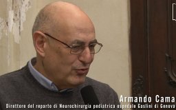 Armando Cama合作论文数Department of Pediatric Neuroradiology, G. Gaslini Children’s Research Hospital3P. Durand合作论文数3Luigi Garibaldi合作论文数Cardinal Glennon Children’s Hospital, St. Louis University Medical School3Antonella Buoncompagni合作论文数3
 Generoso Andria合作论文数Department of Pediatrics, Federico II University3
 Vito Pistoia合作论文数Ospedale Pediatrico Bambino Gesù;Ospedali Pediatrici Istituto Gaslini3A. R. Fantasia合作论文数2Alberto Garaventa合作论文数2F Dagna-Bricarelli合作论文数2A Comelli合作论文数2M Brisigotti合作论文数2M P Fondelli合作论文数2P. Tortori Donati合作论文数2S Canini合作论文数2C. Lombardo合作论文数2G Romeo合作论文数2F CLAUDIANI合作论文数2Cerruti Mainardi P合作论文数2Giustardi A合作论文数2




