Background. Adenocarcinoma with signet ring cell (SRC) features has been reported to be a poor prognostic marker in gastric and colorectal carcinomas. Although uncommon in the esophagus, SRC histology, interestingly, has been correlated with improved survival. Our impression has been that the incidence of esophageal adenocarcinomas with SRC features is increasing and is associated with worse outcomes. We hypothesize that patients with SRC histology present with more advanced disease, respond less well to induction therapy, and have decreased survival after resection compared with patients with non-SRC adenocarcinoma.Methods. The medical records of 151 consecutive patients who underwent resection for adenocarcinoma of the esophagus or gastroesophageal junction in a prospectively maintained database from 1998 to 2011 were reviewed. Outcomes of 23 patients (15%) with SRC histology (21 men, 2 women; average age, 66 years) were compared with 128 patients (85%) with non-SRC adenocarcinoma (116 men, 12 women; average age, 63 years). Overall survival, stage-specific survival, and response to induction therapy were evaluated. Cox regression multivariate analysis was used to identify independent predictors of 3-year survival.Results. SRC and non-SRC patients were evenly matched for clinical and tumor characteristics. Down-staging achieved with induction therapy was 13.3% (2 of 15) in SRC histology patients vs 67.1% (53 of 79) in non-SRC patients (p <= 0.001). Patients with SRC histology who did not respond well to induction treatment demonstrated strong trends toward a worse 3-year survival than patients with non-SRC adenocarcinoma (p = 0.084). The overall 3-year survival was 65.6% in patients without SRC histology vs 34.8% in those with SRC (p = 0.006). Patients with pathologic stage II or III and SRC histology had a 3-year survival of 27.3% compared with 57.4% in patients with non-SRC adenocarcinoma (p = 0.01). Multivariate analysis showed SRC histology trended toward significance as an independent risk factor for poor survival (p = 0.060).Conclusions. Patients with adenocarcinoma of the esophagus or gastroesophageal junction and SRC histology respond less well to induction therapy and have decreased overall survival compared with patients with non-SRC histology. (C) 2013 by The Society of Thoracic Surgeons
Background. Given declining interest in cardiothoracic (CT) training programs during the last decade, increasing emphasis has been placed on engaging candidates early in their training. We examined the effect of supervised and unsupervised practice on medical students' interest in CT surgery.Methods. Forty-five medical students participated in this study. Participants' interest level in surgery, CT surgery, and simulation were collected before and after a pretest session. Subsequently, participants were randomized to one of three groups: control (n = 15), unsupervised training on a low-fidelity task simulator (n = 15), or supervised training with a CT surgeon or fellow on the same simulator (n = 15). After 3 weeks, attitudes were reassessed at a posttest session. Interest levels were compared before and after the pretest using paired t tests, and the effects of training on interests were assessed with multiple linear regression analyses.Results. After the pretest session, participants were significantly more interested in simulation (p = 0.001) but not in surgery or CT surgery. After training, compared with control group participants, supervised trainees demonstrated a significant increase in their interest level in pursuing a career in surgery (p = 0.028) and an increasing trend towards a career in CT surgery (p = 0.060), whereas unsupervised trainees did not.Conclusions. Supervised training on low-fidelity simulators enhances interest in a career in surgery. Practice that lacks supervision does not, possibly related to the complexity of the simulated task. Mentorship efforts may need to involve sustained interaction to provide medical students with enough exposure to appreciate a surgical career. (C) 2013 by The Society of Thoracic Surgeons
Background. Traditionally, cardiothoracic residency programs are 2 or 3 years in length and require the completion of a general surgery residency. Six- year integrated programs (IP) that directly match fourth-year medical students have been recently developed. Our objective was to examine the curricula of traditional 2-year (T2) and 3-year (T3) programs and compare them to the curricula of IP.Methods. We requested curricula from the directors of all IP, T2, and T3 programs participating in the 2011 to 2012 match. We compared the median number of months spent on a cardiothoracic (CT) rotation, an adult cardiac rotation, a thoracic rotation, and a congenital rotation, as well as time spent on "other" nonsurgical rotations. Traditional programs were categorized into 1 of 3 pathways: combined cardiothoracic (CCT), adult cardiac (AC), or general thoracic (GT).Results. Integrated programs spend more time on general thoracic rotations when compared with CCT-T2, CCT-T3, AC-T2, and AC-T3 pathways (p [0.009, p = 0.046, p = 0.001 and p = 0.028, respectively). The IP spend a similar amount of time on CT, adult cardiac, and congenital rotations when compared when 2- and 3-year CCT, AC, and GT pathways. Of note, IP spend significantly more time on "other" nonsurgical rotations than all other pathways (p < 0.001 to 0.008).Conclusions. Integrated programs should not be considered "cardiac pathways" as they spend a significant amount of time on thoracic rotations. Additional nonsurgical rotations provide an opportunity for residents in IP to develop unique skills not currently provided in traditional programs. (C) 2013 by The Society of Thoracic Surgeons
A number of clinical trials have incorporated preoperative chemoradiation therapy (CRT) in an attempt to improve local tumor control, distant recurrence and overall survival for patients with locally advanced but resectable adenocarcinoma of the distal esophagus or gastroesophageal junction (GEJ). This multi-centered, cooperative group study combined cisplatin, docetaxel and panitumumab with radiation therapy preoperatively for patients with locoregionally advanced esophageal adenocarcinoma. Pathologic complete response (pCR) of ≥ 35% was the primary endpoint target goal. From January 15, 2009 to July 22, 2011, 70 patients with distal esophageal or GEJ adenocarcinomas of clinical stage T3N0M0, T2-3N1M0 or T2-3N0-1M1a (M1a with celiac LN ≤ 2 cm) were accrued. Patients received cisplatin (40 mg/m2), docetaxel (40 mg/m2), and panitumumab (6 mg/kg) on weeks 1, 3, 5, 7 and 9 with RT (5040 cGy, 180 cGy/day x 28d) beginning week 5. Pathologic complete response was defined as no viable residual tumor cells. Secondary objectives included near-pathologic complete response (near-pCR), toxicity, and overall and disease-free survival rates. A near-pCR was defined as ≤ 10% of viable residual cancer cells. Adverse events were graded using the Common Terminology Criteria for Adverse Events Version 3.0. Five of 70 patients were ineligible. One patient had a celiac lymph node > 2 cm, and one patient had two primary tumors. Liver lesions were not investigated in one patient. Two patients had proximal gastric cancers. Of the remaining 65 patients (59 M, 6 F; median age 61), 11 patients did not undergo surgery (4 progressions, 3 patient refusals, 3 other, 1 physician decision), leaving 54 evaluable patients. Median follow-up was 20.1 months. The pCR rate was 33.3% and near-pCR was 20.4%. Total doses of cisplatin, docetaxel and panitumumab were administered in 75%, 80%, and 72%, respectively (n = 65). Ninety-four percent of patients received the total radiation therapy dose. Twenty-eight (43.1%) patients had grade 4 or greater toxicity that was possibly related to treatment. Lymphopenia (15 patients) was most common. Operative mortality was 3.7% (2/54). There was one death attributable to multisystem organ failure and one was due to gastric conduit necrosis. ARDS was encountered in 2 patients (3.7%). Median survival was 18.6 mos; 2-yr disease-free survival Kaplan-Meier estimate was 51.6% (95% CI, 36.7% - 72.6%) and 3-yr overall survival was 46.1% (95% CI 34.3 to 62%). This neoadjuvant regimen of cisplatin, docetaxel and panitumumab and radiation is active (pCR + near-pCR = 53.7%) but did not meet the primary outcome. The toxicity is substantial, but manageable. Further evaluation of this regimen in an unselected population is not recommended.
BACKGROUND:Preoperative chemoradiotherapy (CRT) improves outcomes in patients with locally advanced but resectable adenocarcinoma of the esophagus. ACOSOG Z4051 evaluated CRT with docetaxel, cisplatin, and panitumumab (DCP) in this patient group with a primary end point of a pathologic complete response (pCR) ≥35%.PATIENTS AND METHODS:From 15 January 2009 to 22 July 2011, 70 patients with locally advanced but resectable distal esophageal adenocarcinoma were enrolled. Patients received docetaxel (40 mg/m(2)), cisplatin (40 mg/m(2)), and panitumumab (6 mg/kg) on weeks 1, 3, 5, 7, and 9 with RT (5040 cGy, 180 cGy/day × 28 days) beginning week 5. Resection was planned after completing CRT. PCR was defined as no viable residual tumor cells. Secondary objectives included near-pCR (≤10% viable cancer cells), toxicity, and overall and disease-free survival. Adverse events were graded using the CTCAE Version 3.0.RESULTS:Five of 70 patients were ineligible. Of 65 eligible patients (59 M; median age 61), 11 did not undergo surgery, leaving 54 assessable. PCR rate was 33.3% and near-pCR was 20.4%. Secenty-three percent of patients completed DCP (n = 70) and 92% completed RT. 48.5% had toxicity ≥grade 4. Lymphopenia (43%) was most common. Operative mortality was 3.7%. Adult respiratory distress syndrome was encountered in two patients (3.7%). At median follow-up of 26.3 months, median overall survival was 19.4 months and 3-year overall survival was 38.6% (95% confidence interval 24.5% to 60.8%).CONCLUSIONS:Neoadjuvant CRT with DCP is active (pCR + near-pCR = 53.7%) but toxicity is significant. Further evaluation of this regimen in an unselected population is not recommended.CLINICALTRIALSGOV IDENTIFIER:NCT00757172.
Background. Currently, the most accurate staging test for patients with esophageal cancer is endoscopic ultrasound (EUS). At many institutions, patients who have completed neoadjuvant chemotherapy and radiotherapy for esophageal cancer undergo restaging EUS before proceeding to surgical resection. The benefit of this restaging procedure remains controversial.Methods. We retrospectively studied consecutive patients who had pre-resection restaging EUS after receiving neoadjuvant treatment to assess accuracy of EUS restaging and determine whether it predicted survival.Results. Final pathologic data were available for 73 patients who underwent restaging EUS (3 patients had missing T or N stage at one time point). Median time from restaging EUS to resection was 20 days. Restaging EUS accurately predicted pathologic T status in 26 of 72 patients (36%), N status in 44 of 71 (62%), and detected a complete pathologic response in 2 of 19 (10.5%). EUS inappropriately classified 10 patients as TO NO. Agreement between EUS and pathologic staging was poor for T (kappa = 0.14) and N status (kappa = 0.24). Median time from resection to death or last follow-up was 20 months. Pathologic T and N status were each significant predictors of survival (p = 0.049 and p = 0.0004, respectively). There were nonsignificant trends toward better survival for lower EUS T (p = 0.32) and N status (p = 0.0946).Conclusions. Restaging by EUS before resection did not accurately predict pathologic stage in patients with esophageal cancer who received neoadjuvant treatment. As a result of this investigation, our institution no longer routinely performs restaging EUS. (Ann Thorac Surg 2012;93:1855-60) (c) 2012 by The Society of Thoracic Surgeons
This article discusses the incidence, geographic differences, and risk factors for the 2 most common cancers of the esophagus: squamous cell and adenocarcinoma.
Background. Patients often receive induction therapy based on endoscopic ultrasound (EUS)-identified nodal spread (N1) or deep tumor invasion (T3), although controversy exists regarding the role of induction therapy for early stage disease. We aim to evaluate the reliability of EUS in identifying early stage disease and the subsequent impact on treatment and outcomes.Methods. We retrospectively studied 149 patients who underwent EUS and esophagectomy for adenocarcinoma between January 2000 and December 2008. Computed tomography (CT) was performed in all patients, whereas positron emission tomography (PET) was performed in 91%. Clinical stage (c), pathologic stage (p), operative mortality, and survival were recorded.Results. Unanticipated pathologic nodal disease was similar in patients with cT1N0 and cT2N0 tumors (6/25 [24%] versus 7/18 [38.8%]; p = 0.6). Among the 18 cases of cT2N0 disease, 9 (50%) were pathologically staged as T1N0, 8 (44%) were upstaged to pT3N0-1, and 1 (6%) was pT2N0. One case of cT1N0 tumor (4%) was upstaged to pT3N0. Among patients with cT1-2N0 tumors, 5-year disease-free survival for the group that was appropriately staged was 89.8% versus 39.9% for the group that had a higher pathologic stage than their clinical stage (ie, > T2N0) (p < 0.001). Operative mortality for patients with cT1-2N0 tumors was 0/43 (0%), which was no different from that in the higher clinical stage groups with (1/37, 2.7%) or without (2/68, 2.9%) induction therapy (p = 0.5). Multivariate analysis identified marked/intense uptake on staging PET (odds ratio, 5.76, 95%; confidence interval, 1.25 to 26.52; p = 0.021) to be a factor predictive of upstaging of cT1-2N0 tumors.Conclusions. Current staging techniques are inadequate for predicting T1-2N0 disease in esophageal adenocarcinoma. Survival is excellent with operation alone in patients with tumors appropriately staged as T1-2N0, although patients with tumors upstaged to greater than T2N0 have significantly worse survival. Other preoperative factors such as PET uptake may help select patients with cT1-2N0 tumors that will be upstaged at resection. (Ann Thorac Surg 2011; 91: 1509 -16) (C) 2011 by The Society of Thoracic Surgeons
e17510 Background: Epidemiologic studies suggest that MetS increases the risk of lung cancer, yet its effect on cancer mortality remains controversial. We analyzed the effect of MetS/its various components on cancer mortality in large, national cohort of patients with early stage NSCLC, controlling for patient/tumor characteristics and treatment. Methods: Using the Surveillance, Epidemiology, and End Results (SEER)-Medicare linked database, we identified 11,190 patients aged 66 and older with stage I-II NSCLC diagnosed between 1999-2004. Patient/tumor characteristics were obtained from the SEER dataset; data on MetS/its components and cancer treatment was ascertained from Medicare claims. Relationships between the various patient/tumor characteristics and survival were analyzed using chi square tests and logistic regression. Survival in patients with/without MetS/its components was compared using multivariate Cox models. Results: A significant proportion of patients had MetS (19.0%), diabetes (26.6%), hypertension (74.0%) and dyslipidemia (55.7%). Patients with MetS were more likely to be younger, non-white, less educated, and have greater comorbidity (all P<0.001). There was no association between tumor stage, histology, or grade and metabolic syndrome. MetS had no apparent effect on survival. When the effect of its individual components was analyzed, however, diabetes and hypertension were found to be independently associated with worse outcomes (adjusted hazard ratios [aHR] 1.18 [95% confidence internal {CI} 1.11-1.25] and 1.15 [95% CI 1.09-1.22], respectively) after adjusting for other patient/tumor/treatment factors. In contrast, dyslipidemia was associated with significantly improved survival on both univariate (5-year survival: 41% v 29%, P<0.001) and multivariate analyses (aHR 0.82 [95% CI 0.78-0.86]). Conclusions: Diabetes and hypertension have independent adverse effects on survival in patients with early stage NSCLC, whereas dyslipidemia appears to have a protective effect. Trials are needed to determine if use of metformin (in diabetic patients) and/or statins in the adjuvant setting improves long term outcomes in this patient population.
Objective: Using Worldwide Esophageal Cancer Collaboration data, we sought to (1) characterize the relationship between survival and extent of lymphadenectomy, and (2) from this, define Optimum lymphadenectomy.Summary Background Data: What constitutes optimum lymphadenectomy to maximize survival is controversial because of variable goals, analytic methodology, and generalizability of the underpinning data.Methods: A total of 4627 patients who had esophagectomy alone for esophageal cancer were identified from the Worldwide Esophageal Cancer Collaboration database. Patient-specific risk-adjusted survival was estimated using random survival forests. Risk-adjusted 5-year survival was averaged for each number of lymph nodes resected and its relation to cancer characteristics explored. Optimum number of nodes that should be resected to maximize 5-year survival was determined by random forest multivariable regression.Results: For pN0M0 moderately and poorly differentiated cancers, and all node-positive (pN+) cancers, 5-year survival improved with increasing extent of lymphadenectomy. In pN0M0 cancers, no optimum lymphadenectomy was defined for pTis; optimum lymphadenectomy was 10 to 12 nodes for pT1, 15 to 22 for pT2, and 31 to 42 for pT3/T4, depending on histopathologic cell type. In pN+M0 cancers and I to 6 nodes positive, optimum lymphadenectomy was 10 for pT1, 15 for pT2, and 29 to 50 for pT3/T4.Conclusions: Greater extent of lymphadenectomy was associated with increased survival for all patients with esophageal cancer except at the extremes (TisN0M0 and >= 7 regional lymph nodes positive for cancer) and well-differentiated pN0M0 cancer. Maximum 5-year survival is modulated by T classification: resecting 10 nodes for pT1, 20 for pT2, and >= 30 for pT3/T4 is recommended.
Objective: To compare toxicities, disease control, and survival outcomes for patients treated with either cisplatin/irinotecan versus carboplatin/paclitaxel concurrent chemoradiotherapy for locally advanced esophageal cancer. Methods: Single-institution retrospective comparison between treatment groups: the cisplatin/irinotecan group was treated with 2 cycles of induction chemotherapy followed by concurrent chemoradiotherapy, whereas the carboplatin/paclitaxel group began with chemoradiotherapy followed by 2 additional cycles of chemotherapy. Acute toxicities, response rates, disease control, survival outcomes, and patterns of failure were compared between the groups. Results: Between January 2000 and December 2007, 57 patients were identified for inclusion in the present study (38 cisplatin/irinotecan and 19 carboplatin/paclitaxel). Groups were well-balanced by clinical-, pathologic-, staging-, and treatment-related factors. Thirty-five patients (92%) in the cisplatin/irinotecan group and 18 patients (95%) in the carboplatin/paclitaxel group completed the concurrent phase of chemoradiotherapy. There were no significant differences in hematologic or nonhematologic toxicities between the groups. At a median survivor follow-up of 37.6 months (range: 7.3–59.3 months) for the entire population, 22 patients were alive (16 without evidence of disease). The 3-year overall survival estimates was 19.7% for the cisplatin/irinotecan group versus 56.1% for the carboplatin/paclitaxel group (P = 0.022). Estimated 3-year cancer-specific survivals were 24.6% for the cisplatin/irinotecan group versus 59.3% for the carboplatin/paclitaxel group (P = 0.033). Conclusion: Concurrent chemoradiotherapy with carboplatin/paclitaxel is well-tolerated and provided superior overall and disease-specific survival compared with cisplatin/irinotecan chemoradiotherapy in the present study population. Further investigation is warranted.
Infections of the sternoclavicular joint associated with extensive local soft tissue involvement or abscess formation often require surgical debridement and drainage. This condition typically presents with joint pain, local erythema, and swelling. This case demonstrates the simultaneous extension of a sternoclavicular joint infection into the left upper pulmonary lobe and through the overlying skin. (Ann Thorac Surg 2010; 90: 309-11) (C) 2010 by The Society of Thoracic Surgeons
OBJECTIVECisplatin-based chemoradiotherapy is standard treatment for locally advanced esophageal and gastroesophageal cancers; however, the optimal chemotherapy regimen remains to be defined.METHODSRetrospective single institution analysis of toxicities, response rates and survival outcomes in patients with cT3-4 or N1/M1a esophageal squamous cell or adenocarcinoma treated with induction cisplatin and irinotecan followed by concurrent cisplatin, irinotecan and radiotherapy. Secondary analysis for association of disease control and outcomes with demographic, tumor and treatment factors (including histology).RESULTSFifty-three patients were eligible for the present analysis. All patients underwent endoscopic ultrasonography and were either cT3-4 and/or cN1 disease. Fifty patients completed radiotherapy as planned (median dose 50.4 Gy, range 0-61.2), and 35 patients completed four cycles of chemotherapy as planned (range 1-4). Severe acute toxicities included Grade ≥ 3 neutropenia and esophagitis in 13 and 12 patients, respectively. There were no Grade 5 (fatal) toxicities noted. At mean survivor follow-up of 24.5 months (range 2.7-63), 17 patients were alive (8 without disease) and 36 deceased. Forty patients experienced disease recurrence, with initial loco-regional, distant or both failures in 28, 9 and 3 patients, respectively. Estimated 2-year overall survival and freedom from failure were 42 and 9%, respectively, without significant difference by histology.CONCLUSIONSCisplatin/irinotecan chemoradiotherapy is tolerable, demonstrating similar efficacy for squamous cell and adenocarcinoma esophageal cancers.
 Gerard A Silvestri合作论文数Department of Medicine, Medical University of South Carolina29
 Michael B. Wallace合作论文数Division of Gastroenterology & Hepatology, Department of Internal Medicine, Mayo Clinic;Sheikh Shakhbout Medical City26Brenda J Hoffman合作论文数22
 Richard Feins合作论文数University of North Carolina21Robert Wynbrandt合作论文数20Nancy Puckett合作论文数20Walter H. Merrill合作论文数19
 Michael Mack合作论文数Baylor Scott & White Health19Robert J Cerfolio合作论文数18
 Robert H. Hawes合作论文数Orlando Health Digestive Health Institute Center for Advanced Endoscopy, Research and Education;University of Central Florida College of Medicine18Douglas E. Wood合作论文数18
 Keith S. Naunheim合作论文数St. Louis University17
 Bruce Keogh合作论文数National Institute for Clinical Outcomes Research;University College London;National Institute for Clinical Outcomes Research, University College London17John H. Calhoon合作论文数15
 David R. Jones合作论文数Sch Educ, Fresno Pacific Univ14David A. Fullerton合作论文数13Michael Mitas合作论文数Department of Surgery, Medical University of South Carolina13
 Jeffrey B Rich合作论文数13
 Nicholas T. Kouchoukos合作论文数Missouri Baptist Medical Center12
 John W Hammon合作论文数12
 Richard J. Shemin合作论文数David Geffen School of Medicine, University of California, Los Angeles11
 Pedro J. Del Nido合作论文数Department of Cardiac Surgery, Boston Children's Hospital;Harvard Medical School11Meredith L. Scott合作论文数10David J. Cole合作论文数10
 Mark G. Allen合作论文数Department of Electrical & Systems Engineering, School of Engineering and Applied Science, University of Pennsylvania;MicroSensors and MicroActuators Laboratory, Singh Center for Nanotechnology, University of Pennsylvania10Rh Hawes合作论文数10Brenda J. Hoffman合作论文数9
 Jeffrey P Jacobs合作论文数Department of Surgery, College of Medicine, University of Florida Health;Congenital Heart Center, College of Medicine, University of Florida Health;Shands Children's Hospital, University of Florida Health8
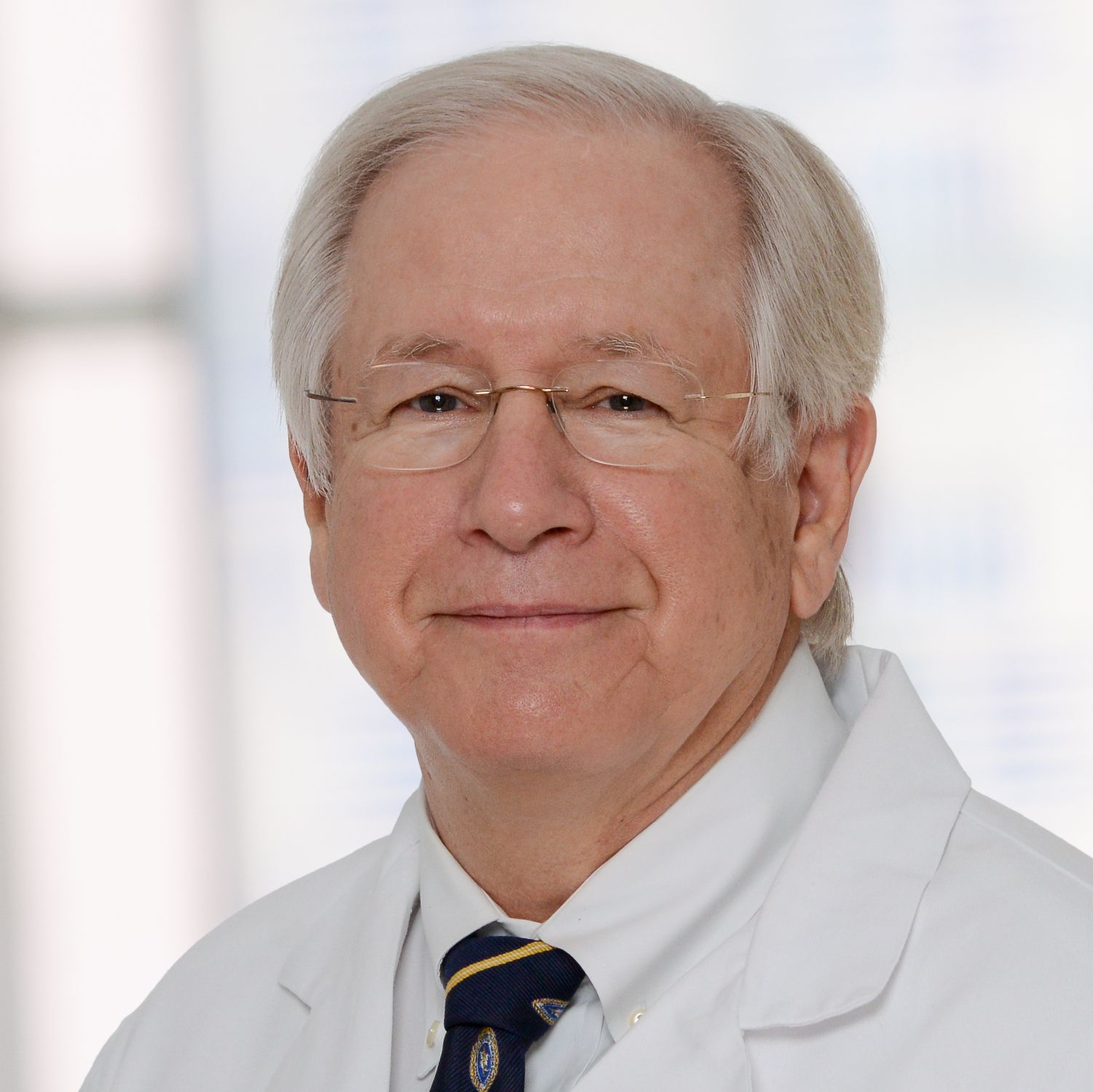 Joseph S. Coselli合作论文数Department of Surgery, Baylor College of Medicine7
 Vinod H. Thourani合作论文数Piedmont Healthcare7Andrea J. Carpenter合作论文数7James G. Ravenel合作论文数Department of Radiology, Medical University of South Carolina7
 John Edmund Mayer Jr.合作论文数Center for Bioethics, Harvard Medical School;Boston Children's Hospital7Daniel Miller合作论文数6
 Frederick Grover合作论文数Department of Surgery, School of Medicine, University of Colorado6Graham Amanda合作论文数6
 Mark J. Krasna合作论文数Jersey Shore University Medical Center6Ross M. Ungerleider合作论文数6
 Mohamad A. Eloubeidi合作论文数University of Alabama at Birmingham6
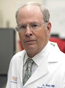 Irving L. Kron合作论文数Department of Surgery, University of Virginia;School of Medicine, University of Virginia6
 Carol A Sherman合作论文数6Harold C. Urschel合作论文数6Kevin Accola合作论文数5L Hoover合作论文数5N Hadzijahic合作论文数5
 Jeffrey P. Gold合作论文数University of Nebraska Medical Center5Joe B. Putnam合作论文数5A. Van Velse合作论文数5Neal D. Kon合作论文数5




