To search the prognostic significance of the neuronatin (NNAT) protein diagnosed with lung adenocarcinoma in the occurence of brain metastasis. Sixty-six of 200 patients diagnosed with lung adenocarcinoma who received radiotherapy (RT) between 2014 and 2017 were included in the study. A total of 66 patients, 33 (50%) with brain pathology and 33 (50%) with lung pathology, were analyzed retrospectively. Furthermore NNAT-immune-reactivity score (IRS) and nuclear positivity were analyzed in terms of pathological factors. In 33 cases where primary tumor pathology was studied, a statistically borderline significant effect was found for the N stage and a statistically significant effect on overall survival (OS) for nuclear positivity. While OS was 7.6 months in nucleus positively stained (PNS) cases, it was 24.4 months in negatively stained (NNS) cases. While the survival of NNAT-IRS positive cases without cranial metastasis was 20.7 months, the survival of the negative group without cranial metastasis was 61.2 months. The survival of cases with PNS without cranial metastasis was 7.1 months, while the survival of cases with NNS results without cranial metastasis was 52.1 months. In cases where intracranial pathologies, the number of intracranial metastases and the presence of extracranial metastases were found to be statistically significant independent factors (p < 0.001, p < 0.001). With NNAT study in initial pathological tissues of patients diagnosed with lung adenocarcinoma, especially nuclear positivity and NNAT-IRS positivity, a strong prediction can be made about the occurrence of brain metastasis.
The place of radiotherapy (RT) for the Central Nervous System (CNS) tumors in elderly patients depends on factors such as tumor type, size, location, and the patient's performance status. The efficacy and tolerability of RT and the dose-fractionation scheme chosen may differ in younger patients. Side effects of RT in elderly patients may have a greater impact, especially on neurological and cognitive functions. It has been shown that alternative treatment approaches, such as hypofractionated RT and temozolomide may offer survival benefits similar to standard treatments for glial tumors in the geriatric population. RT may be considered when surgery is not suitable or in grade II-III histologies in the treatment of meningiomas. Stereotactic radiosurgery (SRS) is generally the method of choice and is well tolerated in elderly patients with low toxicity and high local control rates. Brain metastases are also often seen in elderly and frail patients, and SRS is an effective treatment option in this group of patients. Consequently, it is important to take an individualized approach to the use of RT for CNS tumors in elderly patients.
AIM:To evaluate survival and prognostic factors associated with survival among patients who underwent reirradiation for recurrent/ progressive primary brain tumors. MATERIAL AND METHODS:A multicenter, retrospective study (7 centers, N=236) was conducted by the Neuro-oncology Group of the Turkish Radiation Oncology Association. RESULTS:Median overall survival (OS) was 11 months and 1- and 2-year survival rates were 48% and 22%, respectively. Survival was negatively correlated with cumulative biologically effective dose (BED10) (r=-0.158, p=0.016) and cumulative equivalent dose in 2-Gy fractions (EQD2) (r=-0.158, p=0.016). In univariate analysis, survival was associated with performance status (p < 0.001), histopathology at diagnosis and recurrence (p < 0.001), radiotherapy (RT) method used for recurrence (p=0.025), tumor volume at recurrence (p=0.014), cumulative EQD2 ( < 110 vs. ≥110 Gy, p=0.038), and cumulative BED10 ( < 130 vs. ≥130 Gy, p=0.022). In multivariate analysis, tumor volume at recurrence (HR=1.68, 95% CI=1.06-2.64, p=0.025), Karnofsky Performance Status score (HR=5.7, 95% CI=3.26-9.98, p < 0.001), and histopathology at recurrence (glioblastoma vs. high-grade glioma: HR=0.48, 95% CI=0.26-0.88, p=0.019; glioblastoma vs. low-grade glial tumor: HR=0.16, 95% CI=0.08-0.34, p < 0.001) were found to be independent prognostic factors. Radionecrosis was detected in 25% (n=58) of the patients. Re-resection was associated with a higher rate of radionecrosis (37.7% vs. 18%, p=0.002). CONCLUSION:The prognostic factors most strongly associated with survival in glioma patients undergoing reirradiation were Karnofsky Performance Status score below 70, glioblastoma histopathology, and tumor volume greater than 4.5 cm3. In addition, survival time was negatively correlated with cumulative EQD2 and BED10. The rate of radionecrosis was higher in patients who underwent re-resection compared those who did not.
Amaç: Bu çalışmada kemoradyoterapi ve cerrahi ile birlikte tedavi edilen rektum kanserinde tanı anındaki hemoglobin (Hb) değerinin sağkalım üzerine etkisi olup olmadığı araştırıldı. Gereç ve Yöntemler: Rektum kanseri nedeniyle neoadjuvan/adjuvan radyoterapi (RT) alan 98 hasta çalışmaya dahil edildi. RT neoadjuvan ya da adjuvan pelvik lenf nodları ve primer tümör alanına 45-54 Gy uygulandı. RT ile eşzamanlı 5-FUFA veya kapesitabin kullanıldı. Sağkalımı belirleyen faktörleri değerlendirmek üzere tek değişkenli ve çok değişkenli Cox regresyon analizi yapıldı. Sağkalım açısından Hb düzeyinde sınır değer olup olmadığını bulmak için ROC analizi yapıldı. Bulgular: Medyan genel sağkalım 61 ay (%95 CI 31-90), 1 yıllık sağkalım %82, 5 yıllık sağkalım %50 olarak bulundu. Hb sınır değeri %75 duyarlılık ve %50 özgüllükle 13,45 g/dL olarak belirlendi. Hb düşük grupta genel sağkalım 31±8 (%95 CI:14-48) ay yüksek grupta 81±17 (%95 CI:46-116) aydı (p=0.032). T4 tümörlerde ve neoadjuvan grupta Hb düşüklüğünün mortalite üzerine etkisi daha belirgindi. Tek değişkenli Cox regresyon analizinde T evre, N evre ve Hb düzeyinin 13,75 g/dl’nin altında veya üzerinde olması sağkalım üzerine etkili faktörler olarak bulundu. Çok değişkenli Cox regresyon analizinde ise T evre, N evre ve Hb düzeyi sağkalım üzerinde etkili faktörlerdi Sonuç: Hb düzeyinin 13.45 g/dl'nin altında olması lokal ileri rektum kanserinde, özellikle T4 tümörlü veya neoadjuvan kemoradyoterapi gören hastalarda kötü bir prognostik faktör olduğu görüldü.
ABSTRACT Background: Muscle loss and sarcopenia play a significant role in head and neck cancer. In this study, the value of C3 cross-sectional skeletal muscle index was investigated to evaluate sarcopenia. Methods: Seventy-four patients were included in this retrospective study. Skeletal muscle index (SMI) was calculated using the paracervical muscles at the level of the third cervical vertebra. Survival rates and toxicities were compared. Results: The 3-year overall survival rates were 33.3% in patients with low SMI (≤44.79) and 63.9% in patients with high SMI (>44.79) ( P < 0.01). The 3-year progression-free survival rates were 25.9% in patients with low SMI and 63.2% in patients with high SMI ( P < 0.01). Multivariate analyses found that advanced age (>65) was associated with a 2.9-fold increased risk of death and low SMI was associated with a 3.9-fold increased risk of death. Conclusion: Low SMI is associated with prolonged treatment time, increased toxicity, and decreased survival.
Objective: The current study aims to evaluate the clinical presentation, treatment, and follow-up of children with Wilms Tumor (WT) who had been admitted to Erciyes University, Faculty of Medicine Department of Pediatric Hematology and Oncology hospital, a tertiary center in the central Anatolia region of Turkiye. The study assesses the survival data and features that have had an impact on survival. Materials and Methods: The current study has been planned as a retrospective observational evaluation of patients admitted to the Pediatric Hematology and Oncology Center between 1991-2021. Results: The study retrospectively evaluated a total of 48 patients in terms of demographic characteristics, presentation findings, tumor stages, histopathologies, and survival rates. Patients with an unfavorable histology had a 66.7% chance of both event-free survival (EFS) and overall survival (OS), which is lower than the respective 85.2% and 92.1% odds of EFS and OS for the favorable histology group. However, this is not statistically significant (p = 0.20 for EFS and p = 0.05 for OS). Regarding the impact of stage on survival rates, the EFS and OS for patients with the low-stage disease were 88% and 95.7%, respectively. These rates were significantly superior to those at an advanced stage of the disease, whose EFS and OS were 63.1% and 60.9%, respectively (p = 0.042 for EFS, p = 0.005 for OS). Conclusion: Wilms tumor at an advanced stage and with an unfavorable histology are the major factors resulting in poor survival rates.
BACKGROUND:We aimed to investigate effect of radiotherapy (RT) applications with different dose rates on cytogenetic damages, which focused on micronucleus (MN) formation, and evaluate how this damage varies by cisplatin in rats receiving head-neck RT. MATERIAL AND METHODS:Thirty-six Sprague Dawley rats were divided into five groups. The first and second groups were irradiated at a dose rate of 300 monitor unit/minute (MU/min) and 600 MU/min, respectively. The third group was irradiated at a dose rate of 300 MU/min and given cisplatin. The fourth group was irradiated at a dose rate of 600 MU/min and given cisplatin. The fifth group received neither irradiation nor cisplatin (control group). One thousand polychromatic erythrocytes were scored, and MN frequency in polychromatic erythrocytes was determined for each rat. RESULTS:There was a significant difference among five groups in terms of the number of MN (p: 0.001). The number of MN was significantly higher in the 600 MU/min + cisplatin group (fourth group) compared to the control group [9.5 (1.0-23.0) vs. 1.5 (1.0-2.0), respectively]. It was also significantly higher in 600 MU/min + cisplatin group (fourth group) compared to 300 MU/min group (first group) [9.5 (1.0-23.0) vs. 2.0 (1.0-3.0), respectively]. On the other hand, there was no significant difference among other groups. CONCLUSIONS:Our findings suggest that RT given at a higher dose rate causes more cytogenetic damage, and this damage is increased by concurrent administration of cisplatin.
Objective The aim of this study is to analyze the frequency of lung injury and the sensitivity of the diethylenetriamine penta-acetic acid (DTPA) clearance test in detecting lung injury in patients undergoing radiotherapy (RT) to the thorax.Material and method Twenty individuals scheduled for RT for lung cancer were included as the patient group. The healthy control group consisted of 20 age and gender-matched individuals who were nonsmokers with no history of comorbidities. We conducted follow-up with patients at 0-1-6 months, performing carbon monoxide diffusion test (DLCO), DTPA clearance test (excluding the first month), and high-resolution computed tomography of the thorax. The control group was followed up with DLCO between the baseline and 6th months.Results Ninety percent of the patient group was male, and the median age was 62 years. Seventy percent of the patients had squamous cell carcinoma and adenocarcinoma. Pneumonitis was detected in the patient group in the first month (100%) and fibrosis in the sixth month (%100) Both at the beginning and in the sixth month, the DLCO values of patients who received RT were lower than those of the control group (P = 0.001 and P < 0.001, respectively). While DTPA clearance was similar between irradiated and non-radiated lungs at the beginning, there was a substantial decrease in the irradiated lung in the sixth month(P = 0.001). There was no significant correlation between malignancy type, RT dose, and tumor size(P > 0.05).Conclusion The DTPA clearance test could be an alternative method for demonstrating radiation injury in patients receiving RT.
Amaç: Bu çalışmada da eşzamanlı kemoradyoterapi ile tedavi edilen evre III küçük hücreli dışı akciğer kanseri (KHDAK)’li hastalarda NQO1’in prediktif ve prognostik öneminin tespiti amaçlandı. Gereç ve Yöntemler: Bu çalışmaya eşzamanlı cisplatin+dosetaksel kemoradyoterapisi ile tedavi edilen evre III 64 KHDAK’li olgu dahil edildi. Hastaların patoloji preparatlarında NQO1 immünohistokimyasal (İHK) boyama yapılarak negatif ve pozitiflik açısından karşılaştırıldı. Bulgular: Medyan yaş 63 (sınırlar, 35-83) olup olguların %86 (n=55)’sı erkek idi. Olguların %27 (n=17)’si evre IIIA, %56 (n=36)’sı evre IIIB ve %17 (n=11)’si evre IIIC idi. Olgular histopatolojik olarak %47 (n=30) adenokarsinom, %47 (n=47)’si epidermoid karsinom ve %6 (n=4)’sı alt tipi belirlenemeyen KHDAK şeklinde sınıflandı. Medyan takip süresi 20 ay (sınırlar, 3-7 ay) bulundu. NQO1’in İHK boyaması sonucunda olguların 7 (%11)’si negatif, 11 (%17)’i (+) pozitif, 14 (%22)’ü (++) pozitif ve 32 (%50)’si (+++) pozitif olarak bulundu. Olguların NQO1’e göre medyan genel sağkalımı; (-)’lerde 21 ay, (+)’lerde 19 ay, (++)’lerde 16 ay ve (+++)’lerde 19 ay bulundu. Pozitif NQO1 olan olgularda medyan genel sağkalım sayısal olarak negatif olan olgulardan daha düşük olmasına rağmen istatistiksel olarak anlamlı bulunmadı (p=0.801). Sonuç: Eşzamanlı kemoradyoterapi ile tedavi edilen evre III KHDAK’li hastalarda NQO1 prognostik faktör olabilir ancak çok hasta sayılı çalışmalara ihtiyaç vardır.
Objectives:The treatment of locally advanced laryngeal cancer (LALC) is very challenging.In the last few decades there has been a shift from total laryngectomy towards organ-sparing approaches.The aim of the current study is to compare oncological outcomes between surgery (total laryngectomy) followed by radiotherapy and larynx preservation with chemoradiotherapy (CRT).Materials and Methods: 114 patients with stage III-IV laryngeal cancer were included in the study, between 2009 and 2018.Thirty-six patients (31.6%) were performed total laryngectomy followed by radiotherapy and 78 (68.4%) underwent the larynx preservation approach.Survival differences between the groups were examined with the Kaplan-Meier test and cox-regression tests for factors affecting survival.Results: 5-year overall survival (OS) was found 66.3 months and 74.1 months, in the larynx preservation and the surgical groups, respectively (p=0.29).There was no statistically difference between groups for OS in the patients with T3/N0-N1 (p=0.76), but surgical groups had longer OS in the patients with T3/N2-N3 (p=0.04).There was no statistically difference between groups for OS in the patients with T4/N0-N1 (p=0.47),however CRT groups had longer OS in the patients with T4/N2-N3 (p=0.02).The N2-N3 was the factor associated with poor progression-free survival and distant metastasis free survival in multivariate analysis (p<0.01).Age (≥65) was associated with a 2.1-fold increased risk of death (p=0.01).The trans-glottis tumors were associated with a 3.6-fold increased risk of tracheostomy (p<0.01). Conclusion:The N0-N1 and N2-N3 should also be considered as well as advanced T-category for the treatment of LALC.
Background Access to cancer care is a problem that continues to plague refugees displaced from their home countries. The turbulent political crisis in Syria, which has led to millions of refugees seeking asylum in Turkey, merits further attention. We aimed to study the rate of utilization of radiation therapy among Syrian refugees with cancer living in Turkey in an attempt to identify the contributing factors predictive of non-compliance with prescribed RT. Methods In this retrospective review of 14 institutional databases, Syrian refugee patients in Turkey with a cancer diagnosis from January 2015 to December 2019 who were treated with RT were identified. The demographic data, treatment compliance rates, and toxicity outcomes in these patients were surveyed. Variable predictors of noncompliance such as age, sex, diagnosis, treatment length, and toxicity were studied. The association between these variables and patient noncompliance was determined. Results We identified 10,537 patients who were diagnosed with cancer during the study period, of whom 1010 (9.6%) patients were treated with RT. Breast cancer (30%) and lung cancer (14%) were the most common diagnoses with up to 68% of patients diagnosed at an advanced stage (Stage III, IV). 20% of the patients were deemed noncompliant. Treatment with concurrent chemoradiotherapy (OR 1.61, 95% CI 1.06–2.46, p = 0.023) and living in a refugee camp (OR 3.62, 95% CI 2.43–5.19, p < 0.001) were associated with noncompliance. Age, sex and treatment length were not significantly associated with noncompliance. Conclusions Noncompliance with radiotherapy among Syrian refugees in Turkey remains an area of concern with a multitude of factors contributing to these alarming numbers. Further studies to better ascertain the finer nuances of this intricately complex problem and a global combination of efforts can pave the way to providing a solution.
OBJECTIVE In this study, the effect of endorectal balloon (ERB) on risky organ doses was investigated in patients with low-medium risk prostate cancer treated with Stereotactic body radiotherapy (SBRT). METHODS In the 17 patients, CT simulation and treatment was performed with an empty rectum and no ERB was used (non-ERB group). ERB filled with 100-150 cc air into 15 patients was placed in the rectum and identified with the ERB group. SBRT was performed to be delivered with volumetric modulated arc, with 10 FFF photon beams, using a linear accelerator with a high definition MLC. Dosimetric parameters for the rectum, bladder and penilbulb were compared between the two groups as a retrospective. Independent samples t-tests were performed to examine the differences between the two groups. RESULTS The mean dose given to the rectum was lower in the ERB group than in the non-ERB group, and a statistically significant dose reduction of 149.4 cGy per fraction was found in the ERB group (p<0.01). Similarly, the mean dose given to the bladder was lower in the ERB group than in the non-ERB group, and a statistically significant dose reduction of 178.8 cGy per fraction was found in the ERB group (p<0.01). The mean dose of penilbulb was 373 cGy in the ERB group, while the mean dose of penilbulb was 1314 cGy in the non-ERB group. There was statistically significant differences between groups (p<0.01). CONCLUSION The use of high-volume ERB reduces rectum, bladder and penilbulb doses in prostate cancer patients treated with linac-based SBRT.
Introduction: Three-weekly cisplatin dose is accepted for standard treatment for concurrent chemo-radiotherapy in nasopharyngeal carcinoma. However, different chemotherapy schedules are presented in the literature. Objective: We intend to compare toxicity and outcomes of high dose 3-weekly cisplatin versus low dose weekly-cisplatin and cumulative dose of cisplatin in the patients with nasopharyngeal carcinoma. Methods: 98 patients were included in the study, between 2010 and 2018. Cumulative doses of cisplatin (>= 200 mg/m(2) and <200 mg/m(2)) and different chemotherapy schedules (weekly and 3-weekly) were compared in terms of toxicity and survival. Besides prognostic factors including age, gender, T category, N category and radiotherapy technique were evaluated in uni-multivariate analysis. Results: Median follow-up time 41.5 months (range: 2-93 months). Five year overall survival, local relapse-free survival, regional recurrence-free survival and distant metastasis-free survival rates were; 68.9% vs. 90.3% (p=0.11); 66.2% vs. 81.6% (p = 0.15); 87.3% vs. 95.7% (p= 0.18); 80.1% vs. 76.1% (p = 0.74) for the group treated weekly and 3 weekly, respectively. There was no statistically significant difference between groups. Five year overall survival, local relapse-free survival, regional recurrence-free survival and distant metastasis-free survival rates were; 78.2% vs. 49.2% (p= 0.003); 75.8% vs. 47.9% (p= 0.055); 91% vs. 87.1% (p= 0.46); 80% vs. 72.2% (p= 0.46) for the group treated >= 200 mg/m(2) and <200 mg/m 2 cumulative dose cisplatin. There was statistically significant difference between groups for overall survival and there was close to being statistically significant difference between groups for local relapse-free survival. Age, gender, T category, N category, chemotherapy schedules were not associated with prognosis in the univariety analysis. Radiotherapy technique and cumulative dose of cisplatin was associated with prognosis in uni-variate analysis (HR= 0.21; 95% CI: 0.071-0.628; p= 0.005 and HR =0.29; 95% CI: 0.125-0.686; p= 0.003, respectively). Only cumulative dose of cisplatin was found as an independent prognostic factor in multivariate analysis (HR= 0.36; 95% CI: 0.146-0.912; p = 0.03). When toxicities were evaluated, such as hematological toxicity, dermatitis, mucositis, nausea and vomiting, there were no statistically significant differences between cumulative dose of cisplatin groups (<200 mg/m(2) and >= 200 mg/m(2)) and chemotherapy schedules (3-weekly and weekly). But malnutrition was statistically significant higher in patients treated with 3-weekly cisplatin compared with patients treated with weekly cisplatin (p = 0.001). Conclusion: A cisplatin dose with >= 200 mg/m(2) is an independent prognostic factor for overall survival. Chemotherapy schedules weekly and 3-weekly have similar outcomes and adverse effects. If patients achieve >= 200 mg/m(2) dose of cumulative cisplatin, weekly chemotherapy schedules may be used safely and effectively in nasopharyngeal carcinoma patients. (C) 2019 Associacao Brasileira de Otorrinolaringologia e Cirurgia Cervico-Facial. Published by Elsevier Editora Ltda.
The aim of this study was to investigate the protective effects of erdosteine (ERD) against whole-body irradiation-induced lung injury in rats by assessing biochemical parameters. Thirty albino rats were divided into three equal groups, including control, radiation (RT) alone, and RT + ERD. This study began the day before radiation treatment and continued for four days. Erdosteine was administered orally via a gastric tube at a dose of 50 mg/kg/day, one hour prior to radiation and every day at the same time. Radiation was applied with a total body irradiation dose of 800 cGy in one fraction. Catalase (CAT) and superoxide dismutase (SOD) activities and malondialdehyde (MDA) levels in lung tissue of rats were evaluated three days after radiation treatment. CAT and SOD activities were found to be significantly lower in the RT group than control (p < 0.001 and p < 0.001). They were higher in the RT + ERD group than both RT and control groups (p < 0.001, p < 0.001 and p < 0,001, p < 0,061). MDA levels were significantly higher in the RT group compared to control (p < 0.001), whereas MDA levels decreased in the RT + ERD group compared to RT group (p < 0.001). Erdosteine may prevent radiotherapy-induced lung injury by increasing activities of CAT and SOD and by decreasing levels of MDA.
Aim: The aim of this study was to examine the effect of the prognostic nutritional index (PNI) value on survival in patients with rectum cancer who received postoperative chemoradiotherapy. Material and Method: The study included 65 patients who received adjuvant chemoradiotherapy. Radiotherapy of 5040 cGy and simultaneous 5-FUFA chemotherapy was given to the patients. The patients were divided into two groups as PNI <= 46 and PNI >46 according to the ROC analysis. The differences in survival between the groups were calculated using the log rank test. The univariate and multivariate hazard ratios were calculated using the Cox proportional hazard model. Results: The patients included 22 (33.8%) females and 43 (66.2%) males. The low PNI group comprised 28 (43.1%) patients and the high PNI group, 37 (56.9%). According to the Kaplan-Meier analysis, mean survival was 59 months (95% CI; range, 44.95-73.08 months) in the low PNI group and 80 months (95% CI; range 66.53-94.82 months) in the high PNI group. The 5-year survival rate was 49% in the low PNI group and 65% in the high PNI group. In the univariate analysis, T stage, N stage, tumor diameter, and PNI had an effect on overall survival (p<0.05). In the multivariate Cox regression analysis, T stage (p=0.014), tumor diameter (p=0.023), and PNI (p=0.045) were found to be prognostic factors affecting overall survival, independent of the other variables. Discussion: The results of the study showed that the PNI value before radiotherapy is a poor prognostic factor for rectum cancer.
BACKGROUND: The aim of this study is to search the prognostic value of SPARC expression in rectum cancer cases receiving postoperative radiotherapy. METHODS: Forty three rectal cancer patients are recruited to this retrospective study. All patients received postoperative radiotherapy which the medi an dose was 5040 cGy and concomitant chemotherapy. Samples taken from their paraffin blocks were examined with immunohistochemical procedures. RESULTS: When the association between SPARC expression and the clinicopathological feature was examined, there was a significant association between age and expression levels. Overall survival of patients with low expression was found to be 67 months whereas the overall survival of the patients with high expression was 32 months and the difference was statistically significant. Time to local recurrence of patients with low expression was found to be 74 months whereas time to local recurrence of the patients with high expression was 31 months. Progression free survival of the patients with low expression and high expression were 67 months and 32 months, respectively. In multivariate Cox regression analyses, high expression of SPARC was found to be associated with a statistically significant shorter overall survival and progression free survival. CONCLUSIONS: High expression of SPARC is related to worse prognosis in rectal cancer patients.
 Unal Dilek合作论文数23
 Okan Orhan合作论文数22
 Bunyamin Kaplan合作论文数19Serdar Soyuer合作论文数15
 Neslihan Kurtul合作论文数Medical Faculty, Sutcu Imam University14
 Mete Gundog合作论文数11Metin Ozkan合作论文数Department of Pulmonary Medicine, Gulhane Medical Academy, Turkey11Tasdemir Arzu合作论文数Department of Pathology, Kayseri Education and Research Hospital9Oguz Arzu合作论文数8Oguz Galip Yildiz合作论文数8
 Ali Unal合作论文数Erciyes University;Faculty of Medicine;Dedeman Hospital;Department of Hematology-Oncology;Faculty of Medicine, Erciyes University8
 Serdar Sivgin合作论文数7
 Leylagul Kaynar合作论文数Department of Hematology and Apheresis Unit, Erciyes Medical School7
 Halit Karaca合作论文数Department of Medical Oncology, Erciyes University Medical School7
 Mehmet Eryılmaz合作论文数Department of Emergency Medicine, Gülhane Military School of Medicine7
 Ercan Kurar合作论文数6
 Bulent Eser合作论文数6Kadir Yaray合作论文数6Mevlude Inanc合作论文数Medical Oncology Department, Erciyes University5
 Emre Avci合作论文数Faculty of Science and Arts, Hitit University5Esin Kiraz合作论文数4
 Ahmet Ozturk合作论文数4
 Gökmen Zararsız合作论文数Department of Biostatistics, School of Medicine, Erciyes Üniversitesi4
 Keklik Muzaffer合作论文数Faculty of Medicine, Erciyes University4
 Mustafa Cetin合作论文数SANKO University4
 Huseyin Vural合作论文数Faculty of Medicine, Suleyman Demirel University4B Kaplan合作论文数4
 Sercan Aksoy合作论文数3Erdem Arzu Tasdemir合作论文数3Eda Kaya Pepele合作论文数3Esra Ermiş Turak合作论文数3Hatice Basaran合作论文数3Seher Civcik合作论文数3Fatih Kurnaz合作论文数3
 Ali Firat Inal合作论文数Medical Oncology, Mersin City Hospital3
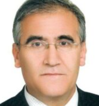 Necati Alkis合作论文数Ankara Oncology Teaching and Research Hospital3Isikdogan Abdurrahman合作论文数3Yetisyigit Tarkan合作论文数3Songül Barlaz Us合作论文数3
 Dodurga Yavuz合作论文数Dept Med Biol, Pamukkale Univ3
 Hatice Karaman合作论文数3
 Arslan Kemal合作论文数Department of General Surgery, Konya Teaching Hospital3Seçme Mücahit合作论文数3Goksu Sema Sezgin合作论文数3
 Mehmet Kucukoner合作论文数Division of Medical Oncology, Dicle University;George Mason University3
 Erkan Arpaci合作论文数3
 Cigdem Pala合作论文数3
 Ummuhan Abdulrezzak合作论文数Department of Nuclear Medicine, Erciyes University School of Medicine3Suleyman Buyukberber合作论文数School of Medicine, Gazi University3




