Disorders of sleep and wakefulness occur in the majority of individuals who have experienced traumatic brain injury (TBI), with increased sleep need and excessive daytime sleepiness often reported. Behavioral and pharmacological therapies have limited efficacy, in part, because the etiology of post-TBI sleep disturbances is not well understood. Severity of injuries resulting from head trauma in humans is highly variable, and as a consequence so are their sequelae. Here, we use a controlled laboratory model to investigate the effects of TBI on sleep-wake behavior and on candidate neurotransmitter systems as potential mediators. We focus on hypocretin and melanin-concentrating hormone (MCH), hypothalamic neuropeptides important for regulating sleep and wakefulness, and two potential downstream effectors of hypocretin actions, histamine and acetylcholine. Adult male C57BL/6 mice (n=6–10/group) were implanted with EEG recording electrodes and baseline recordings were obtained. After baseline recordings, controlled cortical impact was used to induce mild or moderate TBI. EEG recordings were obtained from the same animals at 7 and 15 days post-surgery. Separate groups of animals (n=6–8/group) were used to determine effects of TBI on the numbers of hypocretin and MCH-producing neurons in the hypothalamus, histaminergic neurons in the tuberomammillary nucleus, and cholinergic neurons in the basal forebrain. At 15 days post-TBI, wakefulness was decreased and NREM sleep was increased during the dark period in moderately injured animals. There were no differences between groups in REM sleep time, nor were there differences between groups in sleep during the light period. TBI effects on hypocretin and cholinergic neurons were such that more severe injury resulted in fewer cells. Numbers of MCH neurons and histaminergic neurons were not altered under the conditions of this study. Thus, we conclude that moderate TBI in mice reduces wakefulness and increases NREM sleep during the dark period, effects that may be mediated by hypocretin-producing neurons and/or downstream cholinergic effectors in the basal forebrain.
Traumatic brain injury (TBI) affects millions of people worldwide; however, the immediate impact of TBI and the secondary injury mechanisms are still not fully understood. TBI can cause devastating neuromotor deficits in both acute and chronic stages. Time course studies utilizing animal models of focal TBI have provided essential insight into TBI neuropathology. Here, we describe a surgical technique for creating a mouse model of focal, mild TBI (Dixon et al., 1991; Smith et al., 1995; Bolkvadze and Pitkanen, 2012). Furthermore, we provide protocols for validating TBI models using behavioral tests that examine post-traumatic neuromotor deficits resulting from TBI neuropathology (Fujimoto et al., 2004; Febinger et al., 2015; Smith et al., 1995; Bolkvadze and Pitkanen, 2012).
Disorders of sleep and wakefulness have been reported in up to 73% individuals who have experienced traumatic brain injury (TBI). Specifically, increased sleep need and excessive daytime sleepiness are often reported post-TBI. The etiology of post-TBI sleep disturbances is not well understood, and current pharmacological therapies have limited efficacy. Here, we investigate the effects of TBI on sleep-wake behavior and on hypocretin (Hcrt) and melanin-concentrating hormone (MCH), two neuropeptides important for regulating sleep and wakefulness. Adult male C57BL/6J mice (n = 6–10/group) were implanted with EEG recording electrodes and baseline recordings were obtained. TBI was then induced using controlled cortical impact (CCI). EEG recordings were obtained from the same animals at 7 and 15 days post-surgery. Another group of animals (n = 6–8/group) underwent sham or TBI surgery and was sacrificed 7 or 15 days later. Brains from these animals were used for immunohistochemistry to determine the number of Hcrt- or MCH-producing neurons in the hypothalamus. At 15 days post-surgery, wakefulness was decreased and NREM sleep was increased during the dark period in moderately injured animals. Moderate injury resulted in significantly fewer Hcrt-producing neurons. There were no significant differences among groups in MCH-producing neurons. Because Hcrt is an important promoter of wakefulness, the decrease in number of Hcrt-producing neurons may be one mechanism underlying the observed decrease in wakefulness and increase in NREM sleep after TBI.
Background: Neuroinflammation is an important secondary mechanism that is a key mediator of the long-term consequences of neuronal injury that occur in traumatic brain injury (TBI). Microglia are highly plastic cells with dual roles in neuronal injury and recovery. Recent studies suggest that the chemokine fractalkine (CX3CL1, FKN) mediates neural/microglial interactions via its sole receptor CX3CR1. CX3CL1/CX3CR1 signaling modulates microglia activation, and depending upon the type and time of injury, either protects or exacerbates neurological diseases. Methods: In this study, mice deficient in CX3CR1 were subjected to mild controlled cortical impact injury (CCI), a model of TBI. We evaluated the effects of genetic deletion of CX3CR1 on histopathology, cell death/survival, microglia activation, and cognitive function for 30 days post-injury. Results: During the acute post-injury period (24 h–15 days), motor deficits, cell death, and neuronal cell loss were more profound in injured wild-type than in CX3CR1 mice. In contrast, during the chronic period of 30 days post-TBI, injured CX3CR1 mice exhibited greater cognitive dysfunction and increased neuronal death than wild-type mice. The protective and deleterious effects of CX3CR1 were associated with changes in microglia phenotypes; during the acute phase CX3CR1 mice showed a predominant anti-inflammatory M2 microglial response, with increased expression of Ym1, CD206, and TGFβ. In contrast, increased M1 phenotypic microglia markers, Marco, and CD68 were predominant at 30 days post-TBI. Conclusion: Collectively, these novel data demonstrate a time-dependent role for CX3CL1/CX3CR1 signaling after TBI and suggest that the acute and chronic responses to mild TBI are modulated in part by distinct microglia phenotypes.
Alzheimer's disease is characterized by amyloid plaques, neurofibrillary tangles, glial activation, and neurodegeneration. In mouse models, inflammatory activation of microglia accelerates tau pathology. The chemokine fractalkine serves as an endogenous neuronal modulator to quell microglial activation. Experiments with fractalkine receptor null mice suggest that fractalkine signaling diminishes tau pathology, but exacerbates amyloid pathology. Consistent with this outcome, we report here that soluble fractalkine overexpression using adeno-associated viral vectors significantly reduced tau pathology in the rTg4510 mouse model of tau deposition. Furthermore, this treatment reduced microglial activation and appeared to prevent neurodegeneration normally found in this model. However, in contrast to studies with fractalkine receptor null mice, parallel studies in an APP/PS1 model found no effect of increased fractalkine signaling on amyloid deposition. These data argue that agonism at fractalkine receptors might be an excellent target for therapeutic intervention in tauopathies, including those associated with amyloid deposition.
Neurogenesis occurs throughout life but significantly decreases with age. Human umbilical cord blood mononuclear cells (HUCB MNCs) have been shown to increase the proliferation of neural stem cells (NSCs) in the dentate gyrus (DG) of the hippocampus and the subgranular zone of aging rats (Bachstetter et al., BMC Neurosci 9:22, 2008 ), but it is unclear which fraction or combination of the HUCB MNCs are responsible for neurogenesis. To address this issue, we examined the ability of HUCB MNCs, CD4+, CD8+, CD3+, CD14+, and CD133+ subpopulations to increase proliferation of NSCs both in vitro and in vivo. NSCs were first grown in conditioned media generated from HUCB cultures, and survival and proliferation of NSC were determined with the fluorescein diacetate/propidium iodide and 5-bromo-2′-deoxyuridine incorporation assays, respectively. In a second study, we injected HUCB cells intravenously in young and aged Fisher 344 rats and examined proliferation in the DG at 1 week (study 2.1) and 2 weeks (study 2.2) postinjection. The effects of the HUCB MNC fractions on dendritic spine density and microglial activation were also assessed. HUCB T cells (CD3+, CD4+, and CD8+ cells) induced proliferation of NSCs ( p < 0.001) and increased cell survival. In vivo, HUCB-derived CD4+ cells increased NSC proliferation at both 1 and 2 weeks while also enhancing the density of dendritic spines at 1 week and decreasing inflammation at 2 weeks postinjection. Collectively, these data indicate that a single injection of HUCB-derived T cells induces long-lasting effects and may therefore have tremendous potential to improve aging neurogenesis.
Our view of microglia has dramatically changed in the last decade. From cells being “silent” in the healthy brain, microglia have emerged to be actively involved in several brain physiological functions including adult hippocampal neurogenesis, and cognitive and behavioral function. In light of recent discoveries revealing a role of microglia as important effectors of neuronal circuit reorganization, considerable attention has been focused on how microglia and hippocampal neurogenesis could be an interdependent phenomenon. In this review the role of microglia in the adult hippocampal neurogenesis under physiological condition is discussed.
Aging is characterized by a progressive decline in the efficiency of physiological function and by the increased susceptibility to disease and death. Currently, one of the most plausible and acceptable explanations for the mechanistic basis of aging is the “free radical theory of aging.” This theory postulates that aging and its related diseases are the consequence of free radical-induced damage to cellular macromolecules and the inability to counterbalance these changes by endogenous anti-oxidant defenses. The origin of this explanation has a foundation in the “rate of living theory” [1], according to which the lifespan of an individual depends on its rate of energy utilization (metabolic rate) and on a genetically determined amount of energy consumed during adult life. Pearl [1] proposed that the longevity of an organism is inversely correlated to its mass-specific metabolic rate: increasing an organism’s metabolic rate will decrease longevity, whereas factors that decrease the metabolic rate will increase longevity. The correlation between metabolic rate and longevity has been questioned due to the exception posed by birds, which have a high metabolic rate yet live much longer than mammals [2]. However, despite their high rate of oxygen consumption, it has been shown that birds have a low rate of free radical production in brain and in other tissues; their mitochondria produce up to 10-fold fewer reactive oxygen species (ROS) in vitro [3, 4]. This observation suggests that the mitochondrial rate of free radical production may be more important than the metabolic rate in terms of longevity. Indeed, the mitochondrial rate of free radical production seems to have a much stronger correlation with maximum longevity.Harman [5] originally proposed the “free-radical theory” of aging in the mid-1950s. He suggested that free radicals produced during aerobic respiration have deleterious effects on cell components and connective tissues, causing cumulative damage over time that ultimately results in aging and death. He initially speculated that free radicals were most likely produced through reactions involving molecular oxygen catalyzed in the cells by the oxidative enzymes and enhanced by trace metals such as iron, cobalt, and manganese. The skepticism first spread around this theory was weakened by the discovery in 1969 of the enzyme superoxide dismutase (SOD) [6]. The existence of an intracellular enzyme whose sole function is to remove superoxide anions (O
The chemokine CX3CL1/fractalkine is expressed by neurons as a transmembrane-anchored protein that can be cleaved to yield a soluble isoform. However, the roles for these two types of endogenous CX3CL1 in neurodegenerative pathophysiology remain elusive. As such, it has been difficult to delineate the function of the two isoforms of CX3CL1, as both are natively present in the brain. In this study we examined each isoform's ability to regulate neuroinflammation in a mouse model of Parkinson's disease initiated by the neurotoxin 1-methyl-4-phenyl-1,2,3,6-tetrahydropyridine (MPTP). We were able to delineate the function of both CX3CL1 isoforms by using adeno-associated virus-mediated gene therapy to selectively express synthetic variants of CX3CL1 that remain either permanently soluble or membrane bound. In the present study we injected each CX3CL1 variant or a GFP-expressing vector directly into the substantia nigra of CX3CL1(-/-) mice. Our results show that only the soluble isoform of CX3CL1 is sufficient for neuroprotection after exposure to MPTP. Specifically, we show that the soluble CX3CL1 isoform reduces impairment of motor coordination, decreases dopaminergic neuron loss, and ameliorates microglial activation and proinflammatory cytokine release resulting from MPTP exposure. Furthermore, we show that the membrane-bound isoform provides no neuroprotective capability to MPTP-induced pathologies, exhibiting similar motor coordination impairment, dopaminergic neuron loss, and inflammatory phenotypes as MPTP-treated CX3CL1(-/-) mice, which received the GFP-expressing control vector. Our results reveal that the neuroprotective capacity of CX3CL1 resides solely upon the soluble isoform in an MPTP-induced model of Parkinson's disease.
The protective/neurotoxic role of fractalkine (CX3CL1) and its receptor CX3C chemokine receptor 1 (CX3CR1) signaling in neurodegenerative disease is an intricate and highly debated research topic and it is becoming even more complicated as new studies reveal discordant results. It appears that the CX3CL1/CX3CR1 axis plays a direct role in neurodegeneration and/or neuroprotection depending on the CNS insult. However, all the above studies focused on the role of CX3CL1/CX3CR1 signaling in pathological conditions, ignoring the relevance of CX3CL1/CX3CR1 signaling under physiological conditions. No approach to date has been taken to decipher the significance of defects in CX3CL1/CX3CR1 signaling in physiological condition. In the present study we usedCX3CR1−/−,CX3CR1+/−, and wild-type mice to investigate the physiological role of CX3CR1 receptor in cognition and synaptic plasticity. Our results demonstrate for the first time that mice lacking the CX3CR1 receptor show contextual fear conditioning and Morris water maze deficits. CX3CR1 deficiency also affects motor learning. Importantly, mice lacking the receptor have a significant impairment in long-term potentiation (LTP). Infusion with IL-1β receptor antagonist significantly reversed the deficit in cognitive function and impairment in LTP. Our results reveal that under physiological conditions, disruption in CX3CL1 signaling will lead to impairment in cognitive function and synaptic plasticity via increased action of IL-1β.
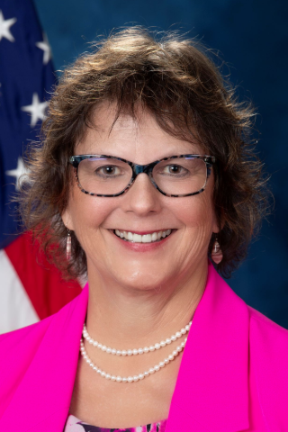 Paula Bickford合作论文数Center of Excellence for Aging & Brain Repair, University of South Florida;Department of Molecular Pharmacology and Physiology, Morsani College of Medicine, University of South Florida63
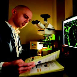 Bachstetter Adam D合作论文数Sanders Brown Ctr Aging, Univ Kentucky30
 Paul R. Sanberg合作论文数Department of Neurosurgery and Brain Repair, Morsani College of Medicine, University of South Florida;Center of Excellence for Aging, University of South Florida19
 Alison Willing合作论文数Department of Molecular Pharmacology and Physiology, Morsani College of Medicine, University of South Florida12
 Morganti Josh M合作论文数Brain and Spinal Injury Center, University of California San Francisco;Departments of Physical Therapy and Rehabilitation Science, University of California San Francisco;University of California San Francisco, University of California San Francisco11Jennifer Vila合作论文数11
 Chikhilupiriro Hudson合作论文数10Charles E. Hudson合作论文数10
 Acosta Sandra A合作论文数Center of Excellence for Aging and Brain Repair, University of South Florida College of Medicine9Mark R. Opp合作论文数9Michael J. Cole合作论文数Rutgers University8
 Cesario Borlongan合作论文数Center of Excellence for Aging & Brain Repair, Department of Neurosurgery and Brain Repair, University of South Florida Morsani College of Medicine7Cyndy Davis Sanberg合作论文数7A. Bachstetter合作论文数6
 Jun Tan合作论文数University of South Florida6
 Kevin Nash合作论文数Department of Pathology and Laboratory Medicine, Children's Hospital of Los Angeles6J. Jernberg合作论文数5Yuji Kaneko合作论文数5
 Heidi Febinger合作论文数Department of Anesthesiology and Pain Medicine, University of Washington5Andrea Schlunk合作论文数5
 Luca Imeri合作论文数Institute of Human Physiology;Centre for Experimental Sleep Research;Medical School, University of Milan;Medical School, University of Milan5C. E. Hudson合作论文数4Jennifer Jernberg合作论文数4Mg Desimoni合作论文数4
 Hannah Thomasy合作论文数4E. J. Weeber合作论文数4M. Cole合作论文数4
 Brent J. Small合作论文数School of Nursing, University of North Carolina at Chapel Hill4
 Naoki Tajiri合作论文数4Ingrid Strömberg合作论文数4Terrence Town合作论文数4
 Michael Mesches合作论文数Department of Veterans Affairs Hospital4
 Huayan Hou (厚华艳)合作论文数4David J. Eve合作论文数Department of Neurosurgery USA, University of South Florida Center of Excellence for Aging and Brain Repair4
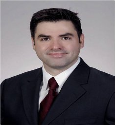 Demian Obregon合作论文数College of Medicine, University of South Florida4John D Sinden合作论文数4Mauro Mancia合作论文数4
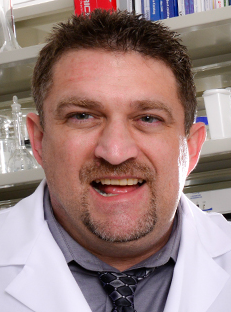 Roland Shytle合作论文数Center for Excellence in Aging and Brain Repair, Department of Neurosurgery, College of Medicine, University of South Florida;College of Medicine Psychiatry and Behavioral Neurosciences, University of South Florida4
 Brian Giunta合作论文数College of Medicine, University of South Florida4C. D. Sanberg合作论文数3f prisco合作论文数3Grimmig Bethany A合作论文数3Pabon Mibel M合作论文数3Pabon Mibel合作论文数3
 Yuyan Zhu (朱育焱)合作论文数The First Hospital, China Medical University3Keith R. Pennypacker合作论文数College of Medicine, University of South Florida3
 Svitlana Garbuzova-Davis合作论文数Center of Excellence for Aging and Brain Repair, Department of Neurosurgery and Brain Repair, Morsani College of Medicine, University of South Florida;Department of Molecular Pharmacology & Physiology, University of South Florida3
 Martina Vendrame合作论文数3




